Note: Descriptions are shown in the official language in which they were submitted.
Luderer-Smith 9-7A
~z9~
LYMPHOCY~E CO~LECTION TUBE
.
; Background of the Invention
5Considerable research has been conducted in recent
years to develop improved means for the separation and
collection of lymphocytes from human blood. An impetus
- for such research has been generated by the need for
histocompatibility determinations in patients requiring
organ transplants. A measure of lymphocyte function is
critical to adjudge the type and level of medication
necessary for immunosuppression.
One well-known method for isolating and collecting
lymphocytes from anticoagulated human blood drawn via
conventional phlebotomy techniques utilizes buoyant
density centrifugation of blood cells. A newtonian
fluid, frequently Ficoll-Paque~, a liquid density
gradient medium having a specific gravity of about
1.077 g/cc marketed by Pharmacia Fine Chemicals AB,
Uppsala, Sweden, constitutes the medium. The method
commonly involYes the four general steps:
la3 a predetermined quantity of the
Ficoll-Paque~ medium is run into the bottom of a
test tube;
25(b) a sample of whole or diluted blood is
carefully pipetted onto the medium;
(c) the test tube is placed in a centrifuge
and the blood-medium combination centrifuged at
.
:,
....,. ,: ,.
~.2~
-2-
about 400-500 G's for about 30-40 minutes to cause
the components of the blood having specific
gravities greater than the medium, viz. >1.077
g/cc, to pass through the liquid; and thereafter,
(d) the lymphocytes, which have a specific
gravity less than 1.077 g/cc, are pipetted off the
medium .
Several problems or concerns have been found to be
inherent in that technigue. For example:
10 ~13 if, during the pipetting of the blood
sample into the separation medium, lymphocytes are
inadvertently diffused below the surface of the
medium, the specific gra~ity of the medium in that
area is so reduced as to become inadequate to
separate the lymphocytes;
. (~) if, during centrifugation, lighter
phases in the blood migrate into the separation
~; medium, they cannot pass upward therethrouyh
because the buoyant force generated by 400-500 G'~
i8 insufficient;
(3) centrifugation forces in excess of about
~00-500 G' 5 cannot be employed with Ficoll-Paque~
medium as it is somewhat watex soluble and higher
centrifugation forces increase this solubility,
thereby leading to a change in its specific
gravity; and
(4) after centrifugation has been completed,
the pipetting of the.lymphocytes off the surface
of the separation medium must be conducted with
substantial care because of the newtonian
character of the Ficoll-Paque~ medium.
., , . . ~ .
- 31 29~
--3--
Numerous suggestions have been proposed ~or
improving upon that technique. Several disclosure~ of
such ~uggestions are recorded below.
U.S. Patent No. 3,852,194 describes a process for
isolating lighter phases from heavier fractions in
human blood utilizing a thixotropic, gel-like material
having a specific gravity which i~ intermediate to that
of the phases to be separated. Upon centrifuging the
gel and blood sample together, the gel exhibits
sufficient flow to form a barxi~r between the lighter
and heavLer phases. That barrier enables the phase
resting thereupon to be easily withdrawn therefxom
using conventional laboratory techniques.
The patent postulates the operability of numerous
gel-like substances; those substances complying with
three general criteria-
(l~ a specific gravity intermediate to that
of the phases to be separated;
~2~ chemical inertness to the phases of
human blood; and
~3) essentially non-flowable when at rest
(thixotropic).
U.S. Patent No. 3,920~549 is asserted to comprise
an improvement upon the disclosure of Patent No.
3,852,194. That improvement involved the use of a
svlid element, termed an ~energizer", having a specific
gravity greater than that of the gel-like substance.
This energizer, during centrifugation, impacts upon the
gel, which is normally placed in the bottom of a blood
collection tube, thereby expediting the upwaxd mo~ement
of the gel along the walls of the tube. In this manner
the energizer accelexates the i~olation of the blood
phases and permits a cleaner separation therebetween.
~,2g~0~
-4-
U.S. Patent No. 4,190,535 is specifically drawn to
a procedure for isolating lymphocytesr monocytes, and
platelets from anticoagulated blood. The process
contemplates three general steps:
S (1) a water-insoluble, thixotropic gel-like
substance having a specific gravity between about
1~065-1.077 g/cc and exhibiting chemical inertness
to blood components is deposited into a sample of
anticoagulated blood;
(2) the gel-blood combination is centrifuged
at a force of at least 1200 G's for a sufficient
length of time that the gel forms a barrier
between the heavier blood cells and the
lymphocytes, monocytes, and platelets; and then
(3) lymphocytes, monocytes, and platelets
; are removed from atop the barrier.
The patent observes that, because a non-newtonian,
water-insoluble gel-like material capable of forming a
barrier at centrifugation forces of in excess of 1200
G's is used, a faster and more complete separation was
possible than with Ficoll-Paque~ medium. The paten~
also observes that the elimination of the liquid
density gradient medium avoids the time-consuming
process of layering two liquids without mixing them.
Other work done in 1983 by Richard J. Carroll,
Albert A. Luderer, and Anthony R. Zine, Jr., and under
the title of SEPARATION OF LYMPHOCYTES AND MONOCYTES
FROM AGED BLOOD, is directed to improving the quality
of the separation of lymphocytes and monocytes from
aged samples of anticoagulated human blood by
inhibiting the shift observed in the buoyant density of
..,
.
.: ,
.
`: .
.299~0~8
--5--
granulocyte whi~e blood cells. The inventive process
involves four genexal steps:
(1~ a ~ample of anticoagulated blood is
mixed with a hypertonic fluid containing an
organic or inorganic ionic substance of relatively
low molecular weight and which i~ chemically
compatible with components of the blood;
(2) a water-insoluble thixotxopic gel-like
substance similar to that described in Patent No.
4,190,535 with a specific gravity between
1.060-1.075 g/cc is deployed into the
blood-hypertonic 1uid mixture;
(3) the gel-blood-hypertonic fluid sample i5
centrifuged at a force of at least 12~0 G's to
cause the gel to form a barrier between the
lymphocytes and monocytes and the heavier cells of
the blood; and then
~4) the lymphocytes and monocytes are
withdrawn from atop that barrier.
Whereas each of the above-discussed disclosures does
indeed modify and improve upon various aspects of the
well-known Ficoll~Paque~ medium technique, none of them
is able to equal or improve upon the performance of the
liquid medium with respect to the purity of the
separated cell population. Because purity is a
critical parameter in cell separation, the
above-discussed disclosures cannot be substituted for
the Ficoll-Paque(~) medium technique in all applicationsO
Consequently, research has continued in an effort to
formulate simpler methods of cell separation which
utilize a liquid medium. More particularly, a process
has been sought which eliminates the time-consuming
procedure necessary to layer blood samples onto the
9~L0~8
--6--
liquid density gxadient ,medium without encountering
mixing at the interface between the two liq~ids. This
layering process generally requires a~out three
minutes/tube to flow the blood sample down the inside
wall of the tube at a rate which will pexmit layering
and avoid turbulence at the interface. Inasmuch as
this procedure is conducted manually and two tubes are
conventionally prepared per sample, the setup time for
readying a group of ten tube~ may require a period of
greater than one hour. The time involved in the
centrifuging step is less critical ~ince many tubes can
be processed at the same time. Further simplification
of the setup procedure could be accomplished if the
patient's blood sample could be drawn directly into the
centrifuge tube, thereby removing the need for
transferring the sample from the collection tube to the
centrifuge tube. In many instances it is desirable, to
add a reagent to the blood sample prior to cell
separation to anticoagulate the blood, dilute the
~lood, or modify physical and/or chemical
characteristics of the blood components.
Therefoxe, a primary ob~ective of the present
invention is to provide a ~eries of devices whi h,
separately or in combination, will not only satisfy the
range of needs of research workers and diagnostic
technicians who may merely wish to eliminate the
layering problem or to minimize 5etup time, but also
will provide a single product wherein all of the
above-described benefits can be enjoyed.
9~ 8
~7--
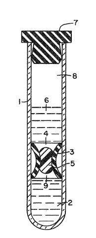
; Brief Desc~pt~on of the Drawin~s
FIGURE 1 comprises a schematic cross section ~n
: side elevation of one non evacuated tube con~iguration
of the inventive lymphocyte separation tube unit. This
tube configuration is employed where a blood sample is
transferred from a blood collection tube to the
inventive sPparation tube.
FIGURE 2 comprises a schematic cross section in
side elevation of one evacuated tube configuratio~ of
the inventive lymphocyte separation tube unit, which
permits blood to be drawn directly from the patient
into the inventive separation tube~
Summary of the Invention
In the most general terms, the present invention
comprises an assembly for centrifugally separating
lymphocytes and monocytes from the heavier phases of a
sample of whole blood or a pretreated cell fraction
thereof and physically partitioning the separated
phases. The inventive assemhly consists of four basic
e1ements:
(1) a container ~customarily a blood
collection tube or a centrifuge tube) having an
open end and a closed end;
(2) a density gradient medium initially
positioned adjacent said closed end;
13) a partition plug initially positioned
above the surface of said medium which seals said
medium therebeneath: and
.,
1.~9~0~3
-8-
,
(4) a free space initially adjacent ~aid
partition plug of sufficient volume to contain
said sample and added blood anticoagulant where
necessary.
A closure means for covering the o~en end of the
container is necessary where a sterile product is
- demanded. Careful practice dictates utilizing a
closure means during centrifugation to avoid aerosoling
of the blood which may be contaminated with pathosenic
materials. For conventional centrifuge tubes t a ~crew
top cap is normally sufficient; for evacuated
collection tube applications, a tight-fitting
elastomeric plug is generally employed to contain the
vacuum during the required storage periods.
FIGURE 1 illustrates a preferred inventive conc~pt
while providing a specific embodiment thereof.
Accordingly, to a conventional centrifuge tube 1 is
added an aliquot of a density gradient medium 2 such as
Ficoll-Paque~. A stationary partition plug 3 having an
aperture therethrough 4 is then inserted into tube 1
and moved to a position immediately above the surface
of medium 2. Plug 3 will advantageously be fashioned
from molded polypropylene having chevron-type sealing
means around its periphery or molded from an inert
elastomeric or plastic material having compression
rings around its periphrey. A hydrophobic gel 5 of a
selected density and viscosity is injected into the
bore 4 of partition plug 3, thereby sealing medium 2
beneath. A free space 9 is re~uired below the yel 5
which is of a ~olume approximately equivalent to that
of gel 5. Thereafter, an aliquot of a suitable reagent
6, e.g., a diluent, may be added above parti~ion plug
3. It will be appreciated that the use of a blood
09~
g
diluent i5 not mandatory but appears to promote better
separation in some cases. A closure 7 is applied to
the open end of tube 1 to maintain ~terility therein.
A free space 8 i5 left between the closure 7 and
reagent 6, that space having a volume somewhat greater
than that of the volume of the blood sample to be
subject to separation. To insure that gel 5 will
remain in bore 4 of partition plug 3 so as to retain
density gradient medium 2 in place during shipment and
storage of tube 1, and where reagent 6 is utilized, to
prevent mixing of the two liquids, a non-newtonian
(thixotropic) gel is preferred. Nevertheless, where
tube 1 is to be used relatively promptly, a newtonian
gel may be used.
FIGURE 2 illustrates the preferred embodiment of
the present invention where the final product is an
evacuated blood collection tube. In this embodiment to
a conventional collection tube 10 is added an aliquot
of a liquid density gradient medium 11. Partition plug
12 consists solely of a mass of a hydrophobic
(thixotropic) yel which is extruded onto the surface of
liquid medium 11, sealing the medium therebeneath. In
like manner to the embodiment described in FIGURE 1, a
thixotropic gel is preferred since it insures long term
sealing of medium 11 therebeneath during shipping and
storage. However, a newtonian gel could self-evidently
be inserted into tube 10 on site, and would perform
quite satisfactorily. The gel is sufficiently tacky to
adhere to the walls of tube 10. The gel has a specific
gravity somewhat greater than that of liquid medium 11,
thereby allowing the yel to move down tube 1 during
centrifugation and so displace liquid medium 11. An
aliquot of a suitable reagent 13 containing a blood
~1
9~
10- ,
anticoagulant, e.g., lith.ium heparin, ~odium heparin,
or EDTA, is added above plug 12. Free space 14 between
reagent 13 and clo~ure means 15 provides the vacuum and
is of sufficient volume to permit the blood sample to
5 be drawn directly from the pa~ent into tube 10.
Closure means 15 will conveniently be a stopper
fabricated from a special butyl rubber.
As has been observed in this inventive embodiment~
the partition plug moves to the bottom of the tube
during centrifugation and, in so doing, displaces the
liquid gradient medium. Thi~ action makes possible the
separation of cell suspensions which have been
previously enriched through prior separation steps. An
example of that situation is the separation of "Buffy
Coat~n. In that protocol whole blood is centrifuged or
merely allowed to stand and settle out. The white cell
population forms a "Buffy Coat",~ i.e., a buff-color~d
layer, on top of the mass of red cells. This layer of
white cells can be removed, diluted, centrifuged, and
partitioned over a gradient density medium to separate
the mononuclear cells~ The procedure is conducted as a
means for reducing the number of separation tubes
required to process an equivalent quantity of cells.
The practice permits the ~eparation of a high
concentratLon of leukocytes utilizing a small amount of
liquid medium which is very expensive. As can be seen,
a mass of red cells to displace the liquid medium is
not necessary in this embodiment of the inventive
method, contrary to the first above-described
embodiment where the partition plug remains stationary
in the ~ube.
Isolation of lymphocytes from blood samples
comprehends three general steps:
2~ 38
~a) a blood sample is ali~uoted or drawn into a
tube employing conventional techniques;
(b) the tube is rocked or otherwise agitated to
mix the blood with any required reagent; and
S (c~ the tube is centrifuged in accoxdance wi~h
~tandard techniques for separating mononuclear cells
~lymphocytes and monocytes) from the heavier phases of
blood utilizing a density gradient liquid procedure.
In the first inventive embodiment the gel ~eal is
moved by the centrifugal force generated~ as the tube
begins to spin, into the space alloted below the
partition plug. Inasmuch as the liquid medium is
incompressible, the plug bore cannot be unsealed if
space is not provided into which the gel can move. In
FIGURE 1, this space is an artifact of the small size
of the bore.
The movement of the gel opens the bore and allows
the red cells to pass downward into the liquid medium
and, being more dense than the medium, displace the
medium upward through the bore to above the partition
plug. Since the volume of cells in whole blood is
approximately 40%, about 8 ml of whole blood will
generally displace a typical aliquot of 3 ml o~ density
gradient medium. Where a diluent is employed, care
must be exercised in practicing this inventive
embodiment to have a sufficient mass of red cells to
displace the liquid medium to at least its minimum
operable heighth above the partition plug.
It will be appreciated that the crux of the
mechanism operating in the first inventive embodiment
resides in the aperture of the partition plug which
confines and controls the interaction of the blood
sample with the liquid medium. Hence, this inventiYe
91 ~ 9
-12-
embodiment can be made operable without the use of a
gel; the gel being a convenient means for ~abricating
tubes with prepackaged medium and reagent.
Furthermore, this inventive embodiment envisions the
use of partition inserts or plugs which fit standard
sizes of centrifuge tubes, thereby enabling users the
option of adjusting the amount of liquid medium desired
for specific applications. This inventive embodiment
also contemplates the design of a separation tube
wherein the partition plug is formed as an integral
part of the tube, e.g., as a raised ring projecting
inwardly from the walls of the tube or a constriction
in the tube. Each of the above operating modes
possesses characteristics which may be of benefit for
particular applications.
It will likewise be appreciated that alternative
devices may be devised to close the bore of the
partition plug where it is desired to ship prepackaged
liquids. The preeminent requirement therefor is that
the unsealing mechanism work unfailingly. For rigid
and semi-rigid sealing mean~ that work by centrifugal
force, very tight control of tolerances and the elastic
properties of the materials is essential. One
dependable alternative comprehends the use of a rod to
seal the bore; the rod extending upward to the closure
of the open end of the tube and having means for
grasping, such that when the closure is removed, the
rod can be manually lifted out. In a variation of that
alternative, the rod is capable of being removably
attached to the closure such that, when the closure is
taken off the tube, the rod is also removed~ The rod
is then detached from the closure prior to the closure
being replaced upon the tube for centrifugation. Those
~1.2~
-13
alternatives mu~t be so dqsigned, however, tha~ they do
nQt lead to contamination of the sterile tube. In
general, non-manual approaches are favored.
In the second embodiment of the invention the gel
pulls away from the walls of the tube upon
cen~rifugation and moves to the bottom of the tube.
This actio~ is sufficiently gradual that the liquid
density gradient medium underlays the blood 6ample
: without appreciable mixing of the two liquids. The two
principal advantages of this inventive e~bodiment are
its ability to be utilized with "Buffy Coats", and the
fact that by out-gassing both the liquid medium and the
gel before assembly, evacuated blood collection tubes
can be pumped down and stoppered on existing evacuation
equipment. Thus, such equipme~t typically has the
topper positioned on top of or above the tube on pump
down, allowing no room for manipulation of partition
plugs or bore sealing at the pump down station.
Finally, the first embodiment of the invention can
also be modified to be operable with ~Buffy Coat~
samplesO This modification involves the use of a
reagent (6 in FIGURE 1) which performs the function of
the red cells, viz., it displaces the liquid medium in
~he bottom of the tube. ~owever, the heavy phase of
this reagent must not be such as to apply additional
sealing pressure to the bore which prevents upward
movement of the liquid medium. One operable reagent
consists of a diluent containing a quantity of heavy
particles, most desirably glass microspheres, having a
mass at least e~ual in volume to hat of the liquid
medium to be displaced and being anext to the medium
and blood components. Because of the inherent large
surface area of the glass particles~ they will be
ol4--
in the blood which can have deleterious effect~, ~uch
as activating platelets in the blood. A silicone
coating applied to glass microspheres operates to
preclude reaction between the blood components and the
medium. The inert particles (glass microspheres) must
be sufficiently small to act as a ~luid and not cause a
bridging action above the partition plug bore. The
~pherical shape of the microspheres also avoids any
su~stantial apparent increase in the viscos~ty of the
reagent. Because the coa~ing of the glass particles
inhibits chemical activation of klood which typically
takes place where blood contacts glass, this practice
is operable in all applications where a stationary
partition plug is utilized along with gel or rigid plug
means ~o seal the bore.
Yet another embodiment of the invention
comprehends the use o~ a ~tationary or moveable
partition plug fashioned from an integral porous ~oam
material. A urethane foam has been particularly useful
in that practice; no attachment of red cells in the
foam was observed.
Where a stationary partition plug i~ employed9 the
diameter thereof will be m~de greater than that of the
centrifuge tube such that, when in~er~ed into the tube,
lt will be under sufficient compression that
centrifugation will not dislodge the partition. The
porosity of the foam chosen is of such ~ineness that,
when positioned atop a density gradient medium, the
~oam will hold the medium in position without movement
due to surface tension. Hence, no mixing of the medium
and blood can be tolerated when whole or diluted blood
samples are poured into the centrifuge tube. During
centrifugation, however, the red cells must pass
L0~8
-15-
.
downward ~hrough ~he ~oam partition to displace ~he
gradient medium upward through the partition. Small
amount~ of medium which inadvertently pass upward
through the partition due to handling, shipping,
- ~ barometric changes, etc., will move back through the
foam as a result o~ capillary action after the tubes
have stood upright for a period of time.
Unlike partitions prepared from solid elastomeric
materials which require displacement thereof und r
compressive forces, i.e., they are incompressible, the
foams are compressible. Spring constants of the foam
material are relatively low and tend to be more linear
due to bending of the matrix rather than through
compression~ Consequently, wide variations in
partition diameters are allowable, which circumstance
makes for easy assembly. Moreover, bodies may be die
cut from a sheet of foam employing very inexpens~ve
tooling compared with such demanded in working w~th
plastics and rubber.
The moveable partition plug can be conveniently
prepackaged in a dry centrifuge tube. The diameter of
the partition is made slightly smaller than that of the
centrifuge tube, permitting it to float upward as the
density gradient medium is poured into the tube~ Two
~5 flotation mechanisms are contemplated. The first
utilizes a foam having a slightly liyhter density than
the medium, and the second employs a foam having a
density slightly greater than the medium.
Where the first mechanism is utilized, the
porosity of the foam will be of such fineness that some
red cells will be entr~pped in the pores during
centrifugation. The entrapped red cells will increase
the apparent weight of the foam, thereby causing it to
~I Z9~8
move downward as the red, cell~ displace khe density
medium upward through the p~rtition.
Where the ~econd mechanism i~ involved, the
porosity of the foam is designed such that all of the
red cells will pass therethrough. The partition wLll
float on the density medium for a period of time
because of the entrapment ot small air bubbles as the
medium is poured into the tube. During centrifugation
~ those air bubbles are displaced and the partition moves
to the bottom of the tube. Care must be exercised to
prevent an excessive quantity of air bubbles which
would hazard mixing o~ the blood sample with the
density medium as the air bubbles release.
In both mechanisms the floating partition must be
of sufficient length that the addition of the blood
sample will not ~pin or tip it. The di~meter of the
partition must be such as to permit ~ree movement, ~ut
not so small as to allow mixing of the blood sample and
the density medium around the perimeter thereof. Most
pre~erably~ the blood sample will be in~roduced from a
pipette at the center of th~ partition and at
sufficiently 810w rate that the blood doe6 not ~orce
the partition rapidly downward into the density medium,
resulting in an upsurge of medium with consequent
mixing with the blood.
Finally, a stationary or a moveahle partition plug
can ~e fashioned of such length and volume of porosity
as to contain the entire amount of the density gradient
medium. The use of such a partition would reduce the
quantity of medium needed and would better retain the
medium during handl ing and shipping. Furthermore,
there would be less tendency for "liquid hammer" to
dislodge the partition during ~hipmentA The partition
-17~
would ~l~o define the foam volume that would be the
interface between the density medium and blood.
DescriPtion of Preferred Embodiments
_ _ . . ..
Lymphocyte separation tube units such as are
depicted in FIGURES 1 and 2 were aseptically prepared
by depositing the density gradient medium,
Ficoll-Paque~, in the bottom of sterile, siliconized
glass or polypropylene centrifuge tubes followed by
placing a silicone-viled, butyl rubber plug having a
bore thr~ugh the center thereof in contact with the
surface of the medium. Polypropylene partition plugs
having chevron seals on the periphery thereof were also
used. A water-insoluble, thixotropic gel chemically
inert to blood constituents, foxmulated as described in
Patent No. 4,l90,535, supra, from a dimethyl
polysiloxane and a methyiated silica wherain the
methylation renders the gel hydrophobic, and containing
fillers to provide specific gravity of 1.085 thereto,
was injected into the bore of the plug, thereby sealing
the Ficoll-Paque~ medium therebeneakh. An air bubble
was left under the partition plug to allow movement o~
the gel upon centrifugation. To avoid mixing, the air
bubble was designed to approximate the volume of gel to
replac it! so that the medium would contact the blood
within the bore of the pluy. To simulate reagent
additions, aqueous solutions were placed in contact
wi~h the gel for pexiods up to several months without
changing the properties of the gel substantially. 10
ml glass centrifuge tubes and 50 ml plastic centxi~uge
tubes were assembled with partition plugs having holes
of various sizes bored therethrough. The resulting
-18-
agsemblies were tested both with the bores open and
with the bores ~ealed wikh the thixotropic gel. Whole
human blood samplPs were pipetted into the tubes
without regard for laminar flow techniques, utilizing
fill times of less than 10 seconds. The tubes were
immediately introduced into an unrefrigerated table top
celltrifuge and centrifuged at about 400 G's for about
30 minutes to achieve equilibrium.
10 ml centrifuge tu~es were aseptically prepared
by depositing Ficoll-Paque~ gradient medium in the
bottom thereof and placing two ml of the hydrophobic
gel described above in contact with and sealing the
medium therebeneath~ A gel of higher specific gravity
was also utilized with some tubes,
Examination of the unsealed bore tube showed no
mixing ~ccurring between the blood and the liquid
medium. Inspection of the tubes with bore~ sealed with
gel found that the gel, under centrifugation, had moved
down to the bottom of the tube and the medium had moved
upward through the bore to assume a position und~rlying
the blood sample. In the tubes utilizing gel alone as
the partition, centrifugation caused the gel to move to
the ~ottom of the tube, therehy displacing the liquid
medium. In each tube design the Ficoll-Paque~ medium
was established as a clear column of liquid above the
plug. Mononuclear blood cells were seen in their
classic position atop the medium. The plasma fraction
of the blood and the platelets were located at the top
of $he centrifuge tube.
Subsequently, the plasma fraction was carefully
withdrawn (pipetted off~ to within a short distance
above the Ficoll-Paque~ medium such that the
lymphocytes and monocytes at the top surface of the
~..29~
--19--
medium were not diskurbed. After careful removal of
the layer of medium containing lymphocytes and
monocytes, those cells were washed and reconstituted in
an isotonic buffer solution. Thereafter, the
percentage of mononuclear cells contained therein was
determined in the conventional manner through
hemotoxylin and eosin staining of the fixed cells.
These separations were compared against the standard
Ficoll-Paque~ medium separating procedure~ The
- 10 performance results with respect to purity, viability,
and yield were essentially identical.
As can be observed from the above, the present
invention offers significant improvements in ease of
use and setup time without sacr~icing cell purity and
recovery.