Note: Descriptions are shown in the official language in which they were submitted.
1~31~ti3
TRANSLUMINAL MICRODISSECTION DEVICE
The present invention relates to a mechanical
device which is used in medical applications and which
is capable of differentially cutting abnormal deposits
from within a patient's vessels.
U.S. Patent No. 4,445,509 entitled METHOD AND
APPARATUS FOR REMOVAL OF ENCLOSED ABNORMAL DEPOSITS
which issued to David C. Auth on May 1, 1984 describes a
rotary mechanical system for differentially cutting
relatively hard intravascular deposits while sparing
relatively soft, normal tissue. In the device described
in that patent, a hollow channel was used for suction
removal of debris generated during the cutting process
in order to prevent the debris from acting as the
nucleus for thrombogenesis or from occluding smaller
vascular channels and thereby inhibiting the normal flow
of life sustaining blood.
Suctioning of debris may not recover all of the
cutting products if vascular flow is present in the
artery being treated, since fluid motion at the cutting
tip will immediately carry some debris downstream. U.S.
Patent No. 4,207,874 entitled LASER TUNNELLING DEVICE
which issued to D.S. Choy on June 17, 1980 describes an
apparatus which removes intravascular deposits by using
a laser to vaporize intravascular obstructions. When
laser energy is used to vaporize debris, the laser may
provide sufficient energy to release each constituent
molecule from the host lattice or it may produce gaseous
X
~Z931~3
2 --
products within the solid matrix, thereby causing a
rupture of the matrix and the release of smaller
constituent particles of the mass. In the former case,
the amount of energy required to uncouple each
individual molecule is relatively large due to the
binding energy of each molecule and to the large number
of molecules per unit volume of obstructing mass. In
the latter case, the released particles can be
relatively large and may be capable of obstructing
smaller vascular branches distal to the site of the
treated obstruction.
In U.S. Patent No, 4,445,509, referred to above,
the preferential cutting of hard deposits vis-a-vis soft
normal tissue is a desirable feature. Unfortunately,
harmful obstructing deposits can, on occasion, be soft.
Frequently, such soft occluding deposits are also
lacking in physical toughness, i.e., they lack the
ability to recover after deformation. Muscular tissue
tends to be rather tough and to be able to recover after
significant elastic deformation. Thus, an additional
physical property which may be considered for
differentiating the cutting efficacy of a particular
device is its ability to distinguish between soft
(compliant), tough tissue, which will not break up as a
result of local deformation, and soft, weak tissue,
which will break up under local deformation. As taught
in U.S. Patent No. 4,445,509, the differential cutting
action derives from the ability of soft tissue to "dive"
out of the way before it is caught in front of the
cutting edge and cleaved off. The process of "diving"
implies deformation which can decimate soft, weak tissue
without seriously damaging soft, tough tissue. However,
even soft, tough tissue can be cleaved if the rate at
which the deformation required to escape cleavage
exceeds the speed with which the tissue can move given
36~,3
- 3
its own inertia. Thus, increasing the surface speed of
the cutting edge can eventually result in the ability to
cleave soft, tough tissue. This distinction can be
useful when it is desirable to cleave obstructive tissue
masses which are soft and weak or soft and tough.
Depending upon the local vessel anatomy, some damage to
normal vessel endothelium or media may occur, and
although less than desirable, that may well be a price
worth paying to relieve the underlying obstructive
condition. Since damage to endothelium and media occurs
routinely in surgical vessel grafts which subsequently
re-endothelize, the prognosis for rehealing of intima
and media damaged adjacent to removed pathological
material is good. Administration of drugs which
suppress normal clotting may be required to inhibit
thrombosis at the damage site during and after
treatment.
When intravascular obstructions have a fibrous
structure, there is a tendency to turn up a "scab" of
material at the base of the cutting zone. Such scabs
grow in size with additional cutting rather than being
clipped off. They can present a problem if left within
the artery, as they may flop across the arterial vessel
and obstruct flow or they may become a nucleating site
for thrombogenesis or regrowth of atheroma.
It has been found that tiny cutting surfaces
which act as shovels operating at surface speeds of
about 40 feet per second (ft/sec) can snip off
microscopic divots before a scab can grow to appreciable
size. These tiny shovels are preferrably comprised of
fragments of diamond crystal or grit. Other sharp grit
could be used, but diamond is inexpensive in this format
and provides good wear characteristics. When these
crystalline fragments (shovels) are very small in size,
X
1~536~3
they necessarily generate very small debris fragments,
If the debris tissue fragments are sufficiently small in
size, they will propagate through the tiniest vascular
channels (capillary beds) without clogging them. Thus
when using 30 micron size diamond fragments, the chip
size of the fragments can easily be less than 5 microns,
i.e., less than the size of a red blood cell, which, of
course, propagates through the capillary network. A 5
micron size debris fragment contains many millions of
constituent molecules. Accordingly, the energy required
to produce such fragments is orders of magnitude less
than would be required if a laser using molecular
evaporation was employed. Releasing many calories of
energy within a blood vessel (using a laser) carries an
attendant high risk of vessel wall damage by thermal
conduction and subsequent thermal necrosis.
In accordance with the present invention, an
elliposoidal cutting head, or burr, similar in shape to
that depicted in U.S. Patent No. 4,445,509 is coated
with tiny diamond chips (shovels). The cutting head is
rotated at a speed which, in conjunction with its
geometrical circumference, provides a surface velocity
of approximately 40 ft/sec. It has been found that a
tip of this type, operated at such a tip velocity, is
able to cut soft material at a high removal rate, while
generating microscopic particles (on the order of 5
microns or less) and leaving behind a tissue base having
a smooth appearance. Such tips can now be fabricated in
sizes ranging from about .5 mm in diameter up to 6 mm
diameter. To achieve a surface speed of 40 ft/sec with
a tip of 1.5 mm diameter requires a rotation rate of
approximately 155,000 revolutions per minute (rpm).
Transmission of such high rates of rotations
through a flexible catheter has recently been shown to
be possible using a .020" trifilar helically wound drive
36~3
5 --
shaft spinning within a thin plastic tube using a solid
steel shaft having a .009" diameter, which tapers down
to less than .005" at the tip, as a stationary core or
rail. Infusion of biocompatible saline through the
plastic sheath provides cooling of the sliding interface
during operation.
Using the same mechanical configuration but
operating at reduced rpm allows the same device to
preferentially cut hard material while sparing soft
material. Operation at high rotation speed will, of
course, cut hard material very well. Indeed, hard
material is usually removed more easily at all speeds
relative to soft material. The point is that at very
high surface speeds (approximately 40 ft/sec and above)
even the soft tissue can be cut, whereas at lower speeds
it is very difficult to remove, but the hard material
can still be dissected. Thus, a single device whose
speed is modulated becomes a multipurpose device capable
of differential cutting or soft tissue cutting. This
device has now been shown to work in a variety of animal
tissues varying from soft to hard while being flexibly
conveyed through a plastic catheter.
In a preferred embodiment of the invention, the
burr is coated with coarse abrasive material at its
distal end and with finer abrasive material at distances
more remote from the distal end, such that the coarse
material acts to quickly abrade obstructions as the
burr is advanced, the finer abrasive material being
adapted more toward polishing the inner surfaces of the
vessel. It is preferable that in the region of the
burr having the widest diameter there be essentially no
abrasive material in order to prevent a rotating burr
which is not being advanced through a vessel from
abrading through the sides of the vessel.
6~3
In the drawing:
FIG. 1 depicts a preferred embodiment of the
present invention;
FIG 2 is an exploded side view of the tip of the
device of FIG. 1;
FIG. 3 is a side view of a version of the
embodiment of the invention which includes a variable
grit angioplasty burr;
FIG. 4 iS a side view of the embodiment of FIG.
10 6 in which the atraumatic tip of FIG. 6 is replaced by a
preformable guide wire; and
FIG. 5 is a cross-sectional view of an
embodiment including radial water spouts.
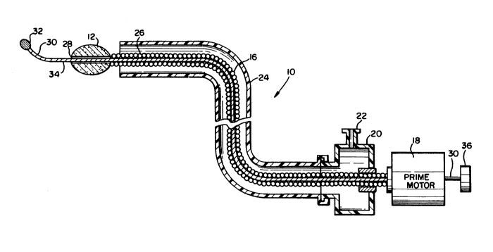
Referring generally to FIGS. 1 and 2, the
15 preferred embodiment 10 of the present invention is
shown. The invention 10 comprises an abrasive tip 12
and an atraumatic tip 32 which is generally of the type
described in U.S. patent No. 4,646,736 entitled
TRANSLUMINAL THROMBECTOMY APPARATUS by the present
inventor which is steerable for accessing branch
vessels. The tip 12 is covered with an abrasive cutting
material, such as diamond grit 14, which is used in the
preferred embodiment of the invention. The tip 12 is
connected via a hollow, flexible drive shaft 16 to a
25 variable speed prime mover 18. In the preferred
embodiment of the invention, the drive shaft 16 is a
. 020" diameter trifilar helically wound drive shaft.
The drive shaft 16 is sealably coupled to a variable
speed rotational prime mover 18, which is capable of
30 high speed rotation. The coupling is accomplished using
a sealed chamber 20 having an injection port 22, so that
injection of drugs or fluids into the lumen which is
formed between the drive shaft 16 and a surrounding
plastic sheath can be accommodated. The distal segment
35 26 of the flexible shaft 16 is preferably passivated
36f~3
with a coating, of a low-friction material, such as du
Pont's Teflon ~ brand tetrafluoroethylene homopolymer,
which will inhibit the winding of intravascular fiber on
the shaft 16 during rotation. The tip 12 includes a
central bore 29 which is aligned with the opening which
extends down the length of the hollow shaft 16. The tip
12 and the shaft 16 are routed into a vessel by using a
central guide rail 30, which may be comprised of a .005"
diameter steel wire. Adjacent the blunt tip 32 at the
distal end of the guide rail 30, there is a preformable
portion 34 of the guide rail 30 which the physician
using the invention may bend to facilitate directing the
invention into branch vessels. The guide rail 30
extends completely through the shaft 16 and through the
prime mover 18 to a rotatable knob 36 which permits the
guide rail 30 to be rotated in order to direct the tip
32 through a patient's vessel in order to perform a
thrombectomy as described in U.S. patent No. 4,679,557.
2n The drive shat 16 and the central rail 30 may be
individually moved with respect to each other and with
respect to the plastic sheath 24 in order to engage a
thrombus or an atheromatous occlusion. The rotational
prime mover 18 for the high-speed helical drive shaft 16
is preferably operable in a range of from 20,000 rpm to
greater than 155,000 rpm. The size of the burr tip 12
is typically in a range of from less than 1 mm diameter
up to about 6 mm, depending upon the vessel size desired
where the lesion is being recanalized.
Such a device provides for transluminal
recanalization of intravascular lesions of soft or hard
constitution consisting of thrombotic or atheromatous
material.
1~36~3 `
-8- WM6a
Referring to FIG. 3, a modified version of
the present invention utilizes a burr 50 having
coated on the distal surface thereof, i.e., the
portion most remote from the prime mover 18 (shown
in FIG. 1), a variety of particles 52 ranging in
size from about 30 mic-ons up to about 150 microns
in diameter. The smaller particles 56 are
preferably located in the area 54 adjacent to the
portion 58 of the burr 50 having the largest
diameter, and the larger particles 60 are
preferably located in the area 62 adjacent to the
distal end 64 of the burr 50. Accordingly, when
the burr 50 is inserted into a patient's vessel
the larger particles 60 adjacent the distal end 64
serve to quickly abrade through any lodged
material, thereby opening the vessel rapidly. The
smaller particles 56 continue to abrade and polish
the inner ~urface of the vessol as the burr 50 is
advanced therethrough. The region 58 having the
wide~t diameter is preferably substantially devoid
of abrasive particles, so that it acts as a
central bearing area. Accordingly, if the burr 50
is allowed to remain in a particular position
within a patient's vessel, the absence of abrasive
material in the central region 58 prevents that
region orm abrading through the wall of the
patient's vessel.
Referring to FIG. 4 the atraumatic tip 32
illustrated in FIGS. 1-3 can be replaced by
preformable spring tip 70 of a type used in the
catheter art. The preformable spring tip 70 can
be bent by a physician, as desircd, prior to
insertion into a patient' 5 vessel, in order that
the unit may be guided through a patient's vessel
to a particular location, genexally under the
assistance of fluoroscopy.
Referring generally to FIG. 5, radial
openings 72 may be formed in the burr 50 to permit
~3~3
WM6a
the outward flow of water (pumped in from the
proximal end), whereby a friction reducing bearing
will be accomplishèd.