Note: Descriptions are shown in the official language in which they were submitted.
1 322307
MEDULLARY NAIL FOR THE TIBIA
FI:E:LD OF TIIE INVENTION
The invention relates to a tibia intramedullary nail for
use in osteosynthesis.
BACKGROUND OF THE INVENTION
The most recent indications for the use of tibia
intramedullary nails are simple horizontal or oblique fractures
of the tibia, multistage breaks of the tibia shaft, and
fragmented breaks of the tibia shaft. Because use of tibia
intramedullary nails is now indicated for more fractures of the
lower leg, a need to change the shape and nature of the nail has
arisen. Formerly, the basic purpose of the nail was to
immobilize the bone internally, with the axial force as well as
torsional moments being borne by the bone itself. The only
function o~ the nail was to maintain the longitudinal axis of
the bone upright. This means that relatively thin, flexible
nails could be used, which have the advantage that they can be
adjusted to the medullary cavity.
This adjustment is particularly important in the insertion
of the nail, since the entry point of the nail is not in line
with the longitudinal axis of the bone. The entry point is
located on the tuberositas tibia, slightly proximal to the
patellar tendon attachment. After the medullary cavity has been
~3223~7
opened with an awl and the medullary cavity has been drilled,
the nail is driven in. The problem with driving in the nail is
that after crossing the medullary cavity, the nail reaches the
posterior cortex, and can be driven in furthex only if its shape
is deformed. With hard bones and young patients, the tip of the
nail sometimes punctures the posterior cortex.
To prevent such a perforation of the posterior cortex, the
method known as bunch-nailing (for example, according to German
Auslegschrift AS 23 41 439) has been proposed. In this method,
wires approximately 2.0 mm thick are pushed into the medullary
cavity. The advantage of this method is that the individual
wires are very flexible and can adjust well to the shape of the
medullary cavity. In this known method, the medullary cavity is
filled up with individual wires, whereupon it becomes possible
to splint the bone. This method has become generally accepted
in treating short oblique ~ractures in the mid-shaft range of
the tibia. The disadvantages of this method become evident,
howeYer, when a fragmented or multistage fracture must be
treated. The individual wires are not sufficient to protect a
fracture in the axial direction or in the rotational directionO
To preserve the length of the bone and to protect against
rotational movements, only a sti~f and rigid implant is
suitable, preferably one which may be locked by screws inserted
proximally an~ distally to the fracture area.
The tibia intramedullary nails known in the prior art,
which create an axial stability and a rotational stability of the
~ - 2 -
: . .,
. . ~
,
: , ,,
1322307
fracture, have a short proximal end segment, or crown hook, and a
distal segment having a straight part with a rounded tip, which
simplifies the stringing together of the fragments. However, as
described above, these nails are rather severely deformed when
inserted, so that the nail can bend plastically in the
longitudinal direction.
If four-fifths of the tibia nail is driven in, the crown
hook penetrates the tuberositas tibia, and thereby relieves the
bending demand made upon the stud~ This relief can be such that
when the stud is fully driven in, it lies loosely in the medullary
cavity.
SUMMARY OF THE INVENI'ION
The invention comprises a one-piece tibia intramedullary
nail having proximal and distal end segments bent towards a center
segment. The proximal end segment is bent at an angle in the
range of about -10 to -20, preferably -20 and the distal end
segment is bent at an angle in the range of about 2 to 4,
preferably +3 from the central axis of the center segment. The
proximal end segment, or crown hook, is much longer than those
known in the art. It enters the medullary cavity much sooner,
whereby it prevents excessive bending or bending pressure on the
intramedullary nail. The distal end segment serves as a gliding
surface when the marrow stud is driven into the posterior cortex.
.,~:.~
~,~
,. ~ ."
~ 322307
DESCRIPTION OF THE DRAWINGS
The invention will be further described with reference to
the drawings wherein:
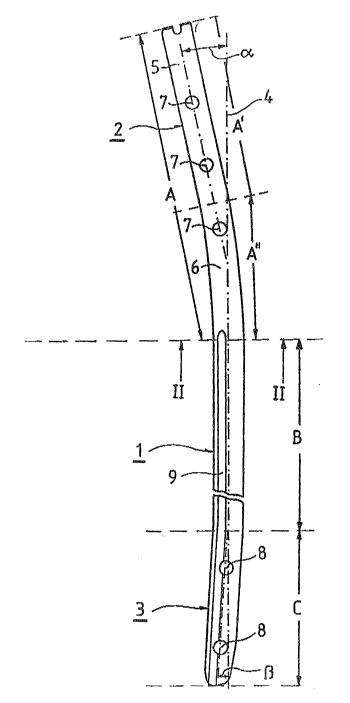
Fig. 1 is an elevational view, partly interrupted of a nail
according to the invention, and
Fig. 2 is a section along line II-II of Fig. 1.
DETAILED DESCRIPTION OF THE INVENTION
The invention provides an intramedullary nail for the
treatment of lower leg fractures of all indications. Its shape
is adapted to the anatomy of the medullary cavity and to the
method of its implantation. Two advantages o~ this tibia nail
are that it can be inserted into the medullary cavity without
plastic d~formation and that it acts as a stable splint after
its insertion.
The tibia nail according to the invention has a proximal
segment, a control segment and a distal segment. The distal end
segment is bent toward the central axis of the center segment.
This distal end segment prov des a sliding surface when the nail
is driven into contact with the posterior cortex. The angle of
impact of the nail is thus more acute, and the nail has a
greater tendency to follow the geometry of the medullary cavity.
The proximal end segment, also called the crown hook, is a
great d~al longer than those of tibia intramedullary nails known
in the art. It penetrates the medullary cavity much earlier and
thereby prevents an excessive bending of or bending pressure on
~ - 4 -
~ . .
:, : . ,: ~
1~2~3~7
the nail. The three-point distortion o~ the nail when it is
driven in is thereby greatly lessened. If the nail is fully
driven in, this exceptionally long crown hook is adjacent to the
anterior cortex. The advantage of this configuration is that
the tibia has the best possible splint, even without
supplementary elements.
When intramedullary nails known in the art are driven in,
they twist, but then lie loose in the marrow space. Because of
its anatomically adapted shape, a nail according to the invention
can be inserted into the medullary cavity relatively simply and
without excessive demands on the implant or the bone, and when it
has been driven in, it forms a positive connection with the bone.
This basic difference has a still greater e~fect when ;mplanta-
tion must be followed by a locking operation, in which the
implant must secure the length as well as the rotation of the
bone. These locking intramedullary nails have thicker walls, and
thus their resistance to bending is approximately 80~ greater.
To implant a prior-art locking intramedullary nail, the
operator had to drill 1.0-2.0 mm further into the medullary
cavity than in normal cases. This procedure has the disadvantage
that a relatively large amount of vital bone must be milled out,
without achievement of greater stability of the osteosynthesis.
An intramedullary nail according to the invention has
considerable advantages over the intramedullary bundle-nailing
method. As mentioned eariler, in marrow bundle-nailing the
splinting of the long bone is exclusively internal. In this
~ - 5 -
,, :, ,:,
, . , , : , . .
, , .: . ~ . , :: . . . ; . .
~322307
method, the marrow area, mostly undrilled, is filled with wiras
approximately 2.0 mm thick. The quality of such an
osteosynthesis is largely dependent on the surgeon's skill,
because it is not easy to insert these wires into the medullary
cavity without getting them crossed and thereby creating
bottlenecks in the cavity through which no more wires can be
fedO The indication for this nailing method is very limited,
since the medullary cavity is hourglass-shaped and thus has only
a short narrow segment that can be sufficiently filled with
wires and can thus be stabilized. When using the intramedullary
bundle-nailing method, it is not possihle to maintain the tibia
length or to prevent rotation in the case of fragmented or
multistage breaks. In caring for a multistage fracture, a
fragmented fracture, or a fracture in a relatively distal or
proximal location, both the length and the rotation can be
secured by means of the tibia intramedullary nails according to
the invention. The nail of the invention is optimally adapted
to 'che anatomy, since it is a relatively stiff implant which can
withstand the full load that bears on the tibia.
The forces operating on the tibia axially, as well as the
torsional moments, are preferably carried via locking screws to
the intramedullary nail. These locking screws are proximal and
distal to the fracture zone, and guarantee optimum
immobilization of the fracture.
In a preferred embodiment of the invention, there are three
holes at the proximal end of the nail, appropriately positioned
~ - 6 -
: .
i
'~ ' ` ; '', '~
.
~322~
in the frontal plane, which receive locking screwsO The
advantage of thi~ configuration is that the drilling for the
insertion of the screw can be done in the medial to lateral
direction. The advantage of this orientation of the screws is
that no blood vessels need be injured. The orientation of the
proximal locking holes, at 90 to the longitudinal axis,
facilitates the use of the same nails for both the right and the
left tibiae.
The distal locking can be done in either the frontal or the
sagittal planes. In very distal fractures, sagittal locking is
preferred to front locking, since tissue coverage of the screw
heads may be problematic.
In the use of short tibia intramedullary nails, tissue
coverage in the sagittal plane is difficult. For this reason,
in this type of locking, distal locking should appropriately be
done in the medial to lateral direction.
Referring to the drawing, Figs. 1 and 2 show an
intramedullary nail according to the invention. The nail may be
made of any customary implant material. It is hollow. In a
preferred embodiment the exterior diameter D at maximum
thickness is 16 mm and the wall thickness d, is 1.2 mm, though
in general the wall thickness can vary ~rom about 0.9 to about
1.3 mm, preferably from about 1.15 mm to about 1~25 mm.
The nail has a center segment 1, a proximal segment 2 and a
distal segment 3.
. -: : , ~
: : : .. :
1 322307
Preferably, proximal end segment 2 is bent at an angle,
alpha, of about ~ 1 from the central axis 4 of center
segment 1, and the distal end segment 3 is bent at an angle,
beta, of about ~3 from central axis 4 of center segment 1. The
angle alpha may vary within a range of about -10 to about -20,
preferahly about -10 to about 12 or from about -13 to about
~17. Angle beta may vary within a range of about -13~ to about
+4, a positive value indicating clockwise displacement from the
axis of the central segment.
In a preferred embodiment, proximal end segment 2 has an
overall length A of about 142 mm, and consists of a straight
section 5 with a length A' of about 80 mm and a bent section 6
with a length A'' of about 66 mm which connects to the center
segment 1. The radius of curvature (not indicated in the
drawings) of bent section 6 is about 200 mm in a preferred
embodiment, but may be varied in the range between about 180 and
about 220 mm, preferably between about 195 and about 205 mm.
Length A of proximal end segment 2, or partial lengths A'
and A'' of sections 5 and 6, are designed for a preferred total
nail length, L, (A' + A'' + B + C) of about 220 mm. For nails
having a different total length L (total lengths of up to about
420 mm are customary) pieces 5 and 6 are extended
proportionally. Otherwise, length A of proximal end segment 2
can be varied, even with an overall length L of about 220 mm,
25 within a range of about 122-162 mm, preferably about 137-147 mm.
~ - 8 -
... ..
,
~: . , . . "~,,
': ; ~
-: ~ ,
, .~, ' : ', " '' ,
~ 132~0~7
The length ratio of the terminal straight piece 5 to the
overall nail length L, A' :L, may vary between about 0.30 and
about 0.40, preferably between about 0.33 and about 0.40 and
most preferably between about 0.350 and about 0~377, while the
length ratio of bent piece 6 to overall nail length L, A'':L,
may vary between about 0.25 and about 0.31, preferably between
about 0.270 and about 0.294.
In a preferred embodiment, the nail has three locking holes
7 in proximal and segment 2, and three locking holes 8 in the
distal end segment 3, two in the frontal plane and one in the
sagittal plane. The number of locking holes 7 and 8 can be
varied according to proposed use and the overall length of the
nail.
The tibia intramedullary nail according to the invention
has a slot shaped recess 9 in the direction of central axis 4.
Recess 9 extends through center segment 1 to the tip of the
distal end segment 3.
,
