Note: Descriptions are shown in the official language in which they were submitted.
132~94~ :
, . .
.~ ~
. ::
TITLE OF THE INVENTION
Balloon Catheter
.,, ,'~'.
BACKGROUND OF THE INVENTION
~ Field of the Invention
Y This invention relates to a balloon catheter. More
particularly, lt relates to a balloon catheter for ;-
expanding and thereby remedying stenosis ln the blood -
vessel for improving the state of the distal side blood
~ stream.
t Description of the Prior Art -
In the event of stenosis or obturations in the
vascular system, such as blood vessels, percutaneous ~-
transuminal angioplasty (PTA) or percutaneous transluminal
coronary angioplasty (FTCA) is performed for enlarging or
recanalizing the narrowed or obturated site of the
vascular system to thereby improve the body liquid stream -
towards the distal side of the vascular system. In PTA or ~ -
PTCA, after a blood vessel ls procured percutaneously, a
fine gu$de wlre is passed through the vessel. A catheter
having an end balloon (expandable member) is introduced
- into the vessel , us~ng this guide wire as the guide,
until the balloon is positioned at the site of lesion
where stenosis or obturation has occurred. A liquid such
as contrast medium is in;ected continuously into the
balloon via an end hub under a pressure of several to ten
atmospheres for dilating the balloon towards the inner ~ ~-
wall of the blood vessel for pressuring and thereby
enlarging the narrowed or obturated sites.
As a balloon catheter employed in PTA or PTCA, there
-~ - is known a bælloon catheter having a coaxial dual tube
system including an inner tube opened at one end and
defining a first lumen and an outer tube surrounding the
inner tube, forming a second lumen with the inner tube and
.
'`'' ' ' . ~ '," ' ' ' ' ~' .. ' "', ' , , ' " '~' ': ' ,', , ' ' . ' '. : '
132~9~1
provided with a distal balloon, or one in which a tubul~r
member defining a lumen is provided with a distal balloon
surrounding the end of the tubular member.
In the case of the former balloon catheter having the
coaxial dual tube system, ring markers formed of an X-ray
opaque material are provid0d at predetermined portions of
the inner tube in the balloon that are substantially in
register with both ends of the balloon., these ring
markers being used as means for identifying the
cylindrical portion of the balloon under X-ray
fluoroscopy. However, considexing that the balloon
catheter is caused to proceed through the inside of the
blood vessel presenting acute bend or bends, the risk is
high that the inner tube disposed within the balloon at
the bends of the blood vessel be broken and collapsed the -
lumen opened at one end the lumen opened at-one end to
obstruct smooth progress of the guide wire.
For overcoming the above difficulties, there is also
known a balloon catheter in which a piping having at both
ends thereof prescribed portions substantially in register
with both ends of the cylindrical portion of the ballo~in
is provided on the inner tube disposed within the balloon.
While it is possible with this known balloon catheter to
prevent the inner tube from being broken and collapsed, it
is difficult for the balloon catheter to proceed through
the inside of the blood vessel presenting acute bend or
bends. Even supposing that the balloon catheter should
have succeeded in proceeding beyond the bend, the risk is ~-
high that the piping remains bent to obstruct the progress
thereof the blood vessel beyond the bend towards the
distal side of the vascular system.
~ .
OBJECT AND SUMMARY OF THE INVENTION
It is a principal object of the present invention to
provide a balloon catheter which is free from the above
- ; . - -
1 ~
problems of the prior art and which is able to prevent
breaking of the inner tube and obstructing of the lumen in
blood vessels presenting an acute bend or bends.
According to the present invention, there is provided
a balloon catheter comprising a tubular body including at
least one lumen, and a foldable balloon provided at a
predetermined forward portion on the outer surface of said
tubular body so that the balloon communicates with at
least one lumen in said tubular body. A coil spring is ~ -
provided at a predetermined portion on the outer surface of
the portion of the tubular body surrounded by the balloon, -;
for reinforcing the tubular body. The coil spring has
opposite end portions and an intermediate portion and is
wound such that the turns thereof are in intimate contact
with each other at both end portions thereof and are sparse
in the intermediate portion ~hereof. -~
According to the present invention, the diameter of a
portion of the balloon catheter which is introduced into -
the patient's body and which is formed by said tubular
body and the folded balloon is not more than 2.7 mm.
It is preferred that the reinforcement be formed of
an X-ray opaque material.
It is preferred that the X-ray opaque material be
platinum, gold, tungstene or alloys thereof.
It is preferred that the opaque material be a silver-
palladium alloy.
It is preferred that the reinforcement be a wire in
the form of a coil and having a circular, rectangular or
an elliptical cross-section.
It is also preferred that the wire of the coil is
formed by a coil spring having turns thereof in intimate
and tight contact with one another. -
Alternatively, turns of the coil spring may be
arranged thick in both end parts and thin in the
intemediate part of the coil spring.
According to the present invention, the portions of
the balloon catheter surrounded by the balloon may be
prevented from being broken and collapsed, even when the
catheter is proceeding through the inside of the blood
132~9~
vessel presenting acute bend or bends, so that the balloon
catheter can be prceeded positively to the site of lesion
in the blood vessel.
BRIEF DESCRIPTION OF THE DRAWINGS
The present invention will now be described with
reference to the accompanying drawings in which
Fig.1 is an enlarged sectional view showing the
; distal end part of the balloon catheter according to a
preferred embodiment of the present invention,
Fig.2 is a sectional view taken along line II-II of
Fig.1,
~ Fig.3 is a sectional view taken along line III-III of ~-
3 Fig.1,
~ Fig.4 is a diagrammatic view showing the proximal end
Z part of the balloon catheter shown in Fig.1,
Fig.5 is an overall side view showing the balloon
catheter of Fig.1, with a portion thereof being broken
away,
Figs.6 to 10 are diagrammatic views for illustrating
~ the operation of the balloon catheter shown in Fig.1, andi Figs.11 and 12 are enlarged sectional views showing
the distal end part of balloon catheters according to
modified embodiments of the present invention.
Fig.13 shows a modification of the embodiment of Fig.l.
.
~5~ 132~941 : -
DESCRIPTION OF PREFERRED EMBODIMENTS
By referring to the accompanying drawings, certain -~
preferred embodiments of a balloon catheter of the present
invention including a tubular member having two lumens
will be explained hereinbelow in detail.
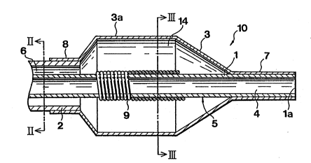
Figs.1 to 4 illustrate a preferred embodiment of the
balloon catheter according to the present invention.
Fig.1 is an enlarged sectional view showing the d~stal
side of the balloon catheter. Figs.2 and 3 are sectional
views taken along line II-II and line III-III of Fig.1,
respectively. Fig.4 is an enlarged sectional view showing
the proximal side of the balloon catheter.
As shown in Figs.1 to 4, the balloon catheter 10 of
the present invention includes an inner tube 1 having a
first lumen opened at one end, an outer tube 2 provided
for encircling the inner tube 1 at a position set back a- `
predetermined distance from a foremost part 1a of the
inner tube 1 and defining a second lumen 6 between it and
the outer surface of the inner tube 1, and a balloon 3
including a distal end 7 attached to the inner tube 1 and
a proximal end 8 attached to the outer tube 2. The
balloon 3 communicates with the second lumen in the
vicinity of the proximal end 8. and has a cylindrical -
section 3a, can be including at least a portion thereof "
substantially cylindrical in contour to permit a
constricted site of the blood vessel to be dilated easily.
A reinforcement 9 is wound about a portion on the outer
peripheral sur~ace of the inner tube which is
substantially in register with the cylindrical section 3a.
The balloon catheter 10 has the inner tube 1 and the
outer tube 2 as a cathether tube or tubular body or member
5, the proximal end of which is provided with a bifurcated ~ -
branched hub 20, as shown in Fig.4. The branched hub 20
has a guide wire port 12 and an injection port 13
132~941
-6-
communicating with the first lumen 4 and with the se~ond
lumen 6, respectively.
The first lumen 4 formed by the inner tube 1 plays
the role of a guide wire passage and a blood route or
channel during use of the balloon catheter. The first
lumen communicates at its proximal end with the guide wire
port 12 formed in the branched hub 20, such that a guide
wire 15 for the balloon catheter as later described is
introduced into an opening end of the guide wire port 12
so as to be guided into the first lumen 4.
., The inner tube 1 is preferably formed of a material
exhibiting certain flexibility, including polyolefins,
such as polyethylene, polypropylene, ethylene-propylene ~ -
copolymers or ethylene-vinyl acetate copolymers,
thermoplastic resins, such as polyvinyl chloride,
polyamide elastomers or polyurethane, silicone rubber or
latex rubber. More preferred are the aforememtioned
thermoplastic resins and most preferred are polyolefins.
The outer tube 2, in the inslde of which the inner -
tube 1 is introduced, is preferably mounted coaxially with
the inner tube 1 and at a position ln which the foremost
part of the suter tube is set back a small distance from
the foremost part 1a of the inner tube 1. The second
lumen 6 is defined between the inner surface of the outer
tube 2 and the outer surface of the inner tube 1. The - -
second lumen 6 plays the role of a channel for injection
of, for example, contrast medium and dischaging of
residual air and communicates at its proximal end with the
injection port 13 of the branched hub 20. The second
lumen ~ also communicates at its distal end with the
inside of the balloon 3 at its rear end. The contrast
medium, for example, are injected and charyed into the
internal space of the balloon 3 via the openin~ end of the -
injection port 13 and the second lumen 6 while the
residual air is discharged simultaneously~
~ . . ~ - ... .: . . . . . . . .. . .
_7_ l 32 ~9
The outer tube 2 is preferably formed of a material
exhibiting certain fexibility, including polyolefins, such
as polyethylene, polypropylene, ethylene-propylene -
copolymers or ethylene-vinyl acetate copolymers,
thermoplastic resins, such as polyvinyl chloride,
polyamide elastomers or polyurethane, silicone rubber or
latex rubber. More preferred are the aforememtioned
thermoplastic resins and mo~t preferred are polyole~ins.
The balloon 3 has its foremost part 7 and rear end 8
secured to the outer peripheral surface of the foremost
part of the inner tube 1 and to the outer peripheral
surface of the foremost part of the outer tube 2 liquid-
tightly, respectively, as with an adhesive or by heat
fusion, for delimiting an expansion space 14 between the
inner surface of the balloon 3 and the outer surface of
the inner tube 1. This expansion space 14 communicates at
its rear end with the second lumen 6 along its overall
periphery so that the contrast medium, for example, may be
charged into the space 14 via the second lumen 6, as ~-~
described hereinabove.
The balloon 3 may be folded in ~uch a manner that,
when the balloon i8 not dilated, it may be folded and -
wrapped about the outer periphery of the inner tube 1. In
order that the constricted site of the blood vessel may be
dilated more easily, at least a portion of the balloon 3
is formed as a substantially equidiametral cylinder for
defining the aforementioned cylincrical section 3a. The
cylindrical section need not be a true cylinder but may be
in the form of a prism having a polygonal cross-section.
It should be noted that the balloon 3 is tapered from
the forward side of the cylindrical section 3a to the
foremost part 7 where it is secured to the inner tube 1
and from the rear side of tha cylindrical section 3a to
the rear end 8 where it is secured to the outer tube 2.
- , . ~ : . - . . .
-8- ~32~941
It should also be noted that, in the state in which
the balloon 3 is folded and wrapped about the inner tube
1, that is, the balloon 3 is wrapped around the catheter
tube 5, the portion of the balloon catheter introduced
into the patient's body be of an outside diameter of not -
more than 2.7 mm at the maximum, since the balloon
catheter can be used satisfactorily in such case in the
body cavity, above all, in a finer vasculum.
The balloon 3 is preferably formed of a material -
exhibiting certain flexibility, including polyolefins,
such as polyethylene, polypropylene, ethylene-propylene
copolymers, ethylene-vinyl acetate copolymers or cross- -~
linked ethylene-vinyl acetate copolymer, thermoplastic
resins, such as polyvinyl chloride, polyamide elastomers
or polyurethane, silicone rubber or latex rubber. More
preferred are the aforememtioned thermoplastic resins and
most preferred are the cross-linked ethlene-vinyl acetate
copolymers
According to the present invention, a reinforcement 9
is wound about a predetermined portion of the outer ~;
surface of the inner tube 1 which is enclosed within the
balloon 3, preferably the portlon thereof in register with
the aforementioned cylindrical section 3a. With the
reinforcement 9 thus wound about the inner tube 1, the
inner tube is rendered more resistant against buckling, so
that there is no risk that the inner tube 1 disposed in
the balloon 3 be broken or the first lumen 4 through which
the guide wire 15 for the balloon catheter is passed be
broken even at a bend of the blood vessel.
The reinforcement 9 is preferably formed of an X-ray
opague material, preferably platinum, gold, tungsten or
alloys thereof and more preferably a silver-palladium
alloy, since a clear contrast image can then be obtained
under X-ray fluoroscopy and thus the cylindrical section
3a of the balloon 3 can be identified more easily.
:
.. . . . . . . --
1325941
g
.
The reinforcement 9 is preferably a wire wound into a
coil. By using the coil as the reinforcement, the inner
tube 1 can be reinforced more strongly against buckling"
The wire of the coil preferably has a circular,
rectangular or an elliptical cross-section for inceasing
reinforcing effects.
The wire in the form of a coil may preferably be a
spring coil so wound that its turns are in intimate
contact with one another. With this dense winding of the
wire, the inner tube can be reinforced more strongly
against buckling. ~-
The branched hub 20 is formed by an inner tube hub 22
and an outer tube hub 23. The inner tube hub 22 -~
communicates with the first lumen 4, has a guide wire port
12 through which the guide wire 15 for the balloon ;
catheter is introduced, and is secured to the inner tube
1. The outer tube hub 23 communicates with the second
lumen 6, has an injection port 13 for injecting contrast
medium, for example, and is secured to the outer tube 2.
The outer tube hub 23 and the inner tube hub 22 are
secured to each other.
The branched hub 20 is preferably formed of
thermoplastic resins, such as, for example, polycarbonate,
polyamide, polysulfone, polyallylate or methacrylate-
butylene-stylene copolymers.
For explaining the operation of the balloon catheter
of the present invention shown in ~igs.1 to 4, the method
of using the balloon catheter in angioplasty (PTA or PCTA)
will be explained by referring to Figs.5 to 10.
It is preferred that, before conducting to
angioplasty by dilating and remedying constrictions
occurred in ths blood vessel, as much air as possible be
removed from the inside of the balloon catheter. To this
end, suction and injection means, such as a in-deflator,
filled with contrast mediaum is attached to the injection
:. :
:, . - . . . : : , ~ .
132~941
. o-- .:
,
port 13 of the catheter and the operation of alternate ~-
injection and suction is repeatedly performed to remove
the air in the second lumen 6 and the balloon 3 to replace
it with the contrast medium.
When the expansion space 14 of the balloon 3 and a
space of the second lumen 6 is filled with the contrast
medium and the residual air is removed completely, a
predetermined amount of the contrast medium filled in the
expansion space 14 is sucked and discharged by an in~ector
fitted with a pressure gauge 24 to cause the balloon 3 to
be wound about the inner tube 1 of the tubular member 5
to reduce the outside diameter of the balloon 3 so that ~`
the outside diameter of the portion of the balloon
catheter 10 introduced into the patient's body is not more
than ~.7 mm, in order to make ready for insertion of the
balloon catheter into the blood vessel in angioplasty.
For angioplasty, a blood vessel 30 is procured, that
is, peirced with the dilater and sheath 25 as shown in -
Fig.6, by relying upon, for example, the Sheath method. A
guiding catheter indwelled gauide wire is prepared. A
guiding catheter 26 is introduced into the blood vessel 30
along the guide wire and left at an inlet 31 to the
coronary artery having a target lesion. The guide wire
for the guide catheter is then removed.
The balloon catheter guide wire 15 is then introduced
at the guide wire port 12 oi the balloon catheter 10 into
the inner tube 1 of the tubular member 5, that is, into
the first lumen 4, until the guide wire is protruded
several centimeters beyond the end opening 1a of the inner - -
tube 1, as indicated in Fig.5. The resulting assembly is
then introduced into the guiding catheter 26 via a balloon
.
catheter port 28 of a Y-shaped connector 27, to the
proximal end of which the guiding catheter 26 is
connected, as shown in Fig.6. The balloon catheter 10 is
then proceeded through the inside of the guiding catheter
-11- 132~941
26 so as to be proceeded via the ~orward end of the
guiding catheter 26 into the blood vessel 30 having the
target lesion 30.
The balloon catheter guide wire 15 is then extended
to the target lesion through the inside of the blood
vessel 30, as shown in Fig.8. The guide wire 15 is left -
in the blood vessel after it has passed through the
constricted site 32.
The balloon catheter 10 is then advanced through the
inside of the blood vessel 30 along the balloon catheter
guide wire 15. The balloon catheter 10 of the present
invention has an increased resistance against bending
since the reinforcement 9 is wound about the portion of
the outer surface of the inner tube 1 disposed within the
balloon 3, so that, even when the blood vessel 30 has an
acute bend, as shown in Fig.8, there is no risk of
obstruction of the progress of the balloon catheter guide
wlre 15 caused by the breaking of the inner tube 1 within
the balloon 3 or the collapse of the first lumen opened at
the end and hence the balloon catheter 10 can be proceeded
smoothly towards the lesion at the dlstal side.
It should be noted that, when the reinforcement 9 is
formed as a wire in the form of a coil, above all, as a
coil spring, which is wound about the outer surface of the
inner tube 1 with the nelghboring turns of the coil in
tight and intimate contact with one another, an increased
resistance is obtained against external forces.
When the co~l wire has an elliptical, rectangular or
a circular cross-section, a further increase is obtained
in the resistance against external forces.
Also, when the reinforcement 9 is formed of an X-ray
opague material, the reinforcement 9 is indicated as a
clsar X-ray contrast image, under X-ray fluoroscopy, such ~ -~
that this reinforcement 9 can be checked visually as an
indicia for the balloon for positively positioning the
-12- 132~941
cylindrical section 3a of the balloon 3 at the stenosis
site 32.
It is preferred that the X-ray opague material be
platinum, gold, tungstene, alloys thereof or a silver-
palladium alloy, since then a clearer X-ray contrast image
is produced and thus the reinforcement may be used more
effectively as the indicia for the balloon 3.
i It is also preferred that, in the state in which the
balloon 3 is folded and wrapped about the inner tube 1,
that is, about the tubular member 5, the portion of the
balloon catheter introduced into the patient's body be of
an outside diameter of not more than 2.7 mm, since then
the balloon catheter can be used more advantageously
within the body cavity, above all, within the vessel
having a narrower inner cavity.
When the balloon 3 reaches the stenosis site 32, as
shown in Fig.g, the contrast medium are in;ected into the
dilated space 14 of the balloon 3, as the contrast medium
are pressurized to several to ten and odds atmospheres, by
the in~ector fitted with a pressure gauge 24, connected to
the ln~ection port 13 o the balloon catheter 10, for
expanding the balloon 3 as shown in Fig.10 for pressuring
and enlarging the diameter of the stenosis site 32.
After termination of this operation, the contrast -
medium are injected into the blood vessel via contrast
medium injection port 29 of the Y-shaped connector 27
connected to the proximal end of the guiding catheter 26
for visual checking of the distal side blood stream by X-
ray fluoroscopy. When it is observed that the blood
stream is improved, the balloon catheter 10 and the
balloon catheter guide wire 15 are removed from the blood
vessel 30. The guiding catheter 26 is then removed and
the pierced portion of the blood vessel is pressed to stop
the hemorrhage to terminate the operation.
- . - -. : . -. , . . ~: - . , .. . : , . :
-13- i32~9~
The above described balloon catheter is formed by
coaxially arranged inner and outer tubes defining two
lumens. However, the present invention may naturally be
applied to a balloon catheter formed by a tubular member
defining a sole lumen.
Referring to Fig.11, showing a second embodiment of
the present invention, a balloon 3' enclosing the distal
part of a tubular member 5' is provided at the distal part
of the tubular member 5' defining the sole lumen 6'. The
reinforcement 9 may be provided at the distal part of the
tubular member 5' surrounded by the balloon 3' for forming
a balloon catheter 10'.
This balloon catheter 10' is used for improving the
state of stenosis of the coronary artery, for example.
The operation of the balloon catheter 10' is basically the
same as that of the above described balloon catheter 10
having the dual tube structure. However, in the present
embodiment, the balloon catheter guide wire 15 is
lntroduced into the lumen 6' at its proximal end and held
so that its distal part does not break the balloon 3'. In
this state, the air inside the balloon 3' is replaced by
the contrast medium in the same way as in the precedlng
embodiment. ~he contrast medium are then sucked and
discharged in a predetermined amount using the ~ -
aforementioned in-deflator to cause the balloon 3' to be
wrapped about the tubular member 5' to reduce the outside ~
diameter of the balloon 3' so that the outside diameter of ~ ~-
the portion introduced into the balloon catheter 10' is
not more than 2.7 mm.
Referring to Fig. 12, illustrating a third embodiment
of the present invention, a tubular member 5" defining a
sole lumen 6" has a distal closed end. At the distal part
of the tubular member 5" is provided a balloon 3"
enclosing the distal part of the tubular member 5". The
lumen 6" of the tubular member 5" may be in fluid
~ .
:: '
-14- 132~941
communication with an expansion space 14" of the balloon
3" through an orifice or orifices formed in the tubular
member 5". The reinforcement 9 may be provided at the
distal part of the tubular member 5" surrounded by the
balloon 3" and have turns therof so wound that they be in
intimate contact with each other. The operation of this
balloon catheter is substantially same as those of the
above-discribed balloon catheters.
In case of the reinforcement being a coil spring, it
may alternatively be so wound that its turns are thick for
example in intimate contact with each other in both end
parts and thin or sparse in the intermediate part of the
coil spring, as shown in Fig. 13.
The balloon catheter in this state is introduced into -
the blood vessel 30, as it is guided by the balloon ~
catheter guide wire 15, until the balloon 3' reaches the ~ -
stenosis site 32 in the same way as in the preceding ~ -
embodiment. The contrast medium are then injected in a
predetermined amount into the expansion space 14' of the
balloon 3', with the aid of the pressure gauge-injector
24, for dilating the balloon 3' for pressuring and
enlarging the stenosis 32.
When it is observed that the state of the blood
stream is improved, the balloon catheter 10' may be -~
removed from the i~side of the blood vessel, as described i
hereinabove.
In the present second embodiment, by providing the
reinforcement 9 at the forward side of the tubular member
5', the tubular member can be reinforced against buckling.
.
EFFECT OF THE INVENITON ~:
As described in detail, the present inveniton
provides a balloon catheter comprising a tubular body ~--
including at least one lumen, and a foldable balloon
provided at a predetermined distal portion on the outer
_A~
132~941
-15-
surface of said tubular body so that the balloon
communicates with at least one lumen in said tubular body,
herein a reinforcement is provided at a predetermined
portion on the outer surface of said tubular body
surrounded by said balloon.
With the above described arrangement of the balloon
catherter, the balloon portion of the balloon catherter
may positively be proceeded to the target lesion, wihtout
breaking and collapsing of the tube portion enclosed in
the balloon even at progress through an acute bend of the
blood vessel. ;`
'`.
' ':,
:',