Note: Descriptions are shown in the official language in which they were submitted.
30,621
t 335527
BIOABSORBABLE SURGICAL DEVICE AND METHOD FOR
TREATING NERVE DEFECTS
BACKGROUND OF THE INVENTION
1. Technical Field
This invention relates generally to medical
devices useful for the repair of nerve defects and,
particularly, to a bioabsorbable surgical device and a
method of its use for spanning a significant nerve gap
where the nerve ends may not be easily pulled and
sutured together.
2. The Prior Art
When a nerve is lacerated or severed it may
be repaired by a common surgical procedure known as
nerve repair or, technically, neurorrhaphy. With the
aid of microsurgical techniques, direct nerve suture
can easily be done without the use of additional
devices when there is no nerve missing between the
severed or lacerated nerve endings. However, when a
portion of the nerve is missing, a nerve gap or nerve
defect exists. This situation may be overcome by
mobilizing the nerve ends, bringing them together and
suturing them if the gap or defect is less than 1.5
centimeters. Fairly good results have been obtained by
suturing the nerve ends together in this fashion.
- 2 - 1 3 3 5 5 2 7
However, problems do still exist. The process of
direct suturing is limited because it is extremely
tedious and time consuming. The use of numerous
sutures can cause trauma to the nerve which stimulates
S the formation of intraneural and extraneural connective
tissue, or scar tissue. Invasion of the repair site by
connective tissue can prevent the regenerating axons in
the proximal stump from entering the microscopic
tubules contained in the distal stump. This situation
often causes formation of painful neuromas at the
suture or nerve graft site. Furthermore, it has been
shown that for defects or gaps greater then 1.5
centimeters, stretching the nerve ends and directly
suturing the ends together creates tension at the
suture line which causes greater scar formation and,
thus, providing poor results.
The technique used to treat nerve gaps is
termed "nerve grafting". Typically, the nerve graft
material is taken from another part of a person's body,
generally a nerve that goes to a sensory area of a
lower extremity, such as the sural nerve. The sural
nerve is taken from the donor site leaving an area of
numbness in the lateral aspect of the patient's foot, a
long scar up the patient's leg and the future potential
for pain at the site at which the sural nerve graft was
taken. It would be desirable to be able to provide a
nerve graft material that could provide for nerve
growth across a significant nerve gap or defect without
using nerve graft material taken from the patient's own
body.
Animal (non-human) nerve graft substitutes
have also been utilized to provide the necessary
spanning of the nerve gap or defect. These nerve
heterografts have been sutured to the nerve ends in the
same fashion as a human graft. However, these types of
~ 3 ~ l 3 3 ~ 5 2 7
substitute nerve grafts suffer from many drawbacks.
First, the chances for success in achieving nerve
regeneration using such grafts has been extremely
unpredictable. Second, there is the potential for an
autoimmune response by the body to the foreign nerve
graft material.
Recognition of this problem has prompted many
researchers to explore alternatives to direct suturing
and the use of nerve grafts in bridging nerve gaps or
defects and a variety of approaches involving the use
of many different types of materials have been
experimented with over the past years. Methods and
devices have been developed which use both suturing and
nonsuturing methods to provide a direct connection
between the nerve ends. All of these alternatives seek
to protect the anastomotic nerve site by wrapping,
tubulizing, or otherwise encasing it with a natural or
foreign substance, either absorbable or nonabsorbable.
However, none of the prior art references disclose a
successful device and method which allows a nerve to
regrow across a significant nerve gap without the use
of a nerve graft or direct nerve end to nerve end
suture line.
U. S. Patents Nos. 4,534,349 and 4,669,474
both to T. H. Barrows disclose a medical device and
method of use for the sutureless repair of lacerated,
severed, or grafted nerves. The device is a
longitudinally-openable, porous, rough-surfaced tube of
a molded natural or synthetic absorbable polymer. This
device was not designed for the treatment of nerve
gaps. It was designed to repair a broken nerve without
the use of sutures by approximating the two nerve ends
together and holding them together within a rough-
surfaced tube. If used in a situation involving a nerve
gap, an autogenous nerve graft would be used. The
_ 4 - l 3 3 5 5 2 7
tubular device would encase both the graft and the two
nerve ends or two separate devices would be required,
one at each end of the graft and respective nerve end.
Furthermore, the Barrows molded tube comes in two parts
which are then hooked together such that the tube would
be fairly rigid which would not permit it to be used in
situations where the repaired nerve would be required
to go around a corner or be subject to bending forces.
Sutureless tubulization techniques are known
to be successful only in the case of very small, single
fascicle nerves. The saphenous nerve in rats (0.3-0.5
mm diameter) was transected and repaired with a
preformed tube or single leaf of collagen membrane as
disclosed by J. M. Rosen, E. N. Kaplan, D. L. Jewett,
and J. R. Daniels, "Fascicular Sutureless and Suture
Repair of the Peripheral Nerves, A Comparison Study in
Laboratory Animals", OrthoPedic Review 8 (4), 85
(1979). This method of repair avoids sutures but
requires a totally tensionless situation to avoid
retraction of the nerve stumps. J. M. Rosen in
Orthopedic Transactions 6(1), 75(1982) reports that the
peroneal nerve in rats (0.5-1.2 mm in diameter) was
transected and repaired with a thin-walled, extruded
tube of polyglycolic acid, cut open longitudinally
along one wall. This method also requires a totally
tensionless situation and is not advisable in the case
of larger nerves since the tight fit required to
maintain adequate nerve stump approximation would not
provide for the release of pressure created by post-
surgical swelling.
U.S. Patent No. 4,662,884 to L. J. Stensaas,et al. discloses a very similar method of nerve repair
(no gap) using a nonabsorbable silicone rubber. The
use of silicone rubber as a tube conduit for nerve
repair is also not without its disadvantages. Since
1 335527
-- 5
the rubber is non-absorbable in the human body, it will
be necessary to perform a second operation to remove
the rubber tube after the nerve ends have regrown
together. Silicone rubber has the further disadvantage
of being impermeable.
There have been many experiments performed on
regrowing nerves across small or insignificant (less
than 1.5 centimeters) nerve gaps or defects. Hakan
Molander, et al., "Regeneration of Peripheral Nerve
Through A Polyglactin Tube", Muscle and Nerve, 5:54-57
(1982), reported satisfactory results in bridging small
nerve gaps (7 to 9 mm in length) by use of a
biodegradable polyglactin suture mesh shaped as a tube
around the nerve defect as a framework for
proliferating cells. Molander, et al. further reported
in "Nerve Repair Using a Polyglactin Tube And Nerve
Graft: An Experimental Study in the Rabbit",
BIOMATERIALS 4: 276-280 (1983), that a method of
bridging a small nerve gap (10 mm in length) with a
polyglactin mesh-tube gave results essentially no
different from a conventional nerve graft. However,
Molander was using his tube only on small or
insignificant nerve gaps (less than or equal to 1 cm).
There is a]so extensive literature reporting
on the use of collagen tubes with or without a laminin
gel to treat nerve defects as disclosed by D. G. Kline
and G. J. Hayes, "The Use Of A Resorbable Wrapper For
Peripheral Nerve Repair, Experimental Studies In
Chimpanzees", J. Neurosurgery, 121, 737 (1946), and by
R. Madison, et al., "Increased Role of Peripheral Nerve
Regeneration Using Bioabsorbable Nerve Guides In a
Laminin-containing Gel, ExPerimental Neurology, 88:
767-772 (1985). However, with the use of collagen
tubes or tubes containing laminin to promote neural
growth, it is noted that collagen and laminin are
1 335527
-- 6
highly immunogenic and that techniques have not been
perfected to allow their use in humans without an
immune response developing. Furthermore, all of these
researchers were using their devices on clinically
insignificant gaps of 1 centimeter (cm) or less on
lower animal forms and not in primates.
Some researchers have found that nerves will
not regenerate across a nerve gap of greater than 10 mm
(1 cm). B. R. Seckel, et al., "Nerve Regeneration
Through Synthetic Biodegradable Nerve Guides:
Regulation by the Target Organ", J. Plast. Reconstr.
Surg. 74: 173-181 (1984), reported that in a rat model
a nerve gap distance of less than 10 mm (1 cm) is
crucial to obtain nerve regeneration across a nerve gap
lS or defect.
However, it has been determined through
discoveries made by the present inventors that nerves
can regenerate across a significant nerve gap greater
than 1 cm. S. E. Mackinnon, A. L. Dellon, et al.,
"Nerve Regeneration Through a Pseudosynovial Sheath in
a Primate Model", Plastic and Reconstructive Surgery,
75: 833-839 (1985), report that the nerve endings in a
baboon grew back together over a 3 cm nerve gap through
a vascularized pseudosynovial sheath. The
pseudosynovial sheath had been grown in the baboon's
own body for a six-week period before use on the
baboon's severed ulnar nerve. For this to work in a
human it would still be necessary to prepare the sheath
in the human body before undertaking repair of the
nerve defect. This would require at least two
operations and include all of the pain and costs
associated with two surgical operations. Therefore, it
would be highly desirable to develop a synthetic
bioabsorbable nerve conduit that could be used in
- - 7 - 1 335527
humans to span significant nerve gaps or defects of 1.5
centimeter or greater.
U. S. Patent No. 3,937,223 to R. W. Roth
teaches a partially-compressed, heat-embossed,
flexible, tissue-absorbable, compacted, surgical
hemostatic felt having specific fiber and density
measurements which is in the form of a thin conformable
mat. Two related patents U.S. Patent Nos. 4,033,938
and 3,960,152, disclose bioabsorbable polymers of
unsymmetrically substituted 1,4-dioxane-2,5-diones
which are broadly stated in col. 9, lines 29-31 and in
the bridging paragraph of cols. 9 and 10 ('938) and in
col. 9, lines 20-23 and lines 51-65 ('152) to be useful
as tubes or sheets for surgical repair such as nerve
and tendon splicing. A similar disclosure in U.S.
Patent No. 4,074,366 to Capozza Col. 6, lines 13-16 and
43-57, relates to poly(N-acetyl-D-glucosamine), (i.e.
chitin). However, there is no enabling disclosure in
the specifications or in their Examples as to how such
tubes are to be prepared, the characteristics required,
or their method of use.
SUMMARY OF THE INVENTION
It is a primary object of the invention to
provide a flexible, bioabsorbable, tube device that can
provide an optimum environment for nerve regeneration
across large or significant nerve gaps of from about 2
millimeters to about 6 centimeters.
Another object of the present invention is to
provide a flexible tube device manufactured from a
synthetic bioabsorbable material such as those listed
in Table I, below, for use as a nerve regeneration
conduit.
1 33~527
8 70557-~8
A further ob~ect of the present lnventlon ls to provlde
a knltted or woven tube manufactured from a synthetlc
bloabsorbable flber whlch ls flexlble enough to be bent through
an arc of up to 180 degrees wlthout plnchlng or crlmplng of the
lnternal dlameter of the tube devlce.
Yet another ob~ect of the present lnventlon ls to
provlde a bloabsorbable, flexlble, knltted or woven tube havlng a
corrugated exterlor and a relatlvely smooth-surfaced lnterlor so
as to promote nerve axon growth wlthln the tube devlce.
And, stlll another ob~ect of the present lnventlon ls
to provlde a flexlble, bloabsorbable, nerve tube devlce and
method for lts use whlch ls tlssue compatlble, mlnlmlzes neuroma
formatlon, accommodates post-surglcal swelllng and provldes an
optlmum envlronment whlch ls nonlmmunogenlc for nerve
regeneratlon across a slgnlflcant nerve gap or defect.
It ls a secondary object of the lnventlon to provlde a
method of uslng a flexlble, bloabsorbable, tube devlce that can
provlde an optlmum envlronment for nerve regeneratlon across
large or slgnlflcant nerve gaps of from about 2 mm to about 6 cm.
Stlll other ob~ects and advantages of the lnventlon
wlll ln part be obvlous and wlll ln part be apparent from the
speclflcatlon.
In accordance wlth one aspect of the present lnventlon
there ls provlded, a medlcal devlce adaptable for use ln the
treatment of a nerve gap or defect comprlslng a flexlble, porous
tube of a bloabsorbable polymer materlal, sald tube havlng a
~'`' .
1 335527
8a 70557-68
plurallty of corrugations on lts exterior surface posltloned so
as to allow sald tube to be bent wlthout crlmplng the lnternal
surface of sald tube and havlng a plurallty of flats provlded on
lts lnterlor surface to provlde a relatlvely smooth lnterlor
surface and havlng a substantlally constant lnternal dlameter to
promote longltudlnal axon growth wlthln the tube devlce and
across the nerve gap, and belng capable of encloslng and
protectlng the ends of a severed or lacerated nerve.
The present lnventlon also provldes a medlcal devlce
adaptable for use ln the treatment of a nerve gap or defect
comprlslng a flexlble, porous tube manufactured from a
bloabsorbable polymer flber whlch ls knltted or woven lnto tube
shape, sald knltted or woven mesh tube havlng a plurallty of
corrugatlons on lts exterlor surface such that sald tube may be
bent wlthout crlmplng the lnternal surface of sald tube and
havlng a plurallty of flats provlded on lts lnterlor surface to
provlde a relatlvely smooth lnterlor surface and havlng a
substantlally constant lnternal dlameter to promote longltudlnal
axon growth wlthln the tube devlce and across the nerve gap, and
belng capable of encloslng and protectlng the ends of a severed
or lacerated nerve.
Also lncluded wlthln the present lnventlon ls the use
of the above descrlbed devlce to treat a nerve gap or defect.
The present lnventlon provldes a devlce and a method of
uslng such a devlce for the repalr of nerve defects or gaps of
one and one half centlmeters or larger comprlslng a flexlble,
, ~
5 2 ~
8b 70557-68
porous, knltted or woven mesh tube of a bloabsorbable polymer
such as those llsted in Table I below. The knitted or woven mesh
structure ls preferred because lt provides a readily flexible
structure having the right porosity to provide an excellent
environment for nerve regeneration wlthin
9 1 3 3 5 5 2 7
the device and at the same time permit oxygen diffusion
into the environment. The tube device is crimped along
its exterior to provide a tube which can be bent
through an arc of up to 180 degrees without pinching or
crimping the internal circumference of the tube device.
The internal surface of the tube is relatively smooth
due to provide an optimum environment for longitudinal
nerve axon growth within the tube device. It is
undesirable to provide a rough internal surface which
may cause the nerve axons to regenerate in an irregular
non-longitudinal fashion within the tube device.
TABLE I
(1) Poly-alpha-hydroxy acids such as
polyglycolic acid (hereinafter PGA), polylactic
acid, copolymers of lactic and glycolic acids, and
said polymers copolymerized with other polyesters
such as epsilon-caprolactone (i.e., U. S. Patent
No. 4,118,470).
(2) Copolymers having a glycolic acid ester
and trimethylene carbonate linkages (U.S. Patent
No. 4,243,775), e.g. the copolymer in the MAXON~
(American Cyanamid Company, Wayne, N.J. 07470,
USA) suture.
(3) Polydioxanone (U.S. Patent No.
4,052,988).
(4) Polyesters formed from diols and
succinic and/or oxalic acid such as U.S. Patent
Nos. 4,032,993 and 3,883,901, isomorphic
copolyoxalates (U.S. Patent No. 4,141,087), and
poly(alkylene oxalates) (U.S. Patent No.
4,140,678).
(5) Polymers made from unsymmetrically-
substituted 1,4dioxane-2,5-diones (U.S. Patent No.
3,960,152).
1 335527
-- 10 --
In one embodiment of this invention the
knitted or woven mesh tube is manufactured from 100
percent PGA. The PGA material is a bioabsorbable
polymer which maintains its tensile strength for
approximately thirty days and then is hydrolyzed slowly
within the body. The known accepted rate of neural
regeneration is approximately one millimeter (1 mm) per
day. Therefore a tube device manufactured from a PGA
polymer would remain in place long enough to allow a
nerve to regenerate across a 30 mm or 3 cm nerve gap or
defect.
In another embodiment of the invention the
knitted or woven mesh tube is manufactured from a
copolymer of glycolide and trimethylene carbonate
lS linkages (MAXON~ suture material). This copolymer is
known to maintain its tensile strength for at least
fifty-six days and is then resorbed slowly in the body.
A tube device manufactured from MAXON~ copolymer fibers
could be used to span nerve gaps or defects of 5
centimeters or more.
The use of the tube device in the present
method of the invention for spanning significant nerve
gaps or defects comprises selecting a device which is a
flexible, porous, bioabsorbable tube device having a
corrugated exterior surface and a relatively smooth
interior surface, placing a small microsuture through a
first end of the tube device and then, through the
epineurium layer of a proximal end of the severed
nerve, pulling and affixing the proximal nerve ending
into the first end of the tube device, placing the
second microsuture through a second end of the tube
device and then through the epineurium layer of a
distal end of the severed nerve, pulling and affixing
the distal nerve end into the second end of the tube
device, allowing the proximal and distal nerve ends to
1 335527
be spaced sufficiently apart such that the proximal
nerve axon will regrow across the nerve gap into the
distal nerve end.
BRIEF DESCRIPTION OF THE DRAWINGS
The invention is illustrated by way of
example in the accompanying drawings which form part of
the specification and in which:
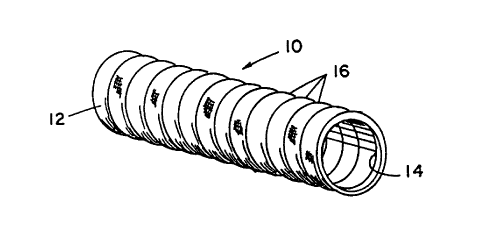
Figure 1 is a perspective view of the crimped
tubular device in accordance with the present
invention;
Figure 2 is a side view of the crimped tube
device shown with proximal and distal nerve ends
affixed within the tube device in accordance with the
present invention;
Figure 3 is a partial cross-sectional view of
the tube device shown in Figure 2 showing the
corrugated exterior surface in relation to the
relatively smooth interior surface and having the
proximal and distal nerve ends sutured in place within
the tube device;
Figure 4 is a side view of the fixture for
crimping the tube including a steel rod and chuck with
an uncrimped tube in place over the rod, a line of
suture material being wrapped around the tube;
Figure 5 is a side view of the crimping
fixture showing the tube being longitudinally collapsed
on the rod with a collar being placed adjacent each end
of the tube to insure the tube holds the desired
configuration; and
Figure 6 is a view of a vacuum oven where the
collapsed tube and crimping fixture are heated under a
vacuum to heat set the externally crimped surface of
the tube.
- 12 - l 3 3 5 5 2 7
DESCRIPTION OF THE PREFERRED EMBODIMENTS
The bioabsorbable device and its method of
use of the present invention is a flexible tube made bv
either the knitting or weaving of bioabsorbable fibers
S into the shape of a tube and, then, dry heat setting of
the tubes to improve the in-vivo strength of the
polymer fibers and and provide a tube with corrugations
along its external surface. The corrugated external
tube surface allows for bending of the tube without
compromising the internal passageway of the tube
device. The tube is used for spanning a significant
nerve gap or defect such as occurs when a nerve is
severed or lacerated and the nerve ends may not be
easily brought back together. The tube is provided
with a relatively smooth interior surface to ease
insertion of the nerve ends into the tube device and to
provide an environment within the tube to promote
longitudinal axon growth across the nerve gap or
defect.
Referring to the accompanying drawings,
Figure l shows a bioabsorbable tube device 10 made in
accordance with the present invention. The tube 10 has
an exterior surface 12 and interior surface 14. The
exterior tube surface 12 is shown having a plurality of
crimps or corrugations 16, thereon. The corrugations 16
on the exterior tube surface 12 allow the tube device
to be bent through an arc of 180 degrees without the
pinching or crimping of the internal surface 14. This
feature is extremely important to the functioning of
the tube device because frequently it is necessary for
the tube device to pass over joints or areas where
bending of the regenerating nerve will occur. If the
interior surface 14 buckles or crimps, the flow of
axonal substances across the nerve gap will be blocked
and the nerve will not fully regenerate across the gap.
- 13 - ~ ~ 3 ~ 5 2 ~
Figures 2 and 3 show the tube device 10 in
place spanning a nerve gap or defect between a proximal
nerve end 18 and a distal nerve end 20. A bioabsorbable
suture material such as a DEXON2 (American Cyanamid
Company, Wayne, N.J. 07470, USA) suture or a MAXON~
(American Cyanamid Company, Wayne. N.J. 07470, USA)
suture is shown at 22 and 24 connecting the proximal
nerve end 18 and distal nerve end 20, to the wall of
the tube device 10.
The suture 22 is threaded through the wall of
the tube device at a point about 5 millimeters away
from an end thereof and, then into the epineural layer
of the proximal nerve end 18. The suture 22 is then
pulled to bring the proximal nerve end into the end of
the tube device 10. The suture 22 is tied to the wall
of the tube in a manner that is known in the art. The
process is then repeated with suture 24 to pull the
distal nerve end 20 into the opposite end of the tube
device.
As shown in Figure 3, the proximal and distal
nerve ends, 18 and 20 are secured within the tube
device 10 such that a gap exists between the nerve
ends. The proximal nerve end 18 will regenerate across
the nerve gap into the distal nerve end 20. Referring
to Figure 3, the corrugations 16 are seen in more
detail as comprising a series of ridges 26 and valleys
28 along the entire exterior tube surface 12. The
interior surface 14 is shown to have a plurality of
flats 30 to provide a relatively smooth surface to ease
insertion of the nerve ends into the tube device and to
provide an optimum environment for axonal growth within
the tube.
The manner of providing the corrugations 16
on the exterior tube surface 12 is shown in Figures
4-6. Figure 4 shows an uncrimped mesh (knitted or
- 14 -
woven) tube placed over a steel rod 32. The diameter of
the rod 32 is appropriately sized so that the tube
slides snugly over the rod. The rod 32 and mesh tube
are mounted on a chuck 34 of a winding device such as a
lathe (not shown) or other commercially available
device to spin rod 32. A braider bobbin (not shown) is
wound with a suture material 36 such as a 4/0 DEXON
(American Cyanamid Company, Wayne, N.J. 07470, USA)
suture material which can be mounted on the cutting
tool holder (not shown) of the lathe. The suture
material 36 is tied to one end of the tube as shown at
38 and then the lathe is rotated to wrap the suture
material about the mesh tube 10. Preferably the suture
material 36 is wound around mesh tube 10 such that
there are approximately twelve (12) wraps of suture
material per longitudinal inch of tube. When the total
length of mesh tube has been wound with suture material
the suture material 36 is cut and tied off around the
opposite end of the mesh tube 10.
Referring to Figure 5, the mesh tube 10 is
shown collapsed or longitudinally compressed on the rod
32 so that the tubes overall length is cut approximately
in half. A collar 40 is inserted on rod 32 to hold the
mesh tube 10 in this collapsed or compressed condition.
The compressed mesh tube 10, rod 32 and chuck 34 are then
placed in a vacuum oven 42 as shown in Figure 6. The
vacuum oven is heated to 130C and a vacuum is pulled to
less than or equal to 1 Torr. The mesh tube 10 is left
in the vacuum oven at < Torr and 130C for two hours.
The use of a vacuum oven on the tube device also improves
the in-vivo properties of the polymer fibres used to make
up the tube device. The heat set process is more fully
described in U.S. Patent No. 3,422,181 to Chirgwin, Jr.
- 1~ - 1 3 3 5 5 2 7
The mesh tube 10, rod 32 and chuck 34 are
removed from the vacuum oven 42 and cooled to room
temperature in a Laminar Flow Hood (not shown). The
suture material 36 is carefully removed leaving a
crimped or corrugated mesh tube. The mesh tube 10
would then have both ends trimmed with scissors and be
inserted into a thermoformed hinged tray. The tray is
placed into a foil pouch for sterilization by known
methods and sealed and sterilized a second time.
The tube device 10 shown in Figures 1-6 is
knitted or woven from a plurality of bioabsorbable
polymer fibers. The preferred polymers and copolymers
are polyglycolic acid (U.S. Patent No. 3,297,033),
polyglycolic acid (U.S. Patent No.3,636,956) and
poly(glycolic-co-trimethylene carbonate) (U.S. Patent
No. 4,243,775). These polymers and copolymers are
preferred because they are known to be well tolerated
by the body upon implantation in addition to being
absorbable within the body.
The polymer and copolymer fibers are
obtainable through methods known in the art. The fibers
are then knitted or woven into tube shape. The various
methods of knitting or weaving such mesh tubes are
further described in the examples below.
In one embodiment of this invention the
knitted or woven mesh tube is manufactured totally from
polymer fibers of 100 percent PGA. The PGA material is
a bioabsorbable polymer which maintains its tensile
strength for approximately thirty (30) days and is then
slowly hydrolyzed within the body. Since the
recognized neural growth rate is approximately one
millimeter (1 mm) per day, a tube device manufactured
from a PGA polymer fiber would remain in place about a
severed nerve long enough to allow a nerve to
regenerate across a 30 mm or 3 cm nerve gap or defect.
1 335527
- 16 -
In another embodiment of this invention, the
knitted or woven mesh tube is manufactured from a
copolymer of glycolide and trimethylene carbonate
linkages (MAXON~ suture material). This copolymer fiber
is known to maintain its tensile strength for at least
fifty-six days before being slowly resorbed into the
body. A tube device manufactured from the MAXON~
copolymer fiber could be used to span nerve gaps of
five centimeters or more.
Objects and advantages of this invention are
further illustrated by the following examples, but the
particular materials and amounts thereof recited in
these examples, as well as other conditions and
details, should not be construed to unduly limit this
invention. For example, while the Examples utilize a
yarn twist in the ''Z" direction, the woven and knit
tube constructions could utilize a twist in the "S"
direction or a combination of fibers with twists in
both the "Z" and "S" directions could be combined in
forming a tube product of the present invention.
EXAMPLE 1
Woven Tube Construction -PGA Polymer Fibers
PGA polymer fibers were woven on a single
shuttle lxl Crompton & Knowles box loom using 16
harnesses. The mesh tube was woven as a double fabric
with selvedge edges attached on both sides. The warp
yarn was 3 ply, 46 denier/21 filament (fiber) PGA yarn
having S turns per inch of twist in the "Z" direction.
The weft (filling) yarn was 3 ply, 46 denier/21
filament PGA yarn having 1.5 turns per inch of twist in
the "Z" direction. The mesh tube construction was a
lxl plain weave having 120 ends per inch per side and
88 picks per inch. The total number of ends in the
mesh tube construction varied from approximately 60 to
- 17 - 1 335527
111 to yield tube sizes of from 2 mm to 6 mm inside
diameter (I.D.). The mesh tube was then crimped, heat
set and cut to the desired length (6 cm) as discussed
above. This construction yields a flexible and porous,
woven mesh tube to be used in accordance with the
present invention.
EXAMPLE 2
Woven Tube Construction - MAXON~ Copolymer Fibers
MAXON~ copolymer fibers were woven into a
mesh tube on the same type of weaving loom as in
Example 1. However, here the warp yarn was 5 ply, 50
denier/25 filament copolymer yarn having 5 turns per
inch of twist in the "Z" direction. The weft (filling)
yarn was 5 ply, 50 denier/ 25 filament copolymer yarn
having 2 turns per inch of twist in the "Z " direction.
The mesh tube construction was a lxl plain weave having
62 ends per inch per side and 68 picks per inch. The
woven mesh tube was crimped and heat set as in Example
1 to provide a tube device in accordance with the
present invention.
EXAMPLE 3
Knit Tube Construction - PGA Polymer Fibers
PGA polymer fibers were knit into a mesh tube
on a tubular weft Lamb Knitting machine using a single
feed jersey stitch construction. The knitting machine
cylinder had a needle density of 25 needles per inch
and the total number of needles in a given cylinder
were varied to yield a mesh tube diameter of from 2 mm
to 6 mm I.D. after fabric finishing. The yarn used was
formed by combining 4 plies of 46 denier/21 filament
PGA fibers, all plied at 2.3 turns per inch of twist in
the "Z" direction. The knitted mesh tubes were
- 18 - l 3 3 5 5 2 7
finished in the same manner as in Example l to provide
a porous, flexible knitted mesh tube to be used in
accordance with the present invention.
EXAMPLE 4
Knit Tube Construction - MAXON~ CopolYmer Fibers
MAXON~ copolymer fibers were knit into a mesh
tube on the same type of knitting machine and knit
construction as in Example 3. However, the cylinder
had a needle density of 33 needles per inch with a
total needle count of about 14 about the perimeter.
The yarn used was formed by combining 3 plies of 50
denier copolymer fibers and 1 ply of 25 denier
copolymer fibers, all plied at 2.3 turns per inch of
twist in the "Z" direction to yield a mesh tube
diameter of about 2 mm I.D. after finishing. The
knitted mesh tube was crimped and heat set as in
Example 1, above.
Various modifications and alterations of this
invention will become apparent to those skilled in the
art without departing from the scope and spirit of this
invention, and it should be understood that this
invention is not to be unduly limited to the
illustrative embodiments set forth herein.