Note: Descriptions are shown in the official language in which they were submitted.
Z~ .fi~- !
-- 1 --
OPHTHALMIC STAPLE AND INSTRUMEN~S FOR IMPLEMENTING U~
The present invention generally relates to surgic~l
staples for implanting in the eye. More specifically,
this invention relates to surgical staples for implanting
in the eye whereby the staples are adjustable while in the
eye to regulate the tension e~erted by the staple on the
eye.
Background of the Invention
In recent years, medical science has made great steps
toward improving vision in the human eye. Among these
improvements, are various forms of surgery on a li~ing
eye. One of the more common forms of surgery i8 the
removal of a cataractus lens, which generally takes the
form of a hardened lens tissue. It is this cataractus
tissue which will cause the blurring of vision.
Freguently, of course, cataracts will appear in the eyes
of the elderly.
In order to remove cataractus tissue, generally the lens
will be removed and replaced with an intraocular lens.
This must be done through an incision made with a scalpel
in the eye. This incision is generally formed in the eye
at the limbus, which is generally the area where the clear
cornea meets the more whiteish-colored sclera. After a
limbal incision has been made, various instruments can be
placed within the eye in order to remove the cataractus -
lens. Sometimes, a phacoemulsification instrument will be
used. After the instrument has removed the cataractus
lens from the eye, and the intraocular lens emplaced `
within the eye, the incision made in the eye must be `
closed. This closing of the incision will present various
problems. For instance, if the incision is closed too
ETH-735
,".: ' ' . ' ' ',
~1 ZS)~ 360
tightly, the resultant healing of the eye will cau8e a
generally ~with the rule~ astigmati8m. On the other hand,
it is also possible that the inci~ion i~ clo~ed too
loosely. This results ~n what i8 known aJ ~against the
rule~ astigmatism. Either type of astigmatism can reaDily
result in about ~ 1 to 2 diopters of change ~n vision
after surgery.
Generally, when sutures are used, the surgeon will keep
these sutures tighter than required to close the
incision. This results in a controlled amount of ~with
the rule~ astigmatism. After surgery, some sutures can be
cut in order to reduce the astigmatism to improve vision
to be near normal. On the other hand, if the sutures are
too loose, it is virtually impossible without re-operation
to correct the ~against the rule~ astigmatism after -
- surgery has been completed.
, . -- ..
In addition, sutures are sometimes adjustable during ~ n-
20 operation by using a slip knot in the sutures. This slip ~-
~; knot can be adjusted so that proper tension is applied to
the wound. Of course, this adjustment of the sutures by a
slip knot is difficult to implement, and reguires much
training and e~pertise.
-~
The reason for introducing an initial ~with-the-rule~
astiqmatism is that it degrades with time, ideally to no
; astigmatis~. However, further degradation will result in
~against-the-rule~ astigmatism. This degradation is ~ ~-
caused by a combination of two factors -- postoperative
stretching of the suture and ~cheese knifing~ of the `~
suture through some of the eye tissue within the suture
loop.
ETH-735
~ ' -`,'`''`.-.`,
Z~n(~3GO
-- 3 --
With this astigmatism problem, as well as the problem of
keeping incision lengths small in order to avo~d
contamination and/or trauma to the open site, it becomes
e~tremely important to find an accurate, reliable and safe
way to close medical cuts made in the eye surface. The
use of staples is indicated.
.,
On the other hand, staples have been difficult to
implement because of the general form of a staple. For
instance, most surgical skin staples are rigidly bo~
shaped. This bo~ shape is not adjustable while in the eye
either during operation or postoperatively because of its
need to be pressured against the eye during forming. Also
because of the small tolerances within the eye, precise
15 and delicate stapling systems are required to implant the ~
staples.
In addition, removal of these staples is difficult because
- the staple should generally be removed through its
insertion point. If a bo~ shaped staple is removed, it
will generally rip tissue on its way out, which is
undesirable. The same holds true for a ~B~ shaped
staple. There, an even greater tissue tear takes place.
What is necessary, therefore, is a staple which is able to
be removed from precisely the same path in which it enters
the tissue, or a staple which can be emplaced within the
eye and cause minimal trauma, so that it can remain in the
eye.
~ummary of the Invention
It is therefore an object of the present invention to
provide a surgical staple emplaceable within the eye. The
surgical staple should be easily emplaceable and capable
of controlling the amount of tension created on the eye
ETH-735
, . ~ ,., .. ,.. ~ . .. . . . .. ~ .
2(~ ;360
surface. In addition, it is an object of the present
invention to provide a surgical staple that will remain in
the eye after surgery.
S It is a further object of the present invention to provide
a surgical staple in which the tension the staple e~erts
on the eye is adjustable while the staple is in the eye.
It is yet another object of the present invention to
provide a surgical staple which will not cause a large
amount of tissue trauma when emplaced within the eye.
It is a further object of the present invention to be able
to apply a surgical staple in the eye with forceps, needle
lS holders, tweezers and~or similar specially formed applying
instruments, instead of the conventional surgical staplers
using drivers and the like. -
It is yet another object of the present invention to
20 provide a surgical staple which is implantable and -
adjustable in the eye with a pair of specially formed -~
pliers and in which the tension can be adjusted while the
staple is implanted within the eye.
It is finally an object of the present invention to
provide a surgical staple in which the staple is
insertable into the eye by inserting one leg into the eye, ~ :
on one side of the incision, then stretching the crown of
the staple, inserting the second leg of the staple into -
the eye on the second side of the incision, and then
adjusting the entire staple in order to create the --
appropriate tension on the eye.
These and other objects of the present invention are - -~-~
accomplished in a surgical staple which has two legs which
ETH-735
zn()~ 60
-- 5 --
are joined by a generally resilient crown. She leg~ of
the staple are situated in a plane perpendicular to the
crown. The crown contains a spring-like member which can
be adjusted, so that the leg8 of the staple are movable
relative to one another by deformation of the spring-like
member. In addition, the staple is insertable within
tissue such as the tissue of the eye by use of a forceps
or needle holder-like device which is able to grip the
staple. One piercing leg is inserted; the staple is ~-
stretched to draw the incision closed; the second staple
leg is then inserted into the far side of the incision;
the staple is then adjusted for appropriate tension on the
eye. The adjusting device is generally pliers-shaped with
mushroom shaped heads, and insertable within the
lS spring-like portion of the crown.
Brief Description of the Drawings
The invention will be more fully described in the
following detailed description of the invention in
conjunction with the accompanying drawings wherein:
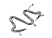
Fig. 1 is a perspective view of a preferred embodiment of
the staple of the present invention.
Fig. 2a is a top plan view of a preferred embodiment of
~; the present invention.
:::
Fig. 2b is an elevation view of a preferred embodiment of
the present invention.
Fig. 3a is a top plan view of a second preferred
embodiment of the present invention.
:
Fig. 3b is an elevation view of a second preferred
ETH-735
i" .,
... ~ - .. ~, ., . . ., ~ - .. ... , - . -.- . . .. . . -
! ~ 6
embodiment of the present invention.
Fig. 4a is a top plan view of a third preferred embodiment
of the present invention.
Fig. 4b is an elevation view of a third preferred
embodiment of the present invention.
Fig. 5a is a top plan view of a fourth preferred
embodiment of the present invention.
Fig. 5b is an elevation view of a fourth preferred
embodiment of the present invention.
: . ,
Fig. 6a is a top plan view of a fifth preferred embodiment
of the present invention.
. ,
Fig. 6b is an elevation view of a fifth preferred ~ ;-
~ embodiment of the present invention.
Fig. 6c is a top plan ~iew of a si~th preferred embodiment
of the present invention.
Fiq. 6d is an elevation view in partial cross section of a
si~th preferred embodiment of the present invention.
Fig. 7 is a perspective view of the application instrument
of the present invention inserting the staple of the -- =--
present invention into tissue. ~ `
i 30
Fig. 8a, Fig. 8b and Fig. 8c are elevation views of the
insertion of the staple of the present invention into -
tissue. ~-
. -
35 Fig. 9 is a partial elevation view of a leg of the staple ~ ~-
ETH-735
27~)()(~3
of the present invention.
Fig. 10 is a perspective view of a preferred embodiment of
an adjusting tool of the present invent~on. ~'~
Fig. lOa is a perspective view of the heads of an
adjusting tool of a second preferred embodiment of the
present invention.
Fig. lla and Fig. llb are perspective views of tensioning
and removal of the staple of the present invention by
using an adjusting tool of the present invention.
Fig. 12 is a perspective view of adjusting staple tension
lS in the staple of the present invention.
Fig. 13 is a top plan view of the staple of the present
invention as inserted into a limbal incision in the eye.
Detailed Description of the Present Invention
:
As can be seen in Figures 1, 2a, 2b, 3a, 3b, 4a, 4b, Sa,
5b, 6a, 6b, 6c and 6d, ophthalmic staples 10, 20, 30, 40,
50 and 60 disclose various preferred embodiments of the
present invention. As more accurately disclosed in Figure
1, with each of these ophthalmic staples there will be a -
pair of legs 12 and a crown 14. The crown 14 lies in a
plane generally perpendicular to the plane of the legs as
can be seen from Figure 2a and 2b. The crown 14 is
spring-like so that it allows the relative motion of the
legs 12 with one another, by deflection of the crown 14.
; This is generally due to the resilient spring-like member
16 of the crown 14 which is able to be deflected depending
on the desired tension in the staple 10. This spring-like
member 16 joins rigid members of the crown 18. These
:~
~ETH-735 -~
.-, - ~,~,..
~: 2()0(1!3fiO
-- 8 --
rigid members 18 connect the generally piercing legs 12 to
the crown 14.
Generally, the spring-like member 16 w~ll conta~n two
cross members 17 which are connected by a generally
fle~ible connecting member 19. At their opposite ends,
the generally rigid cross members 17 are connected to a
receiving member 15. This receiving member 15 is, in
turn, connected to the generally rigid members 18 of the - -
crown portion 14. The use of these receiving members 15
will be e~plained shortly. Thus, as can be seen ~n Figure
3 and 3a, there is an ophthalmic staple 20 having legs 22,
a crown 24, a resilient spring-like member 26, rigid
members 28, cross-members 27, fle~ible connecting members -~
29 and rounded receiving members 25, all corresponding to
the members in ophthalmic staple 10. This staple takes on ~ -
a more rounded shape in order to provide greater
fle~ibility to the staple. ~ -
, ':
As seen in Figures 4a and 4b, the corresponding ophthalmic
staple 30 contains legs 32, crown 34, spring-like member -
36, rigid member 38, cross member 37, and fle~ible
connecting member 39. Receiving member 35 has been made
straight instead of having a rounded appearance. This
25 straight receiving member 35 is useful in minimizing the ~ --
width of the staple.
-:~
On the other hand, Figures 5a and 5b disclose ophthalmic
staple 40 which contains legs 42, crown 44, spring-like
member 46, cross member 47, fle~ible connecting member 49 -~
and rounded receiving member 45. In this type staple
there is no additional rigid member attached to the legs
of the staple. This, of course, reduces the total length
of the staple.
~ ~,
ETH - 7 35
.,
,
.. , ., . , ,, , -
3 fi (~ ~ ~
On the other hand, where more rigidity is desired in the
positioning of the legs, one may use ophthalmic staple 50,
as seen in Figures 6a and 6b. In this device there are
legs 52, crown 54, spring-like member 56, rigid member 58,
cross member 57, fle~ible connecting member 59 and rounded
receiving member 55, all ha~ing a form generally
corresponding to staple 10. In addition, however, there
are provided loops 51 useful in the placement of the legs
without relying escessively on deflection of the
spring-like member 56. These loops 51 are connected to a
rigid member 58 which is now generally perpendicular to
the legs 52 in the plane of the crown 54. This allows for
a greater control of the lateral, or side-to-side motion
of the staple 50 while easily ensuring accurate in-tissue
placement of the staple 50.
Ultimately, the staple of the present invention will take
on the generally preferred embodiment of staple 60
demonstrated in Figures 6c and 6d. In this staple 60,
there are legs 62 which generally estend at about 30-
angles from the crown 64 of the staple. As in all
previously demonstrated ophthalmic staples 10, 20, 30, 40,
50, the plane of the legs 62 is perpendicular to the plane
of the crown 64. The crown 64 contains a spring-like
member 66 which is comprised of a fle~ible connecting
member 69 and rigid cross members 67. These cross members
67 e~tend at about 60 angles away from the peak at the
flesible connecting member 69. The cross members 67 are
connected at their other ends to rounded receiving members
65. In addition, the rounded receiving members 65
describe nearly an entire circle before attachment to
rigid members 68, attached to the legs 62.
:
In general, this staple has a diameter from .0015~ to
.005~, generàlly about .003~, and the length of the staple
~,
ETH-735 ~
2n()(~3fi~
-- 10 --
at the crown is .040~ to .120~, generally about .080~.
The staple legs are roughly .015~ to .050~ in length,
generally about .028~; the legs estend at angles of
between 20 and 55- to the plane of the crown usually
S about 30. The width of the staple is about .010~ to
.080~, usually equally divided on both sides of the plane
of the legs. The legs can either be straight (legs 62 of
Figure 6d) or rounded (legs 62a of Figure 9).
When placement of these staples is desired, the steps in
Figure 7 and Figures 8a, 8b and 8c are followed. A
generally tweezer-like instrument 70 having legs 72 grasps
the staple 10. The grasp is such that the plane of the
tweezer-like instrument lies in the same plane as the
crown 14. First, one leg is embedded into the tissue of
the eye as seen in Figure 8a. This is accomplished by
grasping the tweezer-like instrument 70 near the end of
the crown 14 toward first leg 12a. At that point, the -
tweezer-like instrument 70 is shifted to the other side in
order to insert leg 12b. Intermediate this step the crown
14 is stretched elastically in order to properly embed
both the legs, 12a and 12b. This will avoid deforming the ~ -
; legs when inserting leg 12b. ~
25 At this point, the staple is ready for adjustment. ~ -
Generally, a pliers-like tool 80 as seen in Figure 10 is
used for adjustment. This pliers-like tool contains `
grasping prongs 82 which fit around the legs. As seen in
Figures lla and llb, or Figure 12, these graspinq prongs
82 can be used to adjust the tension in the ophthalmic
staple. By having the grasping prongs pull the staple
together, the tension the legs 12 esert on the tissue can
be increased. By pulling the legs apart, the tension that ~-~
legs 12 esert on the tissue can be decreased. In fact, as -
35 seen in Figure llb, the staple 10 can be removed without ~ - ~
,: , ~
. -
ETH-735
''
~` 2~)()a~;3~
trauma to the eye.
As seen in Figures lOa and lOb, the grasping prong~ 82 are
modified to take on the mushroom 8hape of prongs 84.
These mushroom shaped prongs 84 are more preferable than
the grasping prongs in that they need not be aff~sed
around the estremely small staple 10. In fact, the
mushroom shaped prongs 84 generally fit into the rounded
receivers such as the rounded receiver 65 seen in Figure
6. These mushroom shaped prongs 84 can also ease or esert
more tension on the staple 10.
Generally, the staples are implanted at the end of
ophthalmic surgery. Usually, the conjunctiva is refracte~
from the sclera. The staple is placed on the sclera
surface. Generally, the staple is then left permanently
in the eye. The conjuctiva is pulled over the staple and
the staple becomes embedded in the sclera. The conjuctiva
heals quickly, usually within two to three days. There is
generally no worry about conjunctivitis because the staple
is formed from an implantable metal, often stainless steel
or vitalium. When the staple becomes embedded in the
sclera, the conjunctiva heals smoothly over the staple,
preventing trauma.
While the staple is permanently embedded in the sclera, it
can still be adjustable. If it desired to increase the ~^
tension on the staple, the mushroom shaped pronqs 84 of
the pliers-like instrument 80 are placed within the
conjunctiva and within the rounded receiving member 65.
The staple is eased together to increase or decrease the
tension eserted by the legs 12 on the tissue.
Alternately, mushroom shaped prong 84 can relieve the ~ 9
tension in the staple 10 by easing the crown apart so that
the legs 12 are relased within the sclera.
~ .. . ~ ,.~ . .
ETH-735 -- -
, .. . -
: ,- ~.- , ~,.. .-
. i' ', ~ ,!
- - 12 -
The unique combination of features possessed by the
present invention render them suited for in the eye
implantation. These combinations may be useful for other
forms of tissue implantation outside the eye. Naturally,
while several means are available, the particularly
advantageous embodiments have been chosen to illu~trate
the invention. It will be understood by those skilled in
the art that various changes and modifications may be made - -
therein without departinq from the scope of the invention,
which is defined by the following claims and their
equivalents.
~, ~
~ ~.
, ~
- , ...
~ ~ 25~
- .
j~ 30 i ~ ~ ~
- , ~~- .. .
ETH-735
,
:
,: