Note: Descriptions are shown in the official language in which they were submitted.
2004658
This invention relates to a surgical clip and
insertion tool and, more particularly, to a biodegradable
barbed clip and a tool for inserting the clip that can be
used to repair menisci and soft tissue.
Tears in fibrocartilage and soft tissue,
especially peripheral meniscal tears, are relatively
difficult to repair. Typically, a tear in the vascular
region of the meniscus is sutured using arthroscopic
techniques. The instrument may be inserted through small
incisions which serve as anterior knee portals. Sutures on
long needles are then passed through a meniscal repair
instrument and through the meniscus. An incision is made
in the back of the knee to permit the surgeon to pull the
needles and suture out, and to tie the suture over the
posterior joint capsule. This technique reapproximates the
torn edges of the meniscus and allows for healing.
Although effective, this repair technique
requires a surgeon skilled in arthroscopic meniscal repair.
The technique is also relatively time consuming and more
invasive than it need be, as it requires a second,
posterior incision that increases the risk of infection and
neurovascular damage. As a result, few surgeons will
attempt meniscal repair, choosing instead to simply remove
the damaged portion of the meniscus. The problem with this
approach is that meniscal removal can cause increased
stress on the articular cartilage, which may then lead to
degenerative arthritis.
Surgical clips are often easier to insert than
sutures. However, most clips are not biodegradable, and
occasionally a second operation must be performed to remove
the clip once the tissue has healed. Another disadvantage
of these clips is that they are not well suited for
meniscal and soft tissue repair, as they are typically
metallic, relatively large, and may protrude from the
tissue and cause joint irritation. Thus although
arthroscopic clips can be inserted through a single
incision, they have typically not been used for repair of
, ~
2004658
peripheral meniscal tears nor for arthroscopic repair of
soft tissue.
It is therefore an object of this invention to
provide a surgical clip and insertion tool for operative
arthroscopic repair of menisci and soft tissue.
It is a further object of this invention to
provide an arthroscopic clip and insertion tool which
allows a surgeon not trained in meniscal repair to
reapproximate torn meniscal tissue.
It is a further object of this invention to
provide an arthroscopic clip and insertion tool which
decrease risk of neurovascular damage to the patient.
It is a further object of this invention to
provide an arthroscopic clip and insertion tool which
decrease operative time, by facilitating the operative
procedureO
It is a further object of this invention to
provide an arthroscopic clip and insertion tool that do not
require a second operation to remove the clip.
It is a further object of this invention to
provide an arthroscopic surgical clip with legs that remain
buried in the tissue and do not irritate surrounding
tissue.
It is a further object of this invention to
provide an arthroscopic surgical clip insertion tool that
tightly holds a clip until it is completely inserted.
According to one aspect of the present invention,
there is provided a meniscal clip for arthroscopic repair
of peripheral tears in the meniscus comprising: a pair of
opposed legs formed of rigid biodegradable material, each
of said legs having at least one barb for easily
penetrating the meniscus and opposing withdrawal from it;
and a flexible section interconnecting said legs and formed
of a flexible biodegradable material.
According to another aspect of the present
invention, there is provided an arthroscopic clip for
arthroscopic repair of tears in fibrocartilage and soft
~`-A~
2004658
tissue comprising: a pair of opposed legs formed of rigid
biodegradable material, each of said legs having at least
one barb for easily penetrating the cartilage and soft
tissue, and for opposing withdrawal from it; and a flP~;hle
section interconnecting said legs and formed of a flexible
biodegradable material.
According to yet another aspect of the present
invention, there is provided an arthroscopic clip
comprising: a first curved leg including first anchoring
means for positionally securing said first leg in tissue
without a retainer; a second curved leg including second
anchoring means for securing said second leg in tissue
without a retainer; and a soft flexible connecting member
secured to a first end of each of said legs.
According to still another aspect of the present
invention, there is provided an arthroscopic clip
comprising: a first curved leg including first anchoring
means for positionally securing said first leg in tissue;
a second curved leg including second anchoring means for
securing said second leg in tissue; and a connecting member
secured to a first end of each of said legs, said
connecting member being composed of a soft flexible suture
material.
According to a further aspect of the present
invention, there is provided an arthroscopic clip
comprising: a first curved leg member composed of a rigid
material, said first leg member including at least one
tissue retaining barb; and a second curved leg member
composed of a rigid material, said second leg member
including at least one tissue retaining barb; and a
connecting member composed of a soft flexible material
secured to a first end of each of said legs, said flexible
connecting member being bendable to allow said curved legs
to move toward each other and overlap as said legs are
pushed into tissue, said barbs securing said legs in tissue
without a retainer.
3 --
~ . .~
2004658
According to still a further aspect of the
present invention, there is provided an arthroscopic clip
comprising: a first and a second curved leg composed of a
rigid biodegradable material; and a connecting member
composed of a soft flexible biodegradable suture material
secured to a first end of each of said legs.
According to yet a further aspect of the present
invention, there is provided an arthroscopic clip
comprising: a first leg including first anchoring means
for positionally securing said first leg in tissue without
a retainer; a second leg including second anchoring means
for securing said second leg in tissue without a retainer;
and a soft flexible connecting member secured to a first
end of each of said legs.
According to an additional aspect of the present
invention, there is provided an arthroscopic clip
comprising: a first leg member composed of a rigid
material, said first leg member including at least one
tissue retaining barb; and a second leg member composed of
a rigid material, said second leg member including at least
one tissue retaining barb; and aconnecting member composed
of a soft flexible material secured to a first end of each
of said legs, said flexible connecting member being
bendable to allow said legs to move toward each other and
overlap as said legs are pushed into tissue, said barbs
securing said ~egs in tissue without a retainer.
According to another additional aspect of the
present invention, there is provided an arthroscopic
instrument for applying barbed arthroscopic clips for
repair of tears in fibrocartilage and soft tissue
comprising: a pair of opposed jaws each having at least
one notch for holding the barbs of said clip; biasing means
for separating said jaws in a normally open position; a n
actuating handle having opposed handle members; means for
interconnecting said handle and said jaws; means for
closing said jaws when said handle members are moved a
first way relative to one another and opening said jaws
_ - 4 -
. .. .
,.
2004658
when said handle members are moved the other way relative
to one another; and means for releasing said clip from said
jaws.
This invention results from the realization that
surgical clips for repairing tears in menisci and soft
tissue can be improved dramatically by providing a
biodegradable clip with rigid, barbed legs interconnected
by a flexible section that is inserted in the tissue with
an arthroscopic tool and anchors itself in the tissue to
approximate the tear.
This invention features a surgical clip and
insertion tool for repair of tears in fibrocartilage and
soft tissue. The clip has a pair of opposed legs formed of
rigid biodegradable material interconnected by a flexible,
biodegradable section. Each leg has at least one barb that
allows the clip to be easily inserted into the tissue being
repaired but keeps the clip from working out of the tissue.
The legs of the clip are preferably curved inwardly toward
each other, and each leg preferably has more than one barb
on its outer side. A preferred material for the legs is a
polyglycolic acid polymer. Preferably, the clip is an
arthroscopic clip.
The instrument for applying the barbed clips has
a pair of opposed jaws that are preferably offset to allow
them to overlap when closed. Each jaw has at least one
notch for holding the barbs of the clip. These specialized
jaws tightly hold the clips in place until they are fully
inserted, which allows the physician to place them in
exactly the right position and to exactly the right depth
before they are released. The instrument includes a
biasing means for separating the jaws in a normally open
position, which is the position in which the jaws remain as
the clip insertion begins. The jaws are connected to a
handle by a member such as a tubular member. The
instrument also includes means for closing the jaws when
the two handle members are moved one way relative to one
-A
2004658
another and opening the jaws when the handles are moved
another way.
Preferably, the actuating handle is normally
biased open, and the means for closing the jaws closes them
when the handle members are squeezed together and opens
them when the handle members are spread apart. The means
for closing may include means for pulling the jaws together
as the handle members are moved. The jaws may be disposed
at an angle to the tubular member to facilitate clip
insertion. Additionally, the tips of the jaws are
preferably sharpened to facilitate clip insertion.
Preferably, the instrument further includes means
for releasing the clip from the jaws so it stays in place
when the jaws are opened. The means for releasing may
include means for releasably holding at least one barb of
the clip, and may further include means for selectively
releasing the means for releasably holding the barb from
the barb. Means for actuating the means for selectively
releasing, which may include an actuating member or switch
on the handle of the instrument, are also preferably
included.
In use, the clip is placed in the jaws with the
barbs in the jaw notches. This holds the clip tightly in
place as it is inserted. To insert the clip, the physician
begins pushing the jaws into the tissue, squeezing the
handle members at the same time. The sharpened tips of the
jaws pierce the tissue and, as it is inserted, the legs are
moved together. Preferably, the jaws of the instrument
overlap when they are completely closed. This overlapping
causes the legs of the clip to overlap when it is
completely inserted in the tissue. The switch on the
handle then is moved upward to release the clip from the
jaws. The barbs on the clip legs then hold the clip in
position once the jaws are opened up and pulled away from
the clip. When the instrument is removed from the tissue,
the clip remains with its legs completely embedded within
the tissue with only the flexible, non-irritating
2004658
interconnecting member protruding from the tissue surface.
Since the clip is biodegradable, it slowly dissolves as the
tissue heals itself, and there is no need for a second
operation to remove the clips.
Other objects, features, and advantages will
occur from the following description of a preferred
embodiment and the accompanying drawings, in which:
Figure 1 is an elevational view of a surgical
clip for repairing tears in cartilage and soft tissue
according to this invention;
Figure 2 is a diagram of a meniscular tear
reapproximated by five of the clips of Figure 1;
Figure 3 is a cross-sectional view taken along
line 3-3 of Figure 2;
Figure 4A is an axonometric view of an
arthroscopic tool for inserting the clip of Figure 1
according to this invention;
Figure 4B is a close-up view of the jaws of the
tool of Figure 4A;
Figure 4C is a partial cross-sectional view of
the handle, trigger and actuating member of the tool of
Figure 4A;
Figure 4D is a diagrammatic view of an angled jaw
for the tool of Figure 4A;
Figure 5 is an elevational view of an alternative
surgical clip according to this invention; and
Figure 6 is an elevational view of an alternative
to the jaws of Figure 4A.
A surgical clip for repair of tears in
fibrocartilage and soft tissue which is especially useful
for arthroscopic meniscal repair according to this
invention may be accomplished by providing a clip with a
pair of opposed legs formed of a rigid biodegradable
material. Each leg has at least one barb that allows the
clip to easily penetrate the tissue being repaired and
oppose withdrawal from it. The legs are interconnected by
.~~
2004658
a flexible, non-irritating biodegradable section that
allows the clip to bend.
Preferably, the legs of the clip are curved
inwardly toward each other, and the barbs are on the
outside of the legs. The barbs may alternatively be on the
insides of the legs. These legs are preferably made from
a polyglycolic acid polymer. The flexible interconnecting
section can be formed of suture material or another
relatively soft, flexible material that allows the clip to
bend as it is inserted so that the clip can overlap inside
the tissue being approximated. The clip is ideally suited
for reapproximating peripheral meniscal tears.
The instrument for applying the clips includes a
pair of opposed jaws each having at least one notch for
holding the barbs of the clip. This allows the clip to be
tightly held in place in the jaws until it is completely
inserted in the tissue being repaired. The jaws are biased
apart in a normally open position, and they are attached to
the handle by a tubular member. The actuating handle has
opposed handle members and is also connected to the jaws by
means such as a pair of wires or an actuating member which
close the jaws when the handle is squeezed. The instrument
may have jaws preset at different angles to further
facilitate insertion of the clip. The jaws may also have
sharpened tips to facilitate insertion. In addition, the
jaws are preferably offset so they overlap when closed, so
that the legs of the clip overlap inside the tissue to
better approximate the torn tissue.
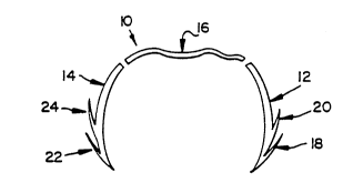
There is shown in Figure 1 one embodiment of a
surgical clip 10 for repair of tears in cartilage and soft
tissue according to this invention. The clip 10 is
especially useful for arthroscopic repair of peripheral
meniscal tears, partial or small rotator cuff tears, labrum
tears in shoulder arthroscopy, and retinacular repair after
patellar dislocations. The clip is also useful as a
replacement for internal sutures, for example, in the
repair of lacerations of the liver or spleen. The clip 10
2004658
has barbed legs 12, 14 with barbs 18, 20, 22, 24,
respectively. The barbs 18, 20, 22, 24 may be on the
inside or outside of the legs 12, 14. The legs 12, 14 are
formed of a rigid biodegradable material which may be a
S polyglycolic acid polymer. The legs 12, 14 are
interconnected by a biodegradable, flexible, non-irritating
section 16 which may be formed of a suture material. The
flexible section 16 bends to allow the legs 12, 14 to move
in toward each other and overlap as they are pushed into
the tissue. Once inserted the clip 10 holds the tissue in
place long enough for it to heal, and slowly dissolve so
the patient does not have to undergo a second operation for
removal of the clip 10.
The peripheral meniscal tear 32 illustrated in
Figure 2 is a relatively small tear that is considered
repairable. Because of the problems to date with meniscal
surgery, a portion 94 of the meniscus 30, encompassed by
the dashed lines, has often been removed when a peripheral
tear 32 is found. Since meniscal removal may cause
increased stress on the articular cartilage and secondarily
lead to degenerative arthritis, repair is far superior to
removal. By using clips such as clips 34, 35 and 38, the
meniscus can be successfully repaired with a single
operative procedure.
In Figure 3, the clip 34 is shown embedded in the
meniscus 30. The clip 34 includes barbed legs 38, 40
interconnected by a flexible section 36. When inserted,
the legs 38, 40 overlap, which causes the legs 34, 40 to
approximate the tissue and minimize the gap 32 through
which fibrous tissue will form and thus further enhance
healing. When the clip 34 is inserted as shown, only the
soft connecting flexible section 36 protrudes from the
tissue and is exposed to the articular cartilage. Since
this soft material does not irritate the surrounding
articular cartilage, the patient does not need to have the
joint rigidly immobilized for a long period of time. This
_ g
1 ~
A
2004658
is an additional advantage of the arthroscopic clip
according to this invention.
An arthroscopic instrument 50 for applying the
barbed clips 10 is shown in Figure 4A. The instrument 50
includes notched jaws 64, 66 made of spring steel formed to
keep them in a normally open position. A tubular member 60
interconnects the jaws 64, 66 to a handle 62. A pin, bolt,
or rivet 79 attaches the jaws 64, 66 to the tubular member
60. The handle 62 includes handle members 52, 54 that are
normally biased apart by a spring 56. A hinge pin 58
allows the handle member 54 to move toward the handle
member 52 as the handle 62 is squeezed.
The action of the opening and closing of the jaws
64, 66 of the instrument can be more clearly seen in Figure
4B. The jaws 64, 66 are formed from a spring steel member
68. Wires 53, 55 are attached to the jaws 64, 66 at points
82, 84, respectively, and are separated by running the
wires 53, 55 through channels 78, 80 attached to the inside
of the tubular member 60. The wires 53, 55 are pulled taut
when the handle 62 is squeezed. This causes the jaws 64,
66 to close. The clip 10 is held firmly in place in the
jaws 64, 66 as it is inserted in the tissue by providing
notches 70, 72 in the jaw 64 and notches 74, 76 in the jaw
66 that are shaped to hold the clip barbs 18, 20, 22, 24.
Insertion is further facilitated by sharpened tips 65, 67,
which pierce the tissue just ahead of tips of the clip 10.
In operation, a clip such as clip 10 of Figure 1,
is inserted in the open jaws 64, 66. The barbs 18, 20 fit
into slots 70, 72 and the barbs 22, 24 fit into slots 74,
76. The tip of the clip 10 is located very close to the
tips 65, 67 of the ~aws 64, 66, respectively. When the
jaws 64, 66 are in place against the two sides of the torn
tissue being repaired, the instrument 50 is moved forward
and the handle 62 is squeezed. This pushes the legs 12, 14
of the clip 10 into the tissue and moves the legs 12, 14
together as the clip 10 is inserted. The jaws 64, 66 of
the instrument 50 are preferably made slightly offset, as
-- 10 --
_
2004658
shown in Figure 4B, so that they overlap when completely
closed. In this case, when the clip 10 is completely
inserted its legs 12, 14 are crossed, as shown in Figure 3.
Whether the clip legs 12, 14 are crossed or not,
they are completely embedded within the tissue. This is
advantageous because the rigid material of the legs 12, 14
could irritate the tissue surrounding the area being
repaired as the prior art surgical clips have done in other
parts of the body. Once the clip 10 is inserted, the jaws
64, 66 are backed out of the tissue and the instrument 50
is removed from the patient. The instrument 50 can then be
used to insert another clip 10 in the torn tissue.
The operation of the handle 62 to open and close
the jaws 64, 66 is shown more clearly in Figure 4C. The
tubular member 60 is attached to the handle member 52 by a
rivet 57. The wires 53, 55 are attached to the handle
member 54, which pivots on a pin 58 when the handle members
52, 54 are squeezed together. As the handle member 54
moves toward the handle member 52, it pulls the wires 53,
55 back, which in turn pulls the jaws 64, 66 together and
causes them to close. Since the jaws 64, 66 are normally
biased apart, when the handle members 52, 54 are released,
the wires 53, 55 are relaxed, and the jaws 64, 66 open to
release the clip 10. A wire 108 is attached to a sliding
switch 92, which pulls the wire 108 when it is slid in the
direction of the arrow (shown in Figure 6) to release the
clip 10 as fully explained below in conjunction with Figure
6.
An alternative way of forming the jaws 64, 66 of
the arthroscopic instrument 50 is shown in Figure 4D. The
jaws 64a, 66a are formed at a 30 angle to the tubular
member 60a. A pin, rivet, or bolt 79a attaches the jaws
64a, 66a to the tubular member 60a.
Preferably, at least three insertion tools 50
with jaws 64, 66 at different angles are available. One
with the jaws 64, 66 aligned with the tubular member 60,
one with the jaws 64, 66 turned down at an angle of
2004658
approximately 15, and one with the jaws 64, 66 turned down
at an angle of approximately 30. This allows the
physician to place the clip 10 exactly as desired,
depending on the location of the tear, utilizing the same
arthroscopic portal during repair.
Another way of forming the arthroscopic clip 10
is shown in Figure 5. A clip lOa includes barbed legs 12a,
14a formed of a rigid biodegradable material, for example
a polyglycolic acid polymer. The barbs 18a, 20a, 22a, 24a
are fully embedded within the tissue being repaired and do
not interfere with joint movement. A pair of tip barbs 96,
98 allow the clip lOa to grip the meniscus so it stays
embedded when the jaws 64, 66 are pulled back and out of
the patient. This is more clearly shown in Figure 6~ A
flexible section 81 is made an integral part of the clip
lOa, but is preferably made from a relatively soft,
flexible biodegradable material which allows the clip lOa
to bend as it is inserted so the legs 12a, 14a can be fully
embedded in the tissue. A preferred material of the
flexible section 81 is 2.0 Dexon suture. Since the
interconnecting flexible section 81 is the only section of
the clip lOa that is exposed from the meniscus after the
clip lOa is inserted, the soft material also provides a
clip that is less irritating to the surrounding cartilage
than the typical stiff or metallic clips would be and which
would not be able to be used intra-articularly.
Another way of forming the jaws to ensure proper
insertion of clip lOa is shown in Figure 6. A pair of jaws
100, 102 are formed to hold the barbs 18a, 20a, 22a, 24a of
the clip lOa while the clip lOa is being inserted and to
release the clip lOa after insertion so that it remains in
place embedded in the meniscus. A pair of barb-holding
members 104 are spring steel members with small
indentations shaped to fit and hold the clip barbs 18a,
20a, 22a, 24a. A wire 106 is attached to the barb-holding
members 104.The wire 108 connects the wire 106 to a
switch 92.
- 12 -
~" ,,.
.....
2004658
When the clip lOa is fully inserted in the
meniscus, the physician operates the switch or lever 92.
The switch 92 operation pulls on the wire 108, as shown in
Figure 6, which in turn pulls the wire 106. The wire 106
is attached to the underside of the barb-holding members
104. As the wire 106 is pulled tight, it pulls the barb-
holding members 104 down away from the clip lOa. This
frees the barbs 18a, 20a, 22a, 24a and leaves them embedded
in the meniscus. The tip barbs 96, 98 also may be included
to help hold the clip lOa in place by gripping the tissue
just enough to allow the clip lOa to separate from the jaws
100, 102 as the jaws 100, 102 are opened and backed out of
the meniscus. In conjunction with the barb-holding members
104, the tip barbs 96, 98 allow the clip lOa to properly
separate from the instrument 50 as it is removed from the
mensicus.
Although specific features of the invention are
shown in some drawings and not others, this is for
convenience only as each feature may be combined with any
or all of the other features in accordance with the
invention .
.:. .