Note: Descriptions are shown in the official language in which they were submitted.
~5;~
203-181
1 ABSO~R~RT~ SURGICAL FASTENER
WITH BONE PENETRATING ELEMENTS
BACKGROUND OF THE lN V~N'l'lON
1. Field of the Invention
This invention relates to surgica~ fasteners ~or
fastening body tissue and moxe particularly relates to an at
least partially absorbable fastener for fastening bone or
hard tissue.
2. Background of the Xelated Art
Bone fastening or fixation devices are well known
in the art. Typically, such fasteners are in the form of
staples, pins, screws, and wires. For example, both Pratt
et al., U.S. Patent No. 4,454,875 and Ellison et al., U.S.
Patent No. 4,570,623 di~close staples for being driven into
bones. Staples of this type are generally fabricated of
biologically inert metal, such as stainless steel, titanium,
cobalt-chromium-molybdenum alloys and the like. The staple
must be relatively strong and hard so that it can be easily
driven directly into bone or hard tissue.
Other metal fasteners are well known cc ?rcial
products used for a wide variety of bone fixation
procedures. Pins and wires are generally constructed from
stainless steel and are grasped in a drill chuck and
self-drilled directly into bone to treat a given traumatic
or pathological condition.
The disadvantage of metal fasteners is that after .
they have completed their function of supporting the bone
while the bone heals, they remain permanently in the body.
Problems can arise after healing, for example, by corrosion
3~ of the metal, or when the pins or staples work loose from
their moorings and migrate through body tissue.
. 35
.
Z~ 3~
-2-
1 Furthermore, permanent metal fixation devices
shield the bone from beneficial stresses after healing. It
has been shown that moderate periodic stress on bone tissue,
such as the stress produced by exercise, helps to pre~ent
decalcification of the bone. Under some conditions, the
stress shielding which results Prom the long term use of
metal bone fixation devices can lead to osteoporosis.
These disadvantages can be mitigated by the use of
bioabsorbable surgical fasteners, which degrade over a
period of time thereby gradually transferring more support
load to the bone as it heals. Such fasteners for bone are
also known in the art. For instance, Tunc, U.S. Patent No.
4,539,981 teaches the use of polymers of L(-)lactide for
fabricating bone fixation devices. Moreover, various types
~f bioabsorbable pin fasteners have been commercialized.
For example, some types of pins are fabricated from poly
(p-dio~Anone) and are indicated for use to fix in place
small bony fragments in the knee and hand, where such
fragments are not in tension. As is characteristic for all
such absorbable pins, holes must be previously drilled into
the bone in order for the pins to be inserted.
Bioabsorbable fasteners are not self-inserting, i.e. they
are not capable of being driven or screwed directly into
bone because the polymeric material they are made of is
relatively soft. The necessity to predrill holes in the
injured bone adds to the surgical procedures and lengthens
the time required to complete the operation.
Up to now, there has been no surgical bone
fastener which retained the advantages of the above
mentioned types of fasteners, without the concurrent
3~ disadvantages
;
~: :
-3-
1 SUMMARY OF THE INV~ ON
Accordingly, it is one object of the present
invention to provide a surgical fastener for bone or hard
tissue.
It is another ob;ect of the present invention to
provide a fastener which can be-implanted directly into bone
or hard tissue.
It is yet another object of the present invention
to provide a surgical fastener which is at least partially
bioabsorbable.
These and further objects are achieved herein by
providing a self-inserting surgical fastener, said surgical
fastener comprising a bioabsorbable fastening body portion
having at least one bone penetrating element, and further by
implanting said surgical fastener into segments of bone or
hard tissue, said bioabsorbable fastening body portion
maintaining said segments in close adjacency for a period of
time sufficient to promote healing.
BRIEF DESCRIPTION OF THE DRAWINGS
Fig. lA illustrates an exploded perspective view
of the staple type bone fastener of the present invention;
Figs. lB, lC and lD illustrate, respectively, top,
side, and botto~ views of the staple type bone fastener o~
the present invention;
Fig. 1~ illustrates the staple type bone fastener
in conjunction with a pusher ~ch~n~ of an applying
instrument:
Fig. 2 illustrates an exploded side view of a pin
type fastener of the present invention;
3o
.
.
.
1 Fig. 3 illustrates an end view of the trocar point
of the present invention;
Fig. 4 shows an alternative embodiment of the
present invention illustrating alternative tip and mounting
fixtures; and,
Fig. 5 illustrates another embo~ nt of the
mounting fixture.
DETAJT~D DESCRIPTION OF THE lNv~NlION
The basis of the present invention is the
attachment of bone penetrating elements to bioabsorbable
polymer implant devices, such as fasteners, thereby enabling
the implant devices to be drilled or driven directly into
bone or other hard tissue. Common types of bone fasteners
include staples and pins, illustrated in the inventive
embodiments by Figs. 1 and 2, respectively.
~ he fast~ ng body portion of the bone fixation
device of the present invention is fabricated ~rom a
biodegradable material such as one or more o~ the several
types of bioabsorbable polymers commonly used in such
applications. Examples include poly (p-dioxanone),
polylactide, polyglycolides, polycaprolactone, poly
~orthoesters~ and the like, as well as copolymers of the
same. Optionally, the biodegradable materi~l used in the
various embodiments of this invention, may contain
;~ reinforcing fibers, so as to produce a high strength
composite. The reinforcing fibers can be, for example,
polymeric, or ceramic materials, and either bioabsorbable or
permanent.
"
'~;
~'~' 35
.,
- ~ , . :
: : ' ::
5~
--5--
1 The terms "biodegradable" and "bioabsorbable" are
used interchangeably herein, and refer to materials which
are chemically broken down and/or assimilated by human (or
~ni~1 ) body tissue.
The bone penetrating elements of the ~astener of
the present invention are preferably in the form of
relatively hard tips ~or initially contacting the bone and
enabling the fastener to penetrate the bone when a suitable
driving force is applied. The tips preferably have means
for cutting bone or hard tissue such as a relatively sharp
point or one or more sharp edges.
The tips should be of sufficient size and mass
relative to the fastening body portion of the surgical
~astener, to have the mechanical strength necessary to
penetrate bone or hard tissue. However, because the
material best suited for fabricating the tips is
non-biodegradable, the optimum size of the tips is the
minimum aize necessary to perform its function of
penetrating hard tissue and bone for those surgical .-
applications in which the tips will ~ in embedded in the
bone. As discussed below, not all surgical applications
require the bone penetrating tips to ,~ embedded in
bone.
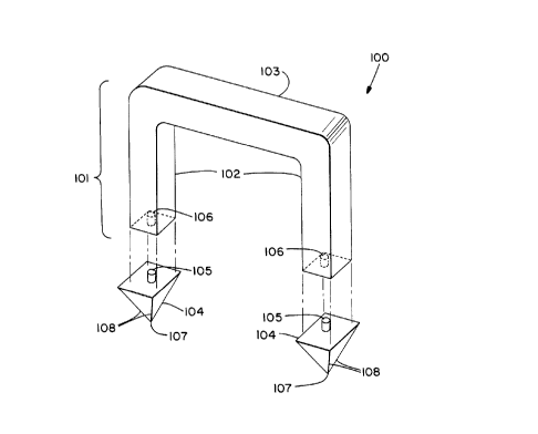
Figs. lA, lB, lC and lD illustrat~ a staple type
fastener 100 of the present invention, which comprises a
~ast~n~ng body portion 101 fnr fastening body tissue. The
fast~n; ng body portion 101 is optimally an integral single
piece construction having a crosspiece 103 and legs 102.
~egs 102 extend substantially perpendicularly from the
crosspiece 103.
3o
.
-6-
1 The fastening body portion 101 as shown in Figs.
lA and lC is U-shaped, with legs 102 ext~n~;ng from the ends
of the crosspiece 103. Alternatively, the legs 102 may be
spaced inward from the ends of crosspiece 103, the invention
can have one or more legs, and although the legs 102 are
illustrated as being of substantially equal length, legs of
unequal length are also contemplated as being within the
scope of this invention.
Unlike prior art fasteners, the fastening body
portion 101 of the pre~ent surgical fastener 100 has one or
more bone penetrating elements such as tips 104 attached to
the distal ends of legs 102, for penetrating bone or hard
tissue. The tips 104 must have a hardness sufficient for
such penetration. Thus, tips 104 are preferably made of a
metal, such as stainless steel, titanium and its alloys,
cobalt-chromium-molybdenum alloys, or other implant grade
metal alloys. Cexamics having appropriate hardness and
toughness may also be used, such as zirconia, aluminum
oxide, carbon/carbon composites, etc. The tips 104 each
have a relatively ~harp point 107 for easier bone
penetration, but any appropriate shape which will perform
the same penetrating function may be employed. Also the
tips 104 may have backward pointing barbs (not shown) to
prevent removal of the fastener.
~- Each tip 104 has a rearward projecting bolt 105
which engages and is received in a corresponding cavity 106
in the distal end of the respectlve leg 102. The tips 104
can be attached to the distal ends of the legs 102 by
various alternative mean~. For example, the bolts 105 may
be threaded, and cavity 106 may be tapped to form a screw
3 ~it. The joint is preferably secured by mounting the tips
,
2~ 3~
--7--
1 104 additionally with a biocompatible adhesive. Or the tips
104 may be fused to the legs 102 by ultrasonic welding, or
induction heating.
Referring to Figs~ lB and lC, tips 10~ have an
impact surface 104A for receiving a driving force for
implanting the fastener. The impact surface 104A is
perpendicular to the direction of the driving force and
extends outwardly beyond the surface of the legs 102 on
three or fewer sides of each leg. The impact surface does
not extend beyond the interior surfaces of the leg 102 into
the space between the two legs. As can be seen from Figs.
lB, lC and lD, the edges of the tips 104 are flush with the
inside surfaces of the legs 102, thereby preserving a tight
fit with in; ~1 latitude for loose movement or play in the
fastener once implanted.
~ ips 104 optionally have sharp points 107 and
edges 108 for cutting through bone and hard tissue, so as to
facilitate implantation of the fastener 100.
Fig. lE illustrates the staple in conjunction with
a pusher 110 for applying the staple to bone or hard tissue.
Pusher 110 has legs 111 which are adapted to apply a driving
force directly onto the projecting impact surface 104A of
tips 104. Surface 112 engages the crosspiece 103 when
impact is applied to keep the tips 104 from being driven
apart from the ends of legs 102. As the tips penetrate bone
this surface pushes the staple into the channels left by the
tips. The driving force may be applied ~nUAlly by the
surgeon, or through the use of a powered instrument.
Figs. 2 and 3 illustrate another embodiment of the
present invention. In this embodiment, surgical pin
3 fastener 200 comprises a bioabsorbable fastening body
. -8- 2 ~ 3 53 g
1portion in the form of a shaft 203, said body or shaft 203
having a bone penetrating element 201 attached thereto.
Bone penetrating e~ement 201 is optimally a hard trocar tip
which is fixed to the shaft 203, and preferably has a
relatively sharp point 205 and one or more relatively sharp
edges 207, for cutting bone.
The trocar tip 201 may be constructed from any
material having a hardness and strength sufficient for
penetrating bone or hard tissue. Examples of appropriate
metals and ceramic materials have been recited herein above.
The trocar tip 201 is preferably fixed to the end
of shaft 203 by a screw mounting. Projecting screw portion
204 of shaft 203 is received into tapped aperture 202 in the
tip 201. Screw portion 204 is preferably an integral part
~f shaft 203 made of the same bioabsorbable material.
Optionally, the joint can be made permanent by adhesively
bonding the threaded joint.
Alternative joining methods can be used both for
the pin 200 and staple 100. For example, the tips 104 and
201 may be fashioned with a sleeve, the sleeve being crimped
onto the fastener structure. Interlocking grooves in the
tips 104 and 201 in conjunction with cooperating grooves in
the legs 102 and shaft 203 respe~tively are also envisioned.
Fig. 4 illustrates alternative embo~; e~ts of the
tip and shaft. Tip 201A can be fluted in order to function
like a drill bit. The shaft 203A may have various types of
connection or mounting fixtures, such as the substantially
cross shaped male fixture 208 pro;ecting perpendicularly
from the end of the shaft, which is adapted to fit into the
corresponding female receptacle 210 in the tip 201A. or, as
3 shown in Fig. 5, trocar tip 201B may have a male mounting
~ '~
.
.
.
9 ~ ~53~
1 fixture 209 adapted to fit into a female receptacle in the
shaft. Male mounting fixture 209 optimally has a base
portion 209A which projects perpendicularly from the bottom
surface of tip 201B, and a pyramid portion 209B which
culminates in a point. These tips and shafts may be mounted
together adhesively or by welding or fusing the tips and
shafts together. Those ekilled in the art will envision
other types of connection fixtures. It should be
realized that the co~necting fixtures 208 and 209 can also
be used with the staple type bone fastener 100.
Typically, prior art metal bone fastening pins are
drilled directly into the bone. Prior art bioabsorbable
pins must have predrilled holes into which they are
inserted. But in accordance with the present invention,
self-inserting pin 200 can be implanted directly ~nto bone
by drilling, yet also has the advantage of being
bioabsorbable. Alternatively, pin 200 may have a tip 201
with an impact surface, such as the impact surface 104A of
the staple fastener 100.
The size of the surgical staple 100 and pin 200
may range from a few milli~eters to several centimeters.
However, the surgical bone fasteners of the present
invention can be made of any size which is appropriate for
its functlon of fastening bone or hard tissue.
Direct implantation of the bioabsorbable fasteners
100 and 200 is possible because the bone penetrating
elements (tips 104 and 201) have a bone cutting means, such
as relatively sharp points and edges which allow the bone
penetrating elements (104 and 201) to pierce the bone when a
driving force is applied to the fastener. The driving force
can be an impulse or pllch~ng force commonly used for drivin~
-10- 2~1~539
1 staple type fasteners such as fastener 100, or a rotary or
drilling force commonly used for pin type fasteners such as
200.
As can readily be ~een, the method for using the
present invention to fasten sel~ ?nts of bone or hard tissue
is relatively simple. Being provided with the ~urgical
fastener of the present invention, the surgeon implants the
device into bone or hard tissue 50 that the bioabsorbable
fastening body portion holds and maintains the segments of
bone or hard tissue in close adjacency for sufficient period
of time to promote healing. Both fasteners 100 and 200 will
degrade over a period of time leaving only the m~tal tips
104 and 201, which, being small, are far less intrusive, and
have little ten~ncy to work loose from the bone and
migrate-
In some surgical methods employing the fastenersof the present invention no hard nonabsorbable piece at all
remains. For example, pin 200 can be driven completely
across a fracture site so that the bone penetrating element
emerges from the far side of the bone. In such a case, the
bone penetrating tip will be cut off and removed, leaving no
portion of the fastener which cannot be absorbed.
While the above description contains many
specifics, these pecifics should not be construed as
limitations on the scope of the invention, but merely as
exemplifications of preferred embodiments thereof. Those
skilled in the art will envision many other variations that
are within the scope and spirit of the invention as defined
by the claims appended hereto.
:
, .