Note: Descriptions are shown in the official language in which they were submitted.
CA 02034394 2000-03-03
70571-32
Field of the Invention
This invention pertains to a dental instrument, and
more specifically to a dental instrument including a laser
device integrally formed with an electronic video dental
camera.
Background
For years, dentists have used dental mirrors for
insertion in a dental patients' mouth for reflecting images of
areas within the patients' mouth for viewing by the dentist.
This technique works, although it has several disadvantages.
First, it is often difficult to hold the dental mirror in an
appropriate position in order to reflect the desired image.
Secondly, it is more difficult to ensure that proper lighting
is available to the area within the mouth to be reflected by
the dental mirror. An even greater disadvantage is that it is
very difficult to use such prior art dental mirrors in a
situation where a dentist wishes to discuss certain regions
within the mouth with other people, be it the patient,
colleagues, dental assistants, or students in. a teaching
institution.
Electronic video endoscopes have been used in recent
years, for example as is described in U.S.
1
CA 02034394 2000-08-02
70571-32
2
Patent 4,667,229 of Cooper et al. Such prior art video
endoscopes use either fiber optics or a miniature camera,
such as a charge coupled device (CCD), in order to
transport an image to a monitor. Such prior art video
endoscopes come in a variety of sizes, but are typi-
cally rather small and tubular in nature in order that
they maybe easily inserted within a body cavity or
surgical opening. Certain prior art endoscopes include
a light source located at their end in order to ensure
proper lighting is available for illumination of the
area of the desired image.
However, such prior art video endoscopes were
not specifically designed for use in dental applica-
tions and are rather clumsy in such applications. As
one example, it is very difficult, if not impossible,
to properly view the lingual aspects of the teeth using
such prior art video endoscopes, due to their tubular
shape.
A number of attempts have been made in the
prior art to provide intraoral camera devices. Such
attempts are illustrated in U.S. Patent Nos. 3,382,781;
4,468,197; 4,479,499; 4,629,425, European Patent
Application No. 0 122 537 A; Offenlegungsschrift
2,208,902; and Offenlegungsschrift DE 304 5162 A1.
It is also known in the prior art to use
lasers in conjunction with dental procedures, as
described, for example, in Myers, "Dental Technology:
Knocking at High-Tech's Door," The Journal of the
American Dental Association (1989) 118:285-294; Myers,
"A Review of Lasers in Dentistry," I1 Dentista Moderno
(1989); Myers, "In vitro caries removal," CDA Journal
(1989) pp. 9-10; Myers et al., "First Soft Tissue Study
Utilizing a Pulsed Nd:YAG Dental Laser," Northwest
Dentistry (1989) pp. 14-16; Myers et al., "The Use of a
laser for Debridement of incipient caries," The Journal
~~3439~
3
of Prosthetic Dentistry (1985) _53:776-777; Myers _et
al., "What Lasers Can Do for Dentistry and You," Dental
Management (1989) _29:26-30; Dunlap, "Is There A Laser
In Your Future," Dental Economics (1988); Laser
Magazine, NR. 1, August (1989) (Various articles and
authors).
However, such prior art laser dental
instruments require the dentist performing the
procedure to rely on viewing the treatment area
directly or via an independent mirror. Direct viewing
is often awkward and does not provide the dentist with
an adequate visual acuity or sufficient clarity to
accurately and efficiently perform the procedure. The
use of prior art viewing tools, such as a standard
dental mirror or even prior art dental imaging devices,
used in conjunction with a prior art dental laser
instrument, is awkward at best, and in most instances,
simply impractical. Furthermore, laser energy is
dangerous to the patient if not applied properly, and
is dangerous to the dentist and his assistant if the
laser energy is accidentally reflected by a mirror,
particularly if the reflected laser energy reaches
their eyes.
Thus there remains the need to provide a
dental practicioner with improved visual acuity,
sufficient clarity, and an appropriate field of view
when performing laser proceedures in a convienient and
confident manner, while providing a safe working
environment for the patient, the practitioner, and
bystanders.
SUMMARY OF THE INVENTION
In accordance with the teachings of this
invention, a novel dental instrument is taught which
includes both a laser device and an electronic video
dental camera. The teachings of this invention
overcome the disadvantages of prior art dental laser
z~:~~3~4
4
instruments which do not provide for other than direct
viewing of the treatment area by the dentist, as well
as the disadvantages of attempting to use such prior
art dental laser instruments together with typical
prior art viewing devices such as dental mirrors and
dental cameras of the prior art. In accordance with
the teachings of this invention, a dental instrument
including a laser device and an electronic video dental
camera is provided having a single handle and a
convenient shape, thereby being readily manipulated by
dentists who are universally familiar with the
manipulation of prior art dental tools. A dental
instrument constructed in accordance with the teachings
of this invention includes a handle to be held by the
user, a distal portion which is to be placed inside the
patient's mouth, a laser light emission port located at
or near the distal end, means for transporting laser
light from an external laser source to the laser light
emission port, and a camera head located at or near the
distal end of the device, with the camera head being
formed at an angle to the handle in order to provide a
field of view which includes the portion of the patient
which is being treated by the laser light emanating
from the laser light emission part.
In one embodiment, means are provided to
cause, as desired, the flow of a selected fluid or gas
over the camera lens in order to defog and/or clean the
camera lens, thereby allowing proper viewing. In one
embodiment, the camera head also includes light sources
for properly illuminating the area to be viewed. As a
feature of one embodiment of this invention, the handle
of the dental camera includes means for communicating
all appropriate signals and fluids to and from the
camera head and the laser light emission port, and, if
desired, valves and switching means located on the
handle for controlling such communication.
~~~~,_~3~4
BRIEF DESCRIPTION OF THE DRAWINGS
Fig. 1 is an external view of one embodiment
of a dental instrument constructed in accordance with
the teachings of this invention;
Fig. 2 is an external view of another embod-
iment of a dental instrument constructed in accordance
with the teachings of this invention;
Figs. 3a and 3b are external views of another
embodiment of a dental instrument constructed in
accordance with the teachings of this invention which
include a flexible light probe;
Fig. 4 is a view depicting an elevator
assembly for manipulating the flexible light probe of
Figures 3a and 3b; and
Figs. 5a through Se depict various embodiments
of this invention which allows light to exit a fiber
optic light guide at an angle which is substantially
perpendicular to the longitudinal axis of the light
guide.
DETAILED DESCRIPTION OF THE INVENTION
In accordance with the teachings of this
invention, a novel dental instrument is provided which
allows the dental practitioner to direct laser energy
2~ to a desired location within a patient's mouth. The
dental instrument of this invention also includes means
for providing a video image of the area to which the
laser energy is directed far viewing on a video screen,
recording on a video tape recorder providing
photographs, and the like. By providing a dental
instrument which not only allows laser energy to be
directed to a desired location within the patient's
mouth but also which provides a magnified view on a
video screen of the operative area, the dental
practitioner is afforded significantly improved imaging
which enables the practitioner to perform the procedure
with a high degree of confidence that the laser energy
~~!'~~~~~~~~
6
is directed to the desired area, and only the desired
area, within the patient's mouth, thereby making the
procedure quicker, more effective, and safer. By
providing a single dental instrument which is used for
both directing the laser energy and providing a view of
the operative area, the practitioner can perform the
procedure with far greater ease than when utilizing
both a laser instrument and a dental mirror for viewing
purposes.
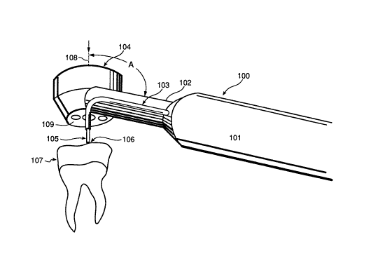
Fig. 1 shows a view of one embodiment of a
dental instrument constructed in accordance with the
teachings of this invention. Dental instrument 100
includes handle 101 suitable for being held by an
operator, and, if desired having forward extension or
neck 102. At the proximal end (not shown) of handle
101 is located one or more connectors for connection,
via a cable assembly (not shown) to a video processor
and control unit and a source of laser energy.
Preferably, means fox providing illumination to the
treatment area is included. The light source can be
located in head 104, or away from head 104 (for example
in neck 102, handle 101, or in the video processor and
control unit) and communicated to the treatment area by
optical fiber, for example. In one embodiment of this
invention, video processor and control unit comprise
the EVE system processor available from Fuji Optical
Systems, Inc. of Los Gatos, California. If desired,
color images can be obtained as described in
"Ultracompact CCD Color Television Camera", Takemura _et
al., Toshiba Review No. 158 Winter (1986), pp. 3-6, and
U.S. Patents 4,727,416 and 4,667,229, assigned to Fuji
Optical Systems, Inc., the assignee of this
application.
In one embodiment of this invention, the laser
source comprises, for example, a typical prior art YAG
laser capable of providing sufficient power to the
treatment area. Preferably, the laser source is
~~~~~39~
capable of providing laser light to the treatment area
over a wide range of power levels, pulse repetition
rates, and pulse widths. Also, preferably the laser
source includes a means for providing not only infrared
energy from a YAG laser, for example, but also visible
light from any suitable source, which is carefully
aligned with the infrared laser energy in order to
serve as a visible targeting or aiming beam to insure
that the operator is aware of where the infrared energy
is being directed.
If desired, neck 102 and handle 101 have
generally circular cross section, although suitable
shapes, such as octagonol, oval, and polygonal can be
used. The important point is that handle 101 is easily
and comfortably gripped by the user, and neck 102 is of
a general size and cross section which is convenient
for insertion into the patient's mouth and, of course,
comfortable for the patient.
Located at the distal end of neck 102 and
mounted on neck 102 at an angle A to neck 102, is
camera head 104. Face 109 of camera head 104 includes
means for receiving an image within a patient's mouth
to be displayed on a video monitor. In one embodiment
of this invention, such means for receiving the image
is fiber optic tubes or one or more rod or relay lens
assemblies or a combination thereof with or without
additional lenses, which transmit the image to an image
sensor (not shown) either within neck 102 or handle
101. In another embodiment of this invention, the
image is transmitted by one or more rod or relay lens
assemblies or fiber optic cable, or a combination
thereof with or without additional lenses, through a
connector (not shown) at the proximal end of handle 101
to an image sensor located in handle 101 or in external
video equipment (not shown). ~iowever, in a preferred
embodiment the image sensor is located directly in
camera head 104 and receives the image from the
2~3~394
8
patient's mouth via a lens, without the need for fiber
optics. By utilizing a video monitor, magnification of
the image of the dental procedure is provided, thereby
enabling the dentist to more easily and accurately
perform a procedure. For example, when utilizing a
monitor having a 13 inch screen (diagonal measure),
magnification of approximately 18X is provided.
Convenient and well framed video recording of laser
dental procedures are also now possible utilizing this
invention.
Of importance, the field of view provided by
the means for receiving an image contained in camera
head 104 is designed to encompass that area where the
laser dental procedure is being performed, i.e, in the
case of Figure 1 the field of view is the portion of
tooth 107 where the laser energy is directed, as well
as a reasonable area surrounding that point, and may
include either the entire tooth, several teeth, or, if
desired, even a full arch. For example, the field of
view is typically within the range of approximately 5
to 50 mm in order to allow the dentist to view not only
the specific point at which the laser energy is
directed, but surrounding areas of the tooth. In
accordance to the teachings of this invention, the
field of view appears highly magnified on a video
scrEen, thereby allowing the laser energy to be
directed with great accuracy, confidence, and safety.
Also shown in Figure 1 is means for
transmitting laser energy 103 from a laser source (not
shown) to the area being treated. In one embodiment of
this invention, means for transmitting laser energy I03
comprises a fiber optic member 105, preferably a single
optical fiber constructed of glass, quartz, or other
suitable material, capable of handling the power and
wavelength of the laser energy being provided. In the
embodiment of Figure 1, fiber optic member 105 is
protected over much of its length by conduit 103, which
~~~43~~
is located external to neck 102. In an alternative
embodiment of this invention, fiber optic member 105 is
formed within neck 102, obviating the need for
additional protective conduit 103. Conduit 103 may be
used as a guide to place optical fiber 105 at the point
of interest.
As shown in the embodiment of Figure 1, the
laser energy is emitted from fiber optic member 105
such that the maximum energy density occurs at the end
of fiber optic member 105, with the laser energy
rapidly dispersing with increasing distance from the
end of fiber optic member 105. In this manner, the
dental instrument of Figure 1 serves as a contact
device, i.e. when the end of fiber optic member 105 is
brought in close contact with an object, such as a
tooth or gum, the laser energy is of sufficient density
to perform the dental procedure. However, as the end
of fiber optic member 105 is moved away from a surface,
the laser light emanating is dispersed such that the
energy density at any particular point is significantly
reduced, thereby preventing the laser energy from
having an effect on other than a particular small area,
as desired. It has been determined that such a contact
device can be provided simply by allowing the laser
energy to emanate from the end of fiber optic member
105. This causes the maximum energy density of the
laser light to be essentially at the termination of the
optical fiber, providing a contact device.
Alternatively, it is possible to provide a
lens at the end of laser energy transmission means 105
so that the focal point is at a distance from the lens,
thereby providing a non-contact device. Figure 2 is a
view of such an alternative embodiment of this
invention in which the instrument is a non-contact
device. In this embodiment, means is provided to
insure that the laser energy emanating from fiber optic
member 105 has a focal point at a predefined distance
2u34~~
from the end of fiber optic member 105. In one
embodiment of this invention, focusing lens 201 is
applied to the end of optical fiber member 105 in order
to provide a desired focal length. Such a focal length
5 can be of any desired dimension and is typically within
the range of approximately 0 to 50 mm.
In one embodiment of this invention, a non-
contact device is provided which includes means for
providing a visible light signal to the area where the
10 operative laser energy is directed or to be directed,
thereby serving as an aiming beam. In one embodiment,
this visible light i5 provided by a visible laser, such
as a laser diode providing visible light, a HeNe laser,
or a non-laser light source. Of importance, the focal
point of the visible light target beam is substantially
the same as the focal point of the operative laser
beam, thereby allowing easy and precise aiming of the
operative laser beam. In one embodiment of this
invention, switch means, such as a foot switch which is
easily manipulated by the dentist is provided for
turning on the operative laser energy. When the
operative laser energy is not engaged, the visible
light signal is engaged in order to allow the dentist
to properly align the dental instrument of this
invention prior to causing the operative laser beam to
be engaged. If desired, the visible light beam can
either remain engaged or be turned off when the
operative laser energy is turned on.
In one embodiment of this invention an
infrared blocking filter is placed in the image path
between the image being viewed and the image sensor
(not shown) in order to prevent infrared energy from
being applied to the image sensor. If not blocked out,
the infrared energy from the laser would be detected by
the image sensor, resulting in a "washed aut" video
signal.
In accordance with the teachings of this
11 z~~~~~~~
invention, a number of possible adapter lenses are
provided, included but not limited to a wide angle
lens, a telephoto lens, a rod lens, a relay lens, or
one or more fiber optic cables serving as a "relay
lens". Such a telephoto adapter lens is very
convenient for viewing very small areas, for example
for use in viewing the capillaries within the gums,
thereby allowing the dentist or oral surgeon to
determine the relative health of the gums by determin-
ing the condition of blood circulation within the gums,
which is useful, for example, when performing
gingevectomy. Such a telephoto adapter lens is also
useful for obtaining a frontal view of the entire
mouth, by holding the telephoto adapter lens at an
appropriate distance from the patient's face. Use of a
telephoto adapter lens for this purpose provides a more
highly magnified image and avoids the frontal image of
the patient's mouth appearing as a "fish eye" view. A
tiny relay adapter lens is highly suitable for viewing
the small spaces between the teeth more readily than
can be viewed using the dental camera itself.
Figures 3a and 3b depict an alternative
embodiment of this invention which includes a flexible
light probe 305 emanating from camera head 304. In one
embodiment, flexible light probe 305 is conveniently
fabricated as the distal end of, or an extension of,
fiber optic member 105 (Fig. 1). Flexible light probe
305 is capable of being manipulated to alter its exit
angle from camera head 304, and thus alter viewing axis
308. Also shown in Figures 3a and 3b is light probe
elevator control 310 which is located on handle 301 for
easy manipulation by the practitioner in order to move
flexible light probe 305 as desired. This allows the
practitioner to hold the instrument in a convenient
position and alter slightly the target area which will
receive the laser energy, thereby making use of the
dental instrument constructed in accordance with the
~~3435~
12
teachings of this invention more convenient than if the
target area cannot be altered with respect the position
of the dental instrument. Also shown in Figs. 3a and
3b are cable assembly 311, camera lens 315,
illuminating lenses 314, and channel opening 316
through which flexible light probe protrudes. While
the embodiments of Figs. 3a and 3b depict the elevator
control located on handle 301 of the dental instrument,
in alternative embodiments the elevator control is
located elsewhere.
Figure 4 depicts one embodiment of an elevator
assembly constructed in accordance with the teachings
of this invention to allow flexible light probe 305 of
Figure 3a to be easily manipulated. As shown in Figure
4, flexible light probe 305 is an extension of an
optical fiber which is held by optical fiber guide
channel 403. Elevator 404 pivots about pivot point 405
in order to cause angular displacement of light probe
305. Elevator 404 pivots in response to movement of
elevator wire 402 which is tied to elevator 404 at tie
point 406. Elevator wire 402 is contained within wire
guide channel 401 in a well known fashion and is
actuated by light probe elevator control 310 of Figures
3a and 3b. It is to be understood that the embodiment
of Figure 4 is but one mechanism for manipulating light
probe 305 and may be housed within the dental
instrument of Figures 3a and 3b or may be conveniently
housed external to such a dental instrument, as
desired.
It is contemplated by this invention that this
elevator technique can also be used to easily
manipulate items other than, or in addition to, light
probe 305. For example, this technique and structure
can be used to manipulate a washing tube, for example,
which serves to provide a fluid or gas jet in order to
cleanse an area where a dental procedure is being
performed.
CA 02034394 2000-03-03
13
In an alternative embodiment of this
invention, a dental instrument is provided in which the
fiber optic member need not be bent about a sh<arp
radius, thereby making manufacture simpler and more
cost effective. As shown in Figure 5a, attachE:d to the
distal end of optical fiber 501 is lens 502 and mirror
503. Mirror 503 serves to reflect the laser energy to
a desired angle from the longitudinal axis of optical
fiber 501, with the angle of redirection being any
desired angle, although an angle within the range of
approximately 40 to 135 degrees is often useful.
In Figure 5b, an alternative embodiment is
shown, including prism 504 used in place of mirror 503
of Figure 5a.
Figure Sc shows an alternative embodiment
where mirror 505 is placed between optical fiber 501
and lens 506.
Figure 5d shows an alternative embodiment
where prism 507 is placed between optical fiber 501 and
lens 508.
Figure 5e shows yet another embodiment in
which a mirror is formed integrally with optical fiber
501 by forming the distal end of optical fiber 501 at
an angle to the longitudinal axis of optical fiber 501,
thereby providing mirror surface 521. A portion of the
outer surface of optical fiber 501 is removed to form a
notch which serves as exit port 522, allowing the light
reflected from mirror surface 521 to exit from optical
fiber 501 at an angle to the longitudinal axis of
optical fiber 501. If desired, mirror surface 521 is
polished and, preferably, coated in order to farm a
highly reflective mirror surface. If desired, lens 535
is used to focus the light exiting optical fiber 501 at
exit port 522. Lens 535, if used, may either be
attached to optical fiber 501 at exit port 522, or may
be mounted at a desired distance from exit port 522.
CA 02034394 2000-03-03
70571-32
The invention now being fully described, it will be apparent to
one of ordinary skill in the art that many changes and
modifications can be made thereto without departing from the
spirit or scope of the appended claims.
14