Note: Descriptions are shown in the official language in which they were submitted.
Uldall-DeBruyne 1-.
i~~~~~''~~c'~
COLLAPSIBLE LUMEN CATHETER FOR
EXTRACORPOREAL TREATMENT
~fechnical Field
The present invention relates generally to
extracorporeal treatments such as hemodialysis in which
blood and its components are treated outside of the body and
which require access to the vascular system of the human
body and, in particular, to mufti-lumen catheters for use in
such treatments.
Background of the Invention
There is an increasing number of hemodialysis patients
in whom creation and maintenance of an arteriovenous fistula
is difficult or even impossible. For these patients, one
prior art long-term catheter hay; provicled a reasonable
solution to their problems. zt provides good blood flow and
can be left in place in the external or internal jugular
vein for many months or even years. The flow
characteristics of this catheter are not ideal, but a plain
tube catheter with an open end appears to maintain patency
better than that of a tapered tube with side ports. The
main problem with this plain tube catheter is its cross-
sectional shape, which is similar to a double-barreled
shotgun, and its' squared off ends. These features make it
unsuitable for percutaneous insertion over a wire guide. As
a result, this plain tube catheter has to be introduced with
a cut-down surgical technique, which requires considerable
time and skill.
A more serious problem with the plain tube catheter is
that once it has been removed, it can not easily be
reinserted into the same site. Therefore, jugular vein
"Express Mail" label number ~~ ~' ~~1 '~ .~'i~ ' ( ,< '
-T, -
Date of Deposit ~~~r' t - J i ~ <. ',
I hereby certify that this paper is being deposited
with the United States Poct~zl Service "E=rpress Mail Post
Office to .4d~fres~;;s" service under 37 Ci=f?~1.10 cn the
date indicated ub;.ve and is addressed io the Commissioner 1
of Patents and Tr:~demarks, Washington, G.C. 20231.
('~ r ~ .- ,~' ...y~Jr ~.< < r.~..-
Signature of person mailin~aper or foe
Uldall-DeBruyne 1-1
sites are soon used up and no longer available to the
patient. One physician has used a peel-away sheath for
percutaneous insertion of this catheter, but a very large 18
French sheath was required to accommodate the largest cross-
sectional dimension of the catheter. Most physicians vJOUI.d
judge the size of: an 18 French introduces sheath to be
undesirable.
Heretofore, it has always been considered necessary for
the positive pressure return lumen to extend beyond the
negative pressure intake lumen of a hemodialysis catheter.
This is to ensure that blood returnincJ from a hemodialysis
treatment machine is delivered downstream from blood heing
extracted for purification. However, a problem with th:i~.
configuration is that clots tend to adhere to the outside
wall of the catheter at the entrance port to the shorter,
negative pressure intake lumen.
To provide modern high efficiency dialysis, it is also
desirable to utilize a catheter having two large diameter
lumens for high blood flow rates and also having an external
cross-sectional dimension which is not too large for
vascular access. One such temporary access or short-term
catheter includes a simple double-D lumen configuration.
The walls of the catheter are thin, and the equal area
lumens make full use of the available space. however, to
insure that this catheter keeps its shape during high flow
rate dialysis, the catheter is made of relatively stiff
material which is unsuitable for long-term placement. If
this temporary catheter .is made of a silastic material, the
intake lumen collapses under the influence of the strong
negative intake pressure. Furthermore, the septum between
the two lumens is pulled into the negative pressure lumen,
thereby adversely changing the cross-sectional area in the
two lumens as well as the blood flow rates therethrough.
Temporary or short-term catheters of the double-D
configuration are used in large numbers all over the world,
but they have a disconcerting tendency to get bloc~:ed.
2
Uldall-DeBruyne 1-1
These catheters are made o.f a relatively stiff material to
prevent the lumens from collapsing. A problem with the
stiff material is that the catheter kinks or buckles when
bent more than 180°. This also leads to lumen obstruction
and the potential risk of cracking or splitting the wall.
Furthermore, catheter stiffness combined with a tapered end
for insertion over a guide wire has been responsible for
many penetrating injuries of the wall. of the superior vena
cava or right atrium. There have been many deaths caused by
such spontaneous perforations. This can occur by erosion,
days er weeks after the catheter is originally inserted.
However, no penetrating vein wall injuries have been
reported with the use of blunt-end silastic catheters.
Silastic catheters with the double-barreled shot-gun
configuration (two cylindrical lumens side by side) are
remarkably resistant to kinking even when bent sharply
through 180°. Also a cylindrical lumen is the theoretical
optimum to achieve maximum flow for the smallest surface
area of the wall. Finally, the cylindrical lumen avoids the
sharp corners in the wall of the double-D configuration
where, at least theoretically, clotting is more likely to
occur.
The side-by-side open-ended design of the long-term
catheter has much less tendency to block, but has not been
used as a temporary catheter since it cannot easily be
introduced percutaneously. The circular intake lumen of the
long-term catheter is similarly recessed back from the
distal end of the return lumen to minimize blood
recirculation. A problem with this is that the wall of the
extended positive pressure return lumen provides a surface
for clots to adhere. In an attempt to salve this blockage
problem, the walls of the negative pressure intake lumen are
provided with side ports. However, it is believed that
these side ports may actually encourage clotting.
The long term catheter typically employs a fixed-
position dacron cuff which may not be conveniently
3
CA 02040708 2001-03-12
positioned to stabilize the catheter. Removal of the
catheter and release of the dacron cuff requires a new
incision and dissection of the cuff by a surgeon.
Dissecting the cuff from ingrown tissue invariably leads
to bleeding, which may be hard to control.
Summary of the Invention
In accordance with one aspect of the present
invention there is provided a hemodialysis catheter for
extracorporeal treatment, comprising: a first elongated
member having a first, negative pressure intake lumen
extending longitudinally therein and a first wall
positioned about and defining said first lumen, said
first lumen having a first predetermined cross-sectional
area, said first wall having a first predetermined
thickness for maintaining said first area when a
predetermined negative pressure is applied to said first
lumen; and a second elongated member attached to said
first member and having a second, positive pressure
return lumen extending longitudinally therein and a
second wall having a second predetermined thickness
positioned about and defining said second lumen and
collapsible on said first wall for insertion through a
narrower introducer sheath and into a blood vessel, said
second lumen having a second predetermined cross-
sectional area, said second wall returning to and
maintaining said second area when introduced into said
blood vessel.
In accordance with another aspect of the present
invention there is provided a hemodialysis catheter for
extracorporeal treatment comprising: a first elongated
segment having first, negative pressure intake lumen and
4
CA 02040708 2001-03-12
second, positive pressure return lumen extending
longitudinally therein and first and second walls, said
first wall positioned about and defining said first
lumen, said first lumen having a first predetermined
cross-sectional area, said first wall having a first
predetermined thickness for maintaining said first area
when a predetermined negative pressure is applied to said
lumen, said second wall positioned about and defining
said second lumen, said second lumen having a second
predetermined cross-sectional area, said second wall
attached to said first wall and having a second
predetermined thickness, said second lumen being
collapsible on said first wall, said second wall
returning to and maintaining said second area when said
first segment is introduced into a blood vessel; and a
second elongated segment extending proximally from said
first segment having said first and second longitudinal
passageways extending therein.
In accordance with yet another aspect of the present
invention there is provided a collapsible dual-lumen
hemodialysis catheter for percutaneous insertion through
a narrower introducer sheath, comprising: an elongated
distal segment having a negative pressure intake lumen
and a positive pressure return lumen extending
longitudinally therein, said lumens having substantially
equivalent cross-sectional circular areas and first and
second walls, said first wall positioned about and
defining said intake lumen and having a first
predetermined thickness for maintaining said cross-
sectional circular area of said intake lumen when a
predetermined negative pressure is applied to said intake
lumen, said second wall positioned about and defining
4a
CA 02040708 2001-03-12
said return lumen and having a second predetermined
thickness, said first thickness being approximately twice
as thick as said second thickness, said first wall being
a predetermined distance longer than said second wall at
a distal end of said distal segment, said distal segment
in a collapsed state having said second wall and said
return lumen being collapsed on said first wall and
having a maximum cross-sectional dimension less than said
narrower introducer sheath for insertion through said
sheath in a blood vessel, said second wall returning to
and maintaining said cross-sectional circular area of
said return lumen when said distal segment is introduced
into said blood vessel, said distal segment also having a
slip coating thereon; an elongated proximal segment
extending proximally from said distal segment and having
a generally elliptical cross-sectional shape, said lumens
extending longitudinally through said proximal segment;
and a moveable collar positioned about said proximal
segment and having a flange with a suture hole therein.
More specifically, the foregoing problems (discussed
in the background) are solved and a technical advance is
achieved with an illustrative extracorporeal treatment
catheter having a collapsible lumen for percutaneous
insertion into a blood vessel through a much smaller
diameter peel-away introducer sheath. This
extracorporeal treatment catheter comprises first and
second elongated tubular members having respective first
and second longitudinal passageways therein, commonly
referred to as lumens. The first and second tubular
members are laterally attached and have respective first
and second walls with different thicknesses about the two
lumens. Advantageously, the second lumen wall is thinner
4b
CA 02040708 2001-03-12
than the first lumen wall and is collapsible about the
second tubular member for inserting them both into a
blood vessel through a smaller diameter introducer
sheath. When inserted in a vein, the collapsed lumen
returns to its original shape. Preferably, the thickness
of the first lumen wall is twice as thick as that of the
second lumen wall. This advantageously permits
percutaneous insertion of the catheter with a stiffening
cannula over a wire guide and through the much smaller
diameter introducer sheath. The distal end of the two
tubular members includes a slippery-when-wet coating for
further easing the percutaneous insertion of the catheter
through the introducer sheath.
The intake lumen wall thickness is thick enough to
withstand the negative pressure associated with
hemodialysis treatment machines without the intake
lumen collapsing. The return lumen wall is thinner
than the intake lumen wall to allow it to
collapse about the first member for percutaneous
4c
Uldall-DeBruyne z-1
insertion through the smaller diameter introduces sheath,
but yet thick enough to withstand the positive pressure of
the returning blood without stretching. The thickness of
the intake lumen wall to that of i:he return lumen wall may
vary in a range of one and a half to three times as thick
and still provide satisfactory blood flow characteristics.
The cross-sectional shape of the two lumens are
substantially equal in area to maintain balanced intake and
return blood flows. Furthermore, the cross-sectional shape
of the two lumens is generally circular to advantageously
maintain maximum laminar blood flow for a given wall surface
area.
As a departure in the art, the negative pressure intake
lumen is longer than the positive pressure return lumen at
the distal ends, which is opposite to that of presently
available hemodialysis catheters. The shorter positive
pressure return lumen advantageously reduces the
accumulation of blood clots and resulting blockage with only
a minimal increase in blood recirculation between the two
lumens.
To eliminate the seepage of blood between the tubular
members where they penetrate the vein wall, the catheter has
been segregated into distal and proximal. segments. For
percutaneous insertion, the distal segment advantageously
includes different thickness walls for collapsing the thin-
walled positive pressure return lumen about the negative
pressure intake lumen and inserting the collapsed distal
segment through a smaller diameter introduces sheath.
Extending proximally from the distal segment, the proximal
segment has a cross-sectional shape of a generally
elliptical character to form a leak proof fit when inserted
into the vein wall. Furthermore, both of the segments are
formed from a biocompatible material such as silastic for
long-term use and have a predetermined durometer for pushing
the catheter through the introduces sheath and blood vessel.
The catheter also advantageously includes a moveable
5
Uldall-DeBruyne 1-~1 ~~~~F~~~
collar or grommet which. can be adjusted in position on the
proximal segment and has a flange extending therefrom to
secure the catheter. to the surrounding tissues. The grommet
can be released by simply pul7.ing on it or by dissenting it
out. In either case, there is no bleeding.
Brief Description of the Drawinct
FIG. 1 depicts a dual lumen hemodialysis catheter having
a collapsible lumen for percutaneous insertion through a
peel-away introduces sheath of the present invention;
FIG. 2 depicts a cross-sectional view of the distal
segment of the catheter. of FIG. 1 along the line 2-2;
FIG. 3 depicts a cross-sectional view of the distal
segment of the catheter of FIG. 1 in a collapsed state and
positioned in an introduces sheath; and
FIG. 4 depicts a cross-sectional view of the proximal
segment of the catheter of FIG. 1 along the Line 4-4.
Detailed Description
Depicted in FIG. 1 is a dual lumen catheter 100 for use
in an extracorporeal treatment such as hemodialysis and the
like. This vascular access catheter is percutaneously
inserted in a blood vessel, such as preferably the jugular
or femoral vein, for either short-term or long-term
hemodialysis treatment of the patient. The jugular access
site is preferable to the subclavian vein because it is much
less likely to cause subclavian vein thrombosis. Subclav.ian
vein thrombosis is a serious long-term disability for a
patient on dialysis if it is not diagnosed and successfully
treated at an early stage, because it interferes with A-V
fistula construction in the ipsilatera7. arm, leading to a
permanently swollen congested arm as long as the fistula is
functioning. Internal. jugular vein thrombosis is probably
not common after internal jugular cannulation, but it causes
no disability even if it occurs and is not 'treated, except
that the patient loses a potential access site.
6
Uldall-DeBruyne 1-1
The catheter basically comprises a dual lumen main body
101 attached to a single lumen, arterial clamping limb 104
and a single lumen, venous clamping limb 105 via
interconnecting manifold 7.06. For connection to
extracorporeal treatment equipment, two female Luer lock
connectors 107 and 108 are connected in a well-known manner
to arterial and venous clamping limbs 104 and 105,
respectively. ThE~ main body of t:ne catheter includes a
distal segment 102 arid a proximal segment 103 extending
proximally therefrom and is comprised of a flexible
biocompatible material such as 70 durometer silicon or
silastic. Distal segment 102 includes a thick-walled,
negative pressure, elongated tubular member 201 and a
shorter, thin-walled, collapsible, positive pressure,
elongated tubular member 202 attached laterally thereto.
The catheter further includes lockable clamps 117 and 118
for clamping arterial and venous clamping limbs 104 and 105,
respectively. One such clamp is the BETA-CAP clamp
available from Quinton Instrument Co., Seattle, Washingtan.
Slide clamps from the Qosina Co. are also acceptable.
Catheter 100 also includes an anchoring grommet 11.6
having a ring-like collar 111 positioned around and slidably
moveable along proximal segment 103. Flange 112 and 113
extend laterally from the collar and have respective
apertures 114 and 115 formed therein to insert sutures
therethrough. The grommet is positioned on the proximal
segment where it crosses the supraclavicular fossa. Sutures
placed through the apertures secure the catheter to the
surrounding tissue. The shape of the grommet permits
capture of the catheter without compressing it. The smooth
rounded flanges allow the grommet to be pulled out with the
catheter when it is removed. The anchoring sutures will
tear out of the flanges and the only thing left inside the
patient will be the sutures 'themselves.
The overall length of the main body of the catheter from
the manifold to the distal tip thereof depends on the
7
Uldall-DeBruyne 1-1
insertion site selected by the physician. When inserted in
the right jugular vein, the main body of the catheter from
manifold to tip is preferably 26cm in length with an llcm
distal segment. For the left jugular site, the main body of
the catheter is approximately 30cm in length with the distal
segment being l5cm. As suggested, the distal segment 102.
includes a collapsible tubular member 202 for inserting the
distal segment with stiffening cannula 109 inserted in
tubular member 201 over wire guide 110 through a well-known
smaller diameter peel-away introduces sheath (not shown).
The introduces sheath which should be no more than l0cm in
length. This will allow the distal segment to be inserted
into the sheath with the distal tip protruding slightly
beyond the distal end of the sheath before it is peeled
away.
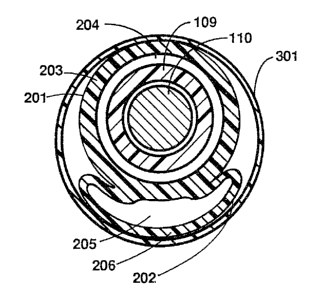
Depicted in FIG. 2 is a cross-sectional view of distal
segment 102 along the line 2-2 of FIG. 1. Distal segment
102 includes thick-walled elongated tubular member 201 and
thin-walled elongated tubular member 202 attached laterally
to member 202 and collapsible thereon. The thick-walled
tubular member includes first wall 203 surrounding first
longitudinal passageway 204 included therein. This first
longitudinal passageway is designated a negative pressure
intake lumen for receiving blood from the vessel o:E a
patient for hemodialysis treatment. By way of example, the
thickness of first wall 203 is approximately .020" with the
cross-sectional diameter of passageway 204 being
approximately .080". The distal end of the negative
pressure intake lumen may be outwardly tapered to prevent
clotting and the collection of blood clots thereon. The
dimensions of tubular member 201 and negative pressure lumen
204 allow for blood flow rates of 350-400m1 per minute
without collapsing.
The second, thin--walled collapsible tubular member 202
includes a second :Longitudinal passageway 205 with second
wall 206 positioned thereabout. The thickness of second
8
Uldall-DF~Bruyne 1-1
wall 206 i_ ap.proxi.matel.y .O:E.O'' with. ioFag:ltL~.dlnal pasSage~~aay
having a cross-sectional diameter of approximately .080",
which is equivalent to that of passageway 204. In an
unco7.3.apsed state, the maxin;um crop=s-~;ectior~al dimens i.on of
distal sec~pmen'c .is approximately .27.0" plus allowances i:or
fabrication and slip coating 207, which will pa:~s thr_oucJh an
18 French (.236") aperture. Second longitudinal passageTaay
205 is designated the positive pressure return lumen f.~,r
retu.rn:ing blood from a hemodialysis ma.c:hine to the veeel of
tile patient. The cross-sectional area of passageways 204
and 205 are substantially equivalent to provide
approximately equal flow rates to and from the patient. The
distal segment also includes slip coating 207 which acts as
a lux~ricant to insert the distal segment through the
1o intrc;ducsr sreath. Une su~oh slip ceatinc3 is a slippery-
when-wet hydrophilic: coating 'that is eommerr.ia.?.ly available
from Hydromer Tnco., Whi.tehouse, N.J. The slip coating i
applied to the outside surface of distal segment 102. This
hydrophilic slip coating is wetted during the insertion
procedure to provide a slipk~nry surface for easier insertion
through the peel-away introdtlcer sheath. Furthermore, the
presence of blood or other fluids in the introduces sheath
furtkler lubricates the c:~llapsed distal segmen;: as it is
being inserted therethrough. This furthcar eases th~~
percutaneous insertion of the catheter. when inserting a
collapsed catheter having an 18 French uncol:lapsed cross-
sectional dimension ttlrough a 12 French introduces sheath.
Another_ lubricious slip coating is Dow Corning medical-grade
silicone fluid spray (a nom-allergenic silicone lubricant)
which is commerc:i.ally available fr~.,m Dow Corning Europe,
Inc., Health Care Group, Brussels, Belgium. Thls JLlirone
spray is applied to the distal segment by the physician just
prior to percutaneous insertion of the catheter.
Experimentally, a 30cm thin-walled, positive pressure
member of a 70 durometer silicon material catheter was able
to tolerate a blood flow of 500m1 per minute and a negative
9
Uldall-DeBruyne 1-1
L
pressure of 300mm/Hg without collapsing when flows were
reversed, and it was used as a negative pressure lumen. In
clinical practice, tt?e ability to reverse the flows is
important ?f on occasion the thick-walled lumen fai7_s to
provide adequate out flow. TY~e dialysis treatment community
has been demanding these flow .rates, but until now has not
been provided with catheters t.o provide these flow rates.
Experiments indicate that blood flow rates of 400m1 per
minute are attainable with arterial and venous pressure
barely exceeding 200mm of mercury.
The cross-sectional shape of the passageways are also
preferably circular to maintain laminar fluid flow. The'
introduction of a smaller radius into the cross-sectional
shape of the passageway typically provides opportunities I:or
the blood flow to become turbulent and increases the risk of
clotting.
A number of competing factors are involved with the
dimensions associated with tine wall thicknesses and lumen
diameters. The tubu7.ar members must be thin and f.lexi.ble
enough for insertion into the vascular system without
kinking or collapsing in operation. Negative pressure lumen
wall 203 must be thick enough to withstand the negative
pressures inwardly exerted thereon by modern hemodialysis
treatment machines without collapsing during intake of blood
from the patient. Thinner, positive pressure lz.zmen wall 206
must be thick enough to withstand the positive pressures
outwardly exerted thereon without stretching. The diameter
of the passageways should be as large as possible to provide
adequate flow rates as demanded by the hemodialysi.s
treatment community. Lastly, the maximum cross-sectional
dimension of the catheter must be minimal for percutaneous
insertion into the blood vessel such as through a 12 French
(.158") peel-away introducer sheath. As a result, the
thickness of negative pressure lumen wall 203 is preferably
twice as thick as that of positive pressure lumen wall 206.
Furthermore, the thickness of negative pressure lumen wall
to
Uldall-DeBruyne 1-1
V
203 may range from one and a half to three times as thick as
that of positive pressure lumen wall 202.
This thin wall construction permits the collapse of
tubular member 202 including positive pressure lumen 205 and
wall 206 about tubular member 201 as depicted in FIG. 3. In
the collapsed state, a hemodialysis catheter typically
having a maximum cross-sectional dimension of 18 French can
be percutaneously inserted with stiffening cannula 109 over
wire guide 110 into a blood vessel through a much smaller 12
French diameter peel-away introducer sheath 301.
Depicted in FIG. 4 is a cross-sectional view of proximal
segment 103 slang the line 4-4 of FIG. 1. The cross-
sectional shape 401 of the proximal segment is formed to
provide a tight fit between the main catheter body and the
vascular access insertion site. Preferably, the cross-
sectional shape is elliptical to prevent the seepage of.
blood from the vascular access site along the outside
surface of the proximal segment of the main catheter body.
Respective negative and positive pressure lumens 204 and 2.05
extend entirely through proximal segment 103.
To insert the dual lumen catheter using the well-known
Seldinger technique, a wire guide 110 is inserted through an
introducer needle into the accessed vein. The introducer
needle is removed, and a 12 French sheath mounted on a
dilator is directly inserted over the guidewire into the
vein. Stiffening cannula 109 is inserted through the
negative pressure lumen of the arterial clamping limb 104,
proximal segment 103, and out the distal tip end of distal
segment 102. The catheter and stiffening cannula are
inserted over wire guide 110 and through the peel-away
sheath with the thin-walled tubular member 202 collapsed.
The peel-away sheath is removed after the distal segment is
inserted through the sheath into the vein. A short distal
portion of the elliptically shaped proximal segment 103 is
then inserted through the venous access site into the vein,
thereby establishing a relatively tight and leak-proof seal.
11
Uldall-DeBruyne 1-1
Grommet 1:16 is mounted onto the catheter by passing it
over the distal tip, aftei th~~ catheter has been pulled up
through the subcutaneous tunnel and before the catheter i.s
inserted through the sheath into the vein. Grommet 116
slides the distal segme:~t and a length of proximal segment
103 and is placed strategical?.y in the supraclavi.cular fossa
and anchored to the subcutaneous tissue before the
supraclav:icular wound is clased. Final position of the
grommet will vary in each patient according to how much
length of the catheter is des.i.red in the blood vessel.
To change the catheter, it will only be necessary to
reopen the supraclavicular incision and remove the
subcutaneous silk sutures which are anchoring the grommet in
place. To remove the catheter without intending to replace
it with another one in that same track, the catheter_ is
subjected to a steady pull. This will tear the sutures out
of the flanges of the grommet.
Of course, it will be understood that the afore
mentioned dual lumen extracorporeal treatment catheter is
merely illustrative of the application of the principles of
this invention and that numerous other arrangements may be
devised by those skilled in the art without departing from
the spirit and scope of the invention. In particular, a
number of other grommets may be slid over or attached to the
proximal segment of the catheter for anchoring 'the catheter
to surrounding tissue. The catheter may also include any
number of other connectors or clamping devices for use with
the arterial and venous clamping limbs. Furthermore, the
shape of the lumens may be varied to an elliptical or even
crescent shape; however, the radii of lumen shapes need to
be maximized to prevent or minimize turbulent blood flow and
the possibility of clotting.
12