Note: Descriptions are shown in the official language in which they were submitted.
~U~~Bf "~
DIRE(:T VISION PROSTATE BALLOON CATHETER
BACKGROUND OF THE INVENTION
The invention relates generally to a device
appropriate for nonsurgical treatment of benign prostate
hyperplasia andl more particularly to a balloon catheter
appropriate for dilating a portion of the urethra constricted
by an enlarged prostate gland.
Benign prostate hyperplasia (BPH) is a disease
characterized bar enlargement of the prostate gland. As the
prostate enlarges, it compresses the urethra, impairs urination
and can lead to urinary tract infection and possible renal
failure. Surgical and non-surgical treatment of BPH have been
proposed.
Surgical treatment of BPH typically involves
transurethral resection of the prostate. This procedure
requires 5 to 6 days of hospitalization and is associated with
some morbidity. Balloon dilation of the prostate is emerging
as an important non-surgical near-term treatment for BPH. It
can be carried out under sedation and local anesthesia in about
20 minutes. In this procedure a balloon catheter is inserted
through the urethra to the prostate and the balloon is inflated
to compress the internal tissue and stretch the outer capsule
of the prostate. The patient can return home with a Foley
catheter in place for two days. The recovery period is usually
3 to 4 days.
An example of a balloon apparatus for treating BPH is
described in U.S. Patent No. 4, 660, 560. FIG. 1 is a cross-
sectional view of a dilating catheter assembly 10 positioned in
the male urinary tract. A multi-channel cystoscope 12 is
received through penile meatus 14 and is positioned in urethra
16 in which dilating catheter 10 is passed through one of its
lumens. An extended Foley-balloon 18 is anchored to bladder
neck 22 while an annular balloon 20 is fixedly positioned with
respect to the prostatic urethra as defined by bladder neck 22
and veru montanum 24. Pressure dilation of the prostatic
urethra by annul;~r balloon 20 continues as long as it is deemed
necessary. U.:c. Patent No. 4,762,128 discloses a single
prostate balloon catheter for imparting an expanded tubular
1
2
stent to extend long term patency. Catheters such as these are
not fully satisfactory since they require multiple
instrumentations of the urethra and multiple, components such as
a sighting lens, sheath and dilation catheter. These are
awkward to simultaneously position properly at the prostate.
Such catheters can lead to improper dilation of the external
sphincter or :improper dilation beyond the bladder neck.
Furthermore, a :>6 F plastic sheath, which is undesirably large,
is required to insert and withdraw the balloon and a lens.
Another example of a balloon catheter for treating
BPH is described in European patent application No. 0,341,988.
A location or positioning balloon is located proximal to a
prostate dilation balloon along the catheter. The location
balloon is positioned to be at the bulbous urethra when the
dilation balloon is at the prostate urethra. This fixes the
location balloon to be intermediate the external sphincter and
bladder to maintain the dilation balloon in proper position
when it is inflated at the prostate urethra. The location
balloon is sized to fit the bulbous urethra on inflation to
prevent undesirable dilation of the external sphincter.
The catheter described in the European patent
application al~;o has drawbacks. To position the dilation
balloon properly, a fluoroscope or lens is required. The
fluoroscope exposes the patient to unnecessary radiation and
the lens must be inserted unguided alongside the catheter
shaft, increasing the likelihood of deleteriously scraping the
urethra. Either option makes the entire procedure undesirably
complex.
Balloons for prostate catheters are commonly
substantially not elastic. Accordingly, when the conventional
balloon is to be removed, it must first be threaded into a
sheath. This; makes the procedure and device unduly
complicated.
Accordingly, it is desirable to provide an improved
balloon catheter for dilating the prostate to reduce
constriction of the urethra which overcomes the shortcomings of
available prostate balloon catheters.
2044867
3
SUMMARY OF THE INVENTION
Generally speaking, in accordance with the invention,
a direct vision catheter for dilating a prostate gland to
reduce constriction of the urethra is provided. The catheter
includes a flexible catheter shaft with an expandable member
such as a balloon and a sighting device such as a fiber optic
lens to assist in properly positioning the expandable member.
The catheter includes a first lumen to accept a telescope and
can include a device to secure the telescope, to provide direct
vision of the proximal end of the balloon and for maintaining
proper balloon position during inflation. Additional lumens
provide for rin:~ing the lens, balloon inflation and guide wire
passage. The balloon is preferably elastic and of a self-
wrapping construction mounted at the distal end of the catheter
shaft. The balloon can be formed with a thickened distal end
to limit expansion and prevent undesirable migration into the
bladder during expansion. Alternatively, the balloon can be
mounted to the ends of two slidable tubes to control balloon
extension durin~~ inflation of the balloon.
Accordingly, the invention provides an improved
catheter for di:Latation of an enlarged prostate.
The invention can also provide an improved balloon
catheter of reduced diameter for dilating an enlarged prostate
and that is les;~ irritating to the urethra.
The unproved balloon catheter of the invention can
dilate the prostate without adversely affecting the external
sphincter of the bladder.
The invention can also provide a catheter with direct
vision of the b~~lloon and anatomical landmarks, e.g. the veru
montanum and external sphincter, during insertion and
inflation.
:.~s
20448 6 7
4
Still other advantages of the invention will in part
be obvious and will in part be apparent from the specification
and drawings.
The invention accordingly comprises the features of
construction, combination of elements, and arrangement of parts
which will be e:~emplified in the constructions hereinafter set
forth, and the ;scope of the invention will be indicated in the
claims.
BRIEF DESCRIPTION OF THE DRAWINGS
FIG. 1 is a cross-sectional view of a conventional
urethral dilating catheter inserted to the male urinary tract;
FIG. 2 is a perspective view of a balloon catheter
with the balloon in the deflated condition constructed in
accordance with the invention;
FIG. 3 is a partial perspective view of the distal
end of the catheter of FIG. 2 with the balloon in an inflated
condition;
FIG. 4 is a cross-sectional view of the catheter of
FIG. 2 taken along line 4-4;
FIG. 5 is a partial side elevational view of the
proximal end of a catheter constructed in accordance with the
invention.
FIG. ~6 is a top plan view of the proximal end of the
catheter of FIG. 5;
FIG. 7 is a partial exploded view showing the
separate elements of the distal end of the catheter of FIG. 2;
FIG. ,~ is a partial side view of the distal end of a
balloon catheter constructed in accordance with another
embodiment of t:he invention; and
FIG. 9 is a bottom plan view of the distal end of a
balloon catheter constructed in accordance with the invention.
DESCRIPTION OF THE PREFERRED EMBODIMENTS
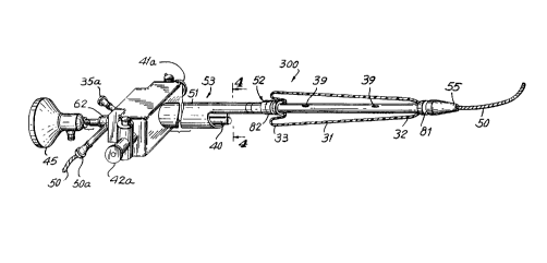
A prostate balloon catheter 300 constructed in
accordance with the invention is shown in FIG. 2. Catheter 300
includes an expandable dilation device such as an expandable
bulb or balloon 31 mounted on a shaft 51. Balloon 31 is
.,i
~U4486"~
positioned in the urethra by threading shaft 51 along a guide
wire 50 to a position determined by assistance from visual
observation through a sighting device such as a telescope or a
fiber optic lens 40 provided within a lumen in shaft 51.
Balloons for balloon catheters and providing fluid to the
distal end of a shaft for inflating the balloon are well known
in the art.
Catheter 300 is advantageous due in part to the one
piece, small diameter construction which permits proper
positioning of balloon 31 without excessive irritation to the
urethra or damage to the external sphincter of the bladder. It
is advantageous to inflate balloon 31 to dilate the constricted
prostatic ureth~__~a without dilating the external sphincter at
the prostate apex. Accordingly, to effectively dilate the
entire prostate length it is necessary to prevent balloon 31
from migrating into the bladder during inflation.
To aid in proper balloon position balloon 31 can be
constructed with a thick wall balloon region 32 distal to a
thin wall balloon region 33. Thick wall region 32 is at the
leading end of ~~atheter 300 and is positioned in and through
the bladder neck:. In the deflated condition, thin wall region
33 is layered over itself so that during inflation, balloon 31
will primarily expand radially and will not be displaced
longitudinally to exert undesirable forces or reposition
balloon 31 with:Ln an improper portion of the urethra. Thick
wall balloon region 32 retards inflation of the distal end of
balloon 31 which significantly retards migration of balloon 31
through the bladder neck into the bladder. Accordingly, during
inflation, balloon 31 substantially maintains its position in
the prostate to prevent undesirable injury to the external
sphincter muscle: and to properly dilate the entire prostrate.
Balloon 31 should inflate to be about 35 mm or at
least about 30 mm and should be available in lengths from about
to 55 mm to .accommodate various prostate urethra lengths.
Balloon 31 should be elastic and able to hold about 6 to 8 atm
until maximum volume, for at least three to four 10 minute
cycles. Pr~aferably, balloon 31 has a polymer
20448 s ~
6
fiber/polyurethane, a glass fiber/silicone or a carbon
fiber/latex construction to provide desirable strength and
expansion characteristics. On deflation, balloon 31 should
return substantially to ifs pre-expansion shape and position.
Shaft 51 i;~ flexible and should be smooth and rigid enough
for proper insertion without the need to utilize a cystoscope
or outer sheath. FIG. 4 shows a cross-section of catheter 300
taken along line 4-4 of FIG. 3. Shaft 51 includes three
separate lumen:. As shown in FIG. 3, shaft 51 includes a
dilation portion 52 of reduced cross-section at the distal end
and a viewing p«rtion 53 proximal to dilatation portion 52. A
guide wire lumen 36 extends through catheter shaft 51 from the
proximal end to the distal tip of shaft 51. The overall
diameter of shaft 51 should be less than 26 F, and more
preferably from about 23.5 to 21 F, or thinner.
Viewing portion 53 of shaft 51 encloses a crescent
shaped lumen 54. Lens 40 slides through lumen 54. When lens
40 is in place, it divides lumen 54 into two rinsing lumens 41
and 42 which arE_ in fluid communication with a pair of rinsing
ports 41a and 42;a, respectively at the proximal end of shaft 51
and provide rin:aing fluid to leis 40 to improve the ability to
see through fiber optic lens 40. Dilation portion 52 is distal
to viewing portion 53 and encloses guide wire 50 and an
inflation lumen 35. Inflation lumen 35 is in fluid
communication with an inflation port 35a to provide fluid for
inflating balloon 31 through a pair of holes 39 in the shaft
wall.
FIG. '7 shows the individual components of the distal
end of catheter 300. A molded tip 55 of dilation portion 52 is
formed with guide wire lumen 36 and seals off inflation lumen
35. Tip 55 is tapered and rounded to avoid irritation to the
urethra during insertion. Preferably a pair of radio-opaque
marker bands 81 and 82 seal the balloon ends and prevent
exposed rough edges. The tip must be constructed to not be
deformed by the radio-opaque bands. Binding, welding, gluing
or other process could also be used for balloon attachment. A
molded spacer 83 is disposed between marker band 82 and shaft
f
.. 3
_ 2044867
7
51. It is preferable to provide spacer 83 with a bright color
to aid in visual positioning of balloon 31.
Referring to FIGS. 5 and 6, a connector:60 is coupled
to the proximal end of catheter 300 and included a~: pair of
rinsing ports 41a and 42a in fluid communication with rinsing
lumens 41 and 42, respectively to provide fluid for rinsing
lens 40. Connector 60 also includes a guide wire port 50a for
insertion of guide wire 50 and a balloon inflation port 35a,
operatively coupled to inflation lumen 35 for providing fluid
for inflating balloon.31 through holes 39.
Connector 60 can accept many commercially available
cystoscopic telescope lens for viewing balloon 31. During
insertion of balloon 31, the physician pears into eye piece 45
and through the telescope lens 40 to insure that balloon 31 is
in proper position prior to inflation. As shown in FIG. 3,
lens 40 is offset and at the bottom of shaft 51. Thus, balloon
31 does not obstruct the view of anatomical landmarks through
lens 40 when it is in a deflated condition. As shown in FIG.
9, shaft 52 can be provided with graduations to assist in
proper positioning. When the user looks into eye piece 45
through lens 40, balloon 31 can be properly positioned and then
inflated to dilate the prostate without injuring the external
sphincter. A dE;vice for clamping telescope lens 40 in position
relative to sha:Et 51 is provided by compression of an elastomer
ring 61 by tightening a threaded cap 62.
In another embodiment, as shown in FIG. 8, a catheter
801 including a dilation bulb 802 may be mounted on the distal
end of a catheter shaft 803 as described by Hanecka and Olbert
in U.K. Patent tdo. 1,566,674. As shown in FIG. 8, catheter 801
includes a tip 805 constructed as tip 55 of FIG. 2. Catheter
801 includes are inner tube 804 slidable within an outer tube
806 having a distal end 807. Tubular elastic expandable
balloon 802 is aealingly mounted to tubes 802 and 806 with the
distal end of balloon 802 sealingly attached to inner tube 804
and the proxima:L end of balloon 802 attached to outer tube 806.
When balloon 832 is inflated, inner tube 804 retracts into
outer tube 806 'with a stop 808 at the distal end of inner tube
=~ r
8
804 at tip 805 contacting distal end 807 of outer tube 806.
Catheter 801 preasents advantageous features because it permits
control of bulb elongation and displacement during inflation by
selectively sliding inner tube 804 within outer tube 806 to
control length and position of balloon 802.
Balloons 31 and 802 may be formed of typical balloon
catheter materials, such as polyester, polyethylene, urethane
or silicone baaed materials. It should be of an elastic
material which c:an be inflated to a diameter of about 30 mm and
be about 15 to 60 mm in length. Preferably, it will deflate
rapidly and hold about 6 to 8 atmospheres of pressure for a
minimum of 3 to 4 cycles of 10 minutes duration. Typically,
such materials are polyurethane elastomers with polyester,
nylon, or aramid reinforcements: silicone resin with
reinforcements such as glass or nylon fibers; and latex
material with a carbon fiber reinforcement.
It will thus be seen that the objects set forth
above, among those made apparent from the preceding
description, are efficiently attained and, since certain
changes may be made in carrying out the above method without
departing from the spirit and scope of the invention, it is
intended that all matter contained in the above description
shall be interpreted as illustrative and not in a limiting
sense.
It is also to be understood that the following claims
are intended to cover all of the generic and specific features
of the invention herein described and all statements of the
scope of the invention which, as a matter of language, might be
said to fall thE~rebetween.