Note: Descriptions are shown in the official language in which they were submitted.
1 2054375 :~
. ::, - .
The present invention relates to a fetal electrode
product for monitoring fetal heart rate. More particularly, ;
the invention relates to an improved connector device for
interconnecting a remote fetal monitoring device and a bipolar
fetal electrode product.
U . S . Patent No. Re 28,990 disclos~s the bipolar
fetal electrode product commonly used to monitor fetal heart ;i~
rate during birth. In the use o~ that product, a doctor
in~erts the forward end of a curved guide tube through the
mother's vagina and cervi~ until the forward end of the guide
tube makes contact with the ~etal head or other portion of the
fetus. Holdi~g the forward end oE the guide tube stationary,
the doctor then pushes the rear end of a flexible driving tube
forwardly until a spiral fetal electrode at the forward end
of one wire of a twisted wire pair makes contact with the
fetal epidermis. The forward end of the other wire is
connected to a spade-like maternal electrode which is
electrically isolated from the spiral fetal electrode~
The doctor ~han rotates the ~lexible driving tube -~ ~;
clockwise about one ~ull turn while maintaining the ~orward
end of the guide tube against the fetal head. This will screw
the spiral electrode into the fetal epidermis. Thereafter,
the doctor removes his fingers from the mother's vagina,
grasps the outer ends of the driving tube and the guide tube,
and slides these tubes as a unit off the wires, leaving only
..``..
2 2 0 5 4 3 7 5
the bipolar electrodes and the two twisted wires within the
mother.
The proximal ends of the wires are then connected
to a suitable apparatus for monitoring fetal heart rate. Such
an apparatus is discussed in U.S. Patent ]No. 4,632,121 which
shows a cable assembly for ef~ecting electrical connection
between the electrodes and the fetal mo]nitor. A galvanic
potential difference may then be measured between the bi~olar
electrodes. The wires connected to the eleetrodes are twisted
about each other so that any induced voltages caused by
external electromagnetic interference will be the same in each
and thereore will not adversely af~ct the measurement o the
galvanic potential difference between the electrodes.
In practice, the ends of the twisted wires are left
uninsulated, e.g. by as much a~ 5/8 inch to 3/4 inch, to allow
connection to the monitor and to enable removal o the guide
and driving tubes from the twisted wires.
Manually connecting the uninsulated ends of the
twisted wire pair to the base plate is ~omewhat cumbersome and
creates the pos~ibility that the wires may unintentionally
short each other. If shorted, the wires will be unable to
transmit correct signal~ from the fetal electrodes.
It would therefore be desirable to effect electrical
interconnection between the fetal and maternal electrodes and
a remote fetal monitoring device without handling uninsulated
ends of wire~
SUMMARY OF THE INVENTI~N
The present invention is directed to an arrangement
for establi~hing electrical connection between fetal and
maternal electrodes and a remote fetal monitoring device. The
arrangement includ~s a connector at the proximal end of a
twi~ted wire pair, fetal and maternal electrodes at the distal
end of the twisted wire pair, and a removal drivin~ tube which
may be pulled over the connector.
It is preferred that the proximal end of the
connector be tapered and that an open ended, cone-like driving
handle ~e arranged at the proximal end of the driving tube to
'~
3 2054375
I facilitate removal of the driving tube by pulling. The driving
j tube may be pulled from the electrodes together with the guide
¦ tube, which surrounds the driving tube.
¦ The connector has elec~rical contacts which are
electrically connected to the proximal ends of the wires o~ the
twisted wire pair. The connector is insertable into an opening -
of a housing of a leg plate so as to establish contact between
the connector contacts and electrical cont:acts in the leg plate
housing.
'~
BRIEF DESCRIPTION OF THE DRAWINGS
For a better understanding of the present invention,
reference is made to the following description and accompanying
drawings while the scope of the invention will be pointed out
in the appended claims.
Fig. 1 is a perspective viaw of the forward end of ; ~
a bipolar fetal electrode product in accordance with the prior ~ ~-
art.
Fig. 2 is a perspective view of the improved
connector incorporated in a fetal monitoring system and
illustrating a leg plate which may be either disposable or -
reusable. ~ -
Fig. 3 is an enlarged cross sectional view taken
along the line 3-3 of Fig. 2 showing a leg plate which has a ;~
reusable housing detachably connected to a disposable adhesive ~ `
pad.
Fig. 4 is a cross-sectional view taken along the line
4-4 of Fig. 3. ---
Fig. 4a is a cross-sectional view taken along the
line 4a-4a of Fig. 4. -i
Fig. 5 is a longitudinal cross-section of the --
electrode assembly showing the guide and driving tubes being
removed from the connected fetal electrode. --
Fig. 6 is a top view of a reusable leg plate with - `
provisions for the attachment of monitor and fetal electrode -~
leads.
4 2 0 5 4 3 7 5
Fig. 7 is an elevation view of the leg plate of Fig.
6.
Fig. 8 is an enlarged cross-sectional view as if
taken along the line 3-3 of Fig. 2, except that for this
embodiment, the housing and adhesive pad are fixed to each
other.
Fig. 9 shows a diagrammatic schematic of another
I embodiment.
i 10 DXSCRIPTION OF THE PRE~ERRED EMBODIMENT
Fig. 1 shows the forward or distal end 2 of a
conventional fetal electrode product in accordance with U.S.
Patent No. Re. 28,990. The fetal electrode product includes
i a guide tube 4, driving tube 6 which is of a smaller diameter
li 15 than that o the guide tube 4, and a twisted pair of wires 8,
! the distal ends of which are connected to respective fetal and
i, maternal electrodes 10, 12. A nonconductive plastic holder 14
electrically insulates the fetal and maternal electrodes 10 and
~! 12 from each other.
The fetal electrode 10 is in the form of a spiral
electrode having a pointed end which is driven into contact
with the fetal epidermis. The maternal electrode 12 is
engageable by slots 16 at the forward end of the driving tube
6 to enable the pointed end of the fetal electrode 10 to rotate
and be driven into the fetal epidermis E (see Fig. 5) by
rotation of the driving tube 6.
After the spiral electrode 10 has engaged the fetus,
the guide and driving tubes 4, 6 may be pulled and removed from
the mother, leaving the electrode head and twisted wire pair
8 in the birth canal. In the prior art, the proximal ends (not
~, shown in Fig. 2) of the twisted wire pair were uninsulated bare
wire for enabling connection to electrical terminals on a leg
plate.
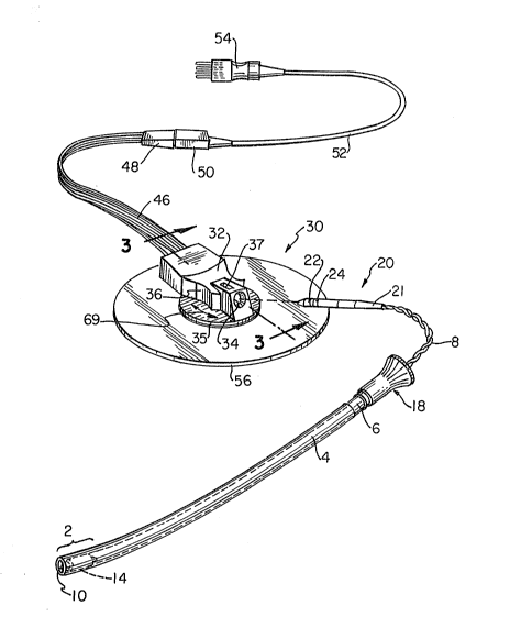
Figs. 2-4 show a connector 20 attached to the
proximal ends of the twisted wire pair 8 in accordance with the
invention. The connector 20 has electrically conductive ring-
shaped contacts 22, 24 which are connected to uninsulated ends
~ ` . ,.
',? ,'.: , .: -:
2054375
26, 28, respectively (see Fig. 4) of the twisted wire pair 8.
The contacts 22, 24 may extend around the entire circumference
of the connector 20 and are axially spaced apart from each
other to avoid electrical contact therebetween.
A drive handle 18 defines a passage 19 (see Fig. 5)
in communication with the inner passage defined by the driving
tube 6. The driving tube 6 is jam fit inlo handle 18 along an
inner taper of handle 18 that extends toward a stepped surface
of the handle 18 (see Fig. 5).
Handle 18, which has a cone-like shape facing rear-
wardly, facilitates pulling of the driving tube 6 off the
twisted wire pair 8. The proximal end 21 of the connector 20
is tapered to fit within the tapered proximal end of handle 18
and thereby travel within the driving tube 6 without jamming.
A leg plate 30 (Fig. 2) includes a housing 32 with
an opening 34, which i9 adapted to receive the connector 20
after the driving and guide tubes are removed. Concavely
curved finger grips 36 are pro~ided on either side of the
housing 32. These grips 36 facilitate holding of the housing
while inserting the connector 20 into the opening 34. The
bottom of the housing 32 includes an integrally formed circular
plate 35.
The outside diameter of the connector body 20 is
smaller than the inside diameter of the driving tube 4. Thus,
the guide and driving tubes 4, 6 may be pulled together over
the connector 20 and thereby removed from twisted wire pair 8
prior to connecting to leg plate 30.
As is seen in Fig. 2, the connector 20 is inserted
into the opening 34 in the direction indicated by the dashed
lines. As better seen in Fig. 3, the opening 34 defines an
elongated path of insertion for the connector 20, which extends
about a centerline. As best seen in Fig. 4a, contacts 38, 40
lie radially outside the path with respect to a radial
direction from the center line.
As an additional feature, an opening 37 may be
provided along the top of the housing 32~ This opening 37
provides access for facilitating cleaning of the electrical
6 2054375 ~
connections and further allows one to visually verify that
~= electrical connection has been made. -~
After insertion of the connector 20 into the opening
34 as shown in Figs. 3 and 4, spring biased electrical
~ 5 contacts 38, 40 within the housing 32 electrically contact th~
'!';.~ ring-shaped contacts 22, 24, respectively. The proximal ends
`~ o~ the leg plate contacts 38, 40 form holding clamps which
~; hold uninsulated ends of leads 42, 44, respectively. The leads
42, 44 extend through ribbon cable 46 ~see Fig. 2) to a signal
receiving socket of a fetal monitoring device Yia inter-
engaging socket and plug connectors 48, 50, cord 52 and plug
54, which i~ adapted to be plugged into the fetal monitor.
~ In a similar manner, leads 41, 43, 45 (see Fig. 6), which will ~ ;
,1, be described later, extend through the ribbon cable 46.
s 15 Figs. 2 and 3 also show a base 56 snapped to the
i~ underside of the housing 32 via a stud 58 within a snap
compartment 60. The base 56 may be an adhesively coated
flexible pad made from foam material. In Fig. 3r the stud 58
is pushed between an opening periphery 62 in the underside of
the snap compartment until its top rounde~ end 64 clears the `~
opening periphery 62. In this position, the stepped
transition area 66 of the stud 58 engages the opening - -
periphery 62. During insertion, the opening periphery 62
bends against the stud 58 into the position shown. The bottom
68 of the stud 58 extends radially outward so as to sandwich
the pad 56 between itself and the underside o~ the snap ^- ;
compartment 60.
~31 The housing 32 is freely rotatable about the stud
58 with respect to the pad 56 in either the clockwise or
~' 30 counterclockwise directions indicated by double arrow 69 (see
,j Fig. 2). This rotation allows the housing 32 to be oriented
into any desired angular orientation. Thi~ rotatable
connection helps to avoid inadvertent disconnection of the
removable connector 20 from the leg plate 30 when, for -
instance, a doctor brushes against the twisted wire pair 8.
Fig. 5 shows the manner of remo~al of the guide and
driving tubes 4, 6 from the connector 20 and twisted wire pair
8. The spiral electrode 10 is in contact with the fetal
'' '' :
,j~;, ' .
7 2054375 ~::
epidermis E. The tubes 4,6 are removed by pulling the driving
handle 18 and guide tube 4 in the direction indicated by the
direction arrows, i.e., in a direction away from the fetal and
maternal electrodes 10, 12. After removal of the tubes, the
connector 20 is inserted into a leg plate.
Fig. 6 shows three leads 42, 43, 44 in electrical
connection with leads 70, 72 and 74, respectively (see also
Fig. 3). Two other leads 41, 45 (not shown in Fig. 3 for sake
of brevity but arranged similar to that shown Fig. 6) are
unterminated to serve as shield wires to help shield against
stray induced voltages from electromagnetic interference and
are electrically connected to a chassis (not shown) of the
monitoring device. The unterminated leads 41, 45 act like an
antenna to pick up stray voltages in the vicinity ancl route
them to the chassis, rather than allow the stray voltages to
follow the path of the other wires. Leads 41, 45 also dis-
tribute capacitance from the other three leads 42, 43, 44 via
ribbon cable 46 to the chassis.
Figs. 6 and 7 also show a leg plate 76, which has the
same housing 32 as that for the leg plate 30 of Fig. 3, except
that the housing is attached to a larger rectangular belt plate
78, rather than to an adhesive pad 56. The belt plate 78 of
Fig. 6 has a ground p}ate 80 welded onto the base 68 (see Fig.
3) of the stud 58.
The belt plate 78 has a slot 82 for holding one end
of a belt 84 as shown on one side and holed pro~ections 86 with
a pin 88 extending through the holes in projections 86 for
holding the other end of the belt 84. The belt plate 78 snaps
into the underside of the housing 32 in the same way as does
the pad 56 so as to enable the housing 32 to rotate about the
stud 58. Since the belt 84 is onIy snugly fit around the
mother's thigh and not too tight as to cause restriction of
blood flow, the ground plate 80 must be larger than the base
68 of the Fig. 3 embodiment needs to be in order to ensure that
contact with mother's leg is maintained.
The belt plate 78 is exemplified by Corometrics
Medical Systems as part of leg plate model no. 2608QOA, C. A
~ ,~
:, ~
: :
8 2 0 5 4 3 7 5 ~ ~
,i . , ,
suitable leg plate belt 84 is exempl:ified by Corometrics
Medical Systems as model no. 202300AA.
Fig. 6 also shows a plug 90 from which extends ribbon
cable 46 and which is fitted into a socket 92 of the leg plate
5 76. This differs from the hard wiring of leads 70, 72, 74 from
ribbon cable 46 of Fig. 3.
Fig. 8 shows a view similar to that of Fig. 3 except
~ that the stud 94 has an undercut 96 in which is resiliently
;~ biased a snap spring 98. Spring 98 is retained in the snap
10 compartment 60 and is ring-shaped to define an opening there- i
through.
In order to snap the stud 94 into place, the top
rounded end 100 of the stud 94 i8 inserted through the opening
defined by the snap spring 98 and into the compartment 60. A i i
15 cap 102 is on the top rounded end 100 of the stud 94. During
this insertion, the snap spring 98 is forced to elastically
expand outward by the cap 102 pressing against the snap spring
98 until the spring 98 clears the downwardly facing end 104 of ",.,".!,~
the cap 102. The snap spring 98 thereafter resiliently closes
20 against the undercut 96 to retain the stud 94 in position.- If
attempt is made to pull the stud 94 out of the compartment 60, ~ ~ -
withdrawal of the stud 94 is prevented by the holding force of -
the spring 98. Thus, a permanent mechanical snapping secure-
ment is obtained.
A conductive gel 105 is applied to the base 106 of
the stud 94. This gel will contact the thigh of the mother. i~
Although not shown in the other embodiments, the gel 105 is
applied to the bases of each of the other embodiments as well. -~
The bases 68, 106 or ground plate 80 serve as ground
electrodes.
In both Figs. 3 and 8 embodiments, the snap
compartment 60 is electrically conductive and is in electrical
contact with the stud 58 or 94, which is also electrically
conductive. ~ead 72 has an uninsulated end which is soldered
to the snap compartment 60.
In the embodiments of Figs. 2-4 and 8, the flexible
adhesive pad 56 may conform to the shape of the mother's leg
., :. .-
:j .
9 2054375
when adhered to the leg. A removable sheet (not shown) may
... .
cover the adhesive on the underside of the pad 56 and then be
removed when the pad 56 is to be applied to the mother~s leg.
The adhesive pad 56 (see Fig. 8) may include a foam material
108 ~ecured to a ~turdier element 110, ~uch a~ vinyl.
As should be apparent, for any embodiment, the ribbon
cable 46 may be either hard wired to the leg plate or may be
plugged into a socket in the leg plate. A180, either
embodiment may employ either the permanent or detachable snap
0 connection. The advantage of using the detachable snap
connection is that the housing may be reused; only the ba~e
which is in direc~ contact with the patient will be di3carded.
Thus, any base is interchangeable with another.
For all the leg plate embodiments, it is de~3irable
to angle the incline of the opening 34 in the housing 32 for
receiving the connector 20 at about eighteen degrees relative
to the horizontal plane (or of the adhesive pad 56 or-belt
plate 78) 90 that in~ertion of the connector 20 will not get
in the way of the mother's leg. The side o~ the leg plates
which receive the connector may be considered an input and the
side which effects connection with the remote monitoring device
may be considered an output.
Preferably, the electrodes and electrical contacts
and conductors are made of nickel or gold plated copper or--else
gold plated over nickel plated copper. High density
polyethylene (such as 12 melt ~rco #7120 or Chevron #9160 or
petrothene LS 606 8 melt, density 0.96) i9 recommended as the
material for the guide and drivi~g tube~ 4, 6, handle 18 and
the housing 32. The conductors are preferably jacketed with
polyvinylchloride. All component~ may be made from th~ same
materials as are de~cribed for their counterparts i~ the
patents previously mentioned.
The outer diameter of the guide tube 4 must be small
enough to avoid harming the mother during its i~ert.ion, such
as about 0.315 inche~. The inner diameter of the driving tube
6 is about 0.146 inches. Thu9, the width of the connector 20
`: `~' '
'. '.
.~ ' ' ~, ;`.
lO 2054375 ~; ~
must be less than 0.146 inches to fit within the driving tube
6. For example, the maximum diameter of connector 20 may be
.125 inches.
Fig. 9 shows a further embodiment of the leg plate
which utilizes a printed circuit board 112 that is fitted in
the bottom of the housing. A11 interconnection leads are then
soldered directly to contacts 114 on the I?rinted circuit board
below. The board is preformed so that all leads will be in
alignment with respective contacts after placing the board in ~-
the housing. The printed circuit board may interconnect the
appropriate contacts.
If desired, the housing may be prevented from
rotating relative to the base by providing projections from the ~;
base which block the housing from rotating.
While the foregoing description and drawings `
represent the preferred embodiments of the present invention,
it will be understood that various changes and modifications
may be made without departing from the spirit and scope of the
present invention. ~ :~
:, ' ., ~,. .:,
: : :