Note: Descriptions are shown in the official language in which they were submitted.
207~3~
- 1 -
DRUG DEIIVERY CATEIETER
BACKGROUN~ OF I~TF INVEN~ON
The present invention relates to a drug delivery device and
method. More particularly, the present invention relates to a drug delivery
device and method that is flexible as to the drug agent employed and the
duration of drug ~ ;.... with an emphasis on physician control over
d~,uluy~ of both the drug and the device.
Obstructive a~h~los~ u~ic disease is a serions health problem
facing our society today. This disease is the result of the deposit of fatty sub-
stances on the interior of the walls of the alteries. The build-up or
- ' of such deposits results in a narrowing of the inside diameter of
the artery which in turn restricts the blood flow through the artery. This
disease, wherein the opening or lumen of the artery is narrowed, is known as
a~ .uscl~,lu~i~ and the :a~ ' is known as a lesion.
One commonly used procedure for treating an obstruction caused
by a~h~u~ulclu~;~ is a procedure Icnown as coronary artery bypass graft
surgery ("bypass surgery"). Although bypass surgery has been used with
moderate success in the treatment of a~.luscl~u~ it can be extremely
invasive and traumatic to the patient.
-1-
2~74~
- 2 -
One less invasive and traumatic procedure developed more
recently is coronary ~A~,iu,ulaa~y. Coronary angioplasty, and angioplasty in
general, is a procedure in which a balloon is positioned in the inside of the
artery at the site of the ? I~t~ or lesion and inflated in order to dilate
the a~ .u~k,lu~ic lesion and thus open the restricted area of the artery. In
order to advance the balloon to the lesion, the balloon is attached to the distal
end of a small diameter catheter, which includes means for inflating the
balloon from the other end of the catheter. The catheter is v~ d or
"steered" through the patient's vessels to the site of the lesion with the balloon
in an un-inflated form. When the un-inflated balloon is properly positioned at
the lesion, the balloon is then inflated to dilate the restricted area.
While al~;iulJlaa~y has been relâtively successful in treating
coronary artery disease, restenosis of the treated site often occurs
a,u,u~ / 3 to 6 months following the procedure. It is believed that the
primary factor in developing restenosis is the healing that takes place after the
injury caused by the il~t~ .. iOII of balloon dilation procedure. The restenosis
has close analogy to scar formation in that the histologic result has a similar
.liol~ ,lo~y. The histologic response is called ir~tirnal fibrous hy~erplasia. A
main result of the intimal fibrous hyperplasia consists of smooth muscle cells
from the vessel wall that proliferate and migrate im the vessel wall. The net
result is a thickening of the vessel wall. Over time, this thickening reoccludes
or restenosis the vessel to a point where it is clinically significar~t. That is, the
blood flow through the vessel is diminished to a rate similar to the rate ~efore
3 2~743~
the angioplasty procedure. The occurrence of this seems to happen approxihna-
tely 30-35 % of the time following an af,~,iu~ ly to that specific site.
Several alternative procedures have been attempted to try to
affect the occurrence or rate of the restenosis following ill-~ liiUIl to the
lesion site in the coronary altery. These procedures have included the use of
lasers, mechanical ~ ~Lv~y devices, heated baUoons, and metal , ' ' '
stents. While each of these procedures has shown some success in dealing
with the initial lesion, aU have the similar problem of restenosis at a similar or
even greater occurrence. Current estimates of restenosis of the lesion site
using these alternative procedures ranges between 40-50%. The time frame of
restenosis of all of these is generally from 3-6 months after the procedure.
Therefore, it appears that this restenotic healing lesion area is
', ' of the type of ~ lLiv,~dl procedure used. Rather, it is a
iologi~ response to any type of injury brought to that lesion site. Because
of this intervention ', ' ,UII,~;Vl(J~ response, it is felt by many
physicians that potentially the best way to deal with restenosis would be by a
' ~ means, such as a drug agent, targeted at the 1.: - h ~1 events
that take place after injury.
To date, most 1' ' "i trials involve either an oral or
Ju~l~ injected drug that is delivered throughout the whole body in
hopes of trying to effect this small site in the arteries. This type of
~ , treatment is known as a "systemic treatment." Some agents that
have been tried in human clinicals include heparin, calcium channel blockers,
4 207~3~-~
inr~n~in converting en;~yme inhibitors, fish oil, and growth peptides. Other
agents that may not have been tried in clinicals but are of interest include
~I~UIIII)U~ synthetase inhibitor, serotonin ...-t~.6. , HMGCoA reductase
inhibitors, platelet derived growth factors, r~ ~ cell factors, platelet
L~ Liun inhibitors, and thrombm inhibitors such as hirudin or its analogs.
The indication for use of most of these has been either in vitro
cell culture studies or animal studies. These studies have shown some effect
on the smooth muscle cell ~,-uii~ Lion which is a major component of the
intimal fibrous hyperplasia that takes place in the restenotic lesion. However,
none of the systemic human trials to date has shown a major effect on the
occurrence of restenosis.
Even though none of these agents have been completely
successful in the in vivo human clinical trials, it is still generally felt that one
of these agents or some other new agent, if delivered locally and site
specifically to the lesion, would still be able to reduce the proliferative
response. One of the problems with systemic techniques is the inability to
deliver a high enough, - of the agent locally at the lesion in order
to effect the pl-y~iul~g;~, response. In the in vitro studies which have shown
some success, a high ~ of the agent was used. Thus, it is believed
that if the agent was delivered specifically to the site as opposed to
systemically, the agent may be delivered at a high enough I to
truly effect the ~Jh.~ h)~;;c res,oonse.
- 4 -
` 5 2a~3~
The reason many of these agents have not been used in a higher
r"nrpntr~tinn in vivo in humans is that many of the agents may exhibit
"",lr~:".l~lr-. side effects. Thus, if a high ~r~nr,rn~r~tif1n of the agents is given
systemically, they may have unwanted pl.~,;ul~,~;ic effects. Therefore, if the
drug can be given with lugh locally to the vessel wall while
1" the systemic amount of drug, the desired result of . ~ ;. g the
restenotic growth while preventing any unwanted systemic effects may be
achieved.
There are other ways known to date in trying to create a site
speciflc local delivery of drug to a site. One approach presently ~
is the use of a perforated or sweating balloon. For example, a drug delivery
device is disclosed by Wolinsky, H., et al. in the article entitled, Use of a
Perforated Balloon Catheter to Deliver C'~l ' Heparin Into the Wall of
a Normal Canine Artery, 15 JACC 475 (Feb. 1990). This device is a per-
cutaneous t ~ 1 coronary ~u~gio~ "y (PTCA) balloon with several
microholes in the balloon for delivery of an agent during balloon dilatation.
The drug is A ' ~ into the same fluid which is used to inflate the
balloon.
A major concern with any device with perforated holes is the
effect of a jet stream on the wall of the blood vessel. If a balloon with holes is
Di~Uli~, a relatively high velocity and/or pressure jet stream may be
~t ~ttt th ugh the holes thett ue ~ h gcahons tt the~ j t st t~ts
- ~ 207~304
- 6 -
may cause tissue damage resulting in very severe dissection of the vessel waU
after the dilatation procedure.
Another d;~lv ~ of available devices, such as the one
disclosed by Wolinsky et al., is that these devices cause a substantial blockage
of blood flow in the subject vessel during the procedure. Thus, such devices
may only be used for the fairly short time frame (typically, from one to two
minutes), similar to the time frame of the actual v . ' ~/ dilatation.
Other available drug delivery devices are disclosed, for example,
in United States Patent Numbers 4,824,436 (Wolinsky) and 4,636,195
(Wolinsky). These devices are directed to a dual occlusion catheter in which a
balloon is inflated proximally and distally of the - ' . or lesion
creating a space for infusion of a drug. This dual balloon catheter creates a
space for infusion of drug separate from the blood flow. This device,
however, also can only be used for a short period of time because it occludes
blood flow.
Perfusion is very important in developing a suitable type of
delivery device. It is necessary that the device provide a la~e latitude in time
over which the agent could be delivered and therefore, devices which occlude
blood flow may not provide the necessary latitude. Because the basic research
into the 1,;~ y and 1' ~,;olc~ic events indicate that the initial events
begin ' l~ after mjury and continue intensely for several hours, it is
desirable for the drug delivery system to aDow drug delivery for several hours
to a day or two beginning - " '~, after intervention. This research also
- 6 -
2~7430~
- 7 -
points out that the initial events ~ create a cascade of events that
ultimately lead to intimal thickening. While these .. ' or lesions do
not become apparent for several months, it is felt that if these initial events can
be modulated, blocked, or even ~c~ ~t~A then the subsequent cascade can
be altered and a diminished overall thickening could be achieved.
Even in devices where the shaft tubing provides a lumen for
blood flow, the blood flow is limited by the diameter of the tube. In fact,
when the shaft tubing is used for a blood flow lumen, there is a competing
balance of making the shaft large enough to ;.~cr~-~ the larger volume of
blood flow while still trying to minimr~e the size so that the catheter can be
Illàll~ v~.~tid through the patient's ~la~culail~. This limited blood flow, while
providing obvious advantages to no blood flow, may not be completely
aali~rd~uly in providing the necessary blood flow to the heart (or other part of
the body).
Catheters with expandable devices are also used im other
procedures. For example, United States Patent Number 4,183,102 (Guiset)
discloses a device which includes a series of toroidal shaped sleeves which
conform to a vessel wall~ The device disclosed in this patent is for the
treatment of some aortic aneurysms and does not include any means for drug
delivery.
Therefore, it is desirable to have a drug delivery device capable
of providing the necessary blood flow to the heart for restenosis treatment.
Further, such a device may also be extremely desirable in other procedures
- 7 -
2~7~g
- 8 -
where a d~ug is to be delivered to a specific site in a vessel. For example,
drug delivery devices may be useful in procedures where a drug or agent is
used to dissolve the stenosis in an effort to avoid the use of ,~A.giv~kl~ly or
~Lh~ I~L~J..,~ procedures altogether or to deliver a i' ~ bolyLic agent to
dissolve a clot at the lesion site.
It will be recognized from tbis discussion that there is a need for
a generic type of drug delivery system which ~ i~, physician control
over the deYice and agent. The device should have flexibility as to the agent
that is to be delivered and should be capable of delivering any number of
agents (either separately or at the same time), or possibly also allow a change
in the protocol of the delivery. It should also be flexible with respect to the
time frame over which these agents would be delivered. In order to effect this
time frame of delivery, the device should also allow a large amount of blood
flow by or through the device in order to maintain adequate distal perfusion of
cardiac or other muscle during the delivery time.
Therefore, it is a primary object of the present invention to
provide a device and method wbich can contain a relatively high ~~
of a drug agent in a selected portion of a vessel, such as a blood vessel.
It is another object of the present invention to provide a device
which can be used in a flexible time frame.
It is a further object of this invention to provide a drug delivery
catheter which permits a relatively high fluid flow rate through the vessel in
which it is inserted while the device is employed.
- 8 -
-
~ 2~743~
It is a still further object of this irlvention to provide a device
which is flexible as to the drug and the number of drugs which can be
delivered as well as the time frame over which they can be delivered.
Sl~MMARY OF THE lNVEN~ON
To achieve these and other objects, the present invention
provides a new and unique drug delivery catheter which may be inserted into a
vessel, such as a blood vessel. The drug delivery catheter of the present
invention comprises an elongated tubular shaft which includes a drug lumen for
delivering a drug to the treatment site and a uniquely configured inflatable
balloon assembly. The balloon assembly is disposed at the distal end of the
shaft and includes an inflatable balloon member. The balloon member has a
....i~"..,.l;..,. such that when the balloon member is uninflated, the fluid in the
vessel (such as blood) may flow around the balloon assembly. This provides
an All~...,!j~...l...l~ which may be easily inserted and . 1 1 through the
vascular system. When the balloon member is an inflated state, part of the
balloon member contacts the vessel wall defining a . pocket between
the vessel wall and the balloon assembly. The balloon assembly includes
apertures in the . pocket which are in fluid ~ with a
drug lumen in order to provide the drug to the . pocket. A flow
lumen is also defined through the balloon member when it is inflated in order
to allow the fluid in the vessel, such as blood, to flow through the balloon
g
2074304
asgembly. The catheter also includes an inflation lumen which
i5 used to inflate the balloon member. The catheter may also
include a plurality of drug cr~nt~i -nt pocket6 either in fluid
communication with each other or isolated from each other.
The present invention also F-n~ 'lq a method of
using the drug delivery catheter to deliver drugs to a
treatment site in desired concentrations.
The present invention provides a time frame for drug
delivery in relatively high concentrations and which can be
used in a relatively flexible time frame. For example the time
frame may be greater (or less) than several minutes to possibly
several hours or a day to two days.
This time frame of delivery is important in procedures
intended to abate the occurrence of restenosis because the time
frame needed to favour~bly modulate the restenotic response is
variable .
Further, the device and method of the present invention
may be advantageously used to deliver a drug agent intended to
dissolve a lesion or to dissolve a clot at a lesion site.
BRIEF DESCRIPTION OF THE DR~WINGS
FIGURE 1 shows an ~ of the drug delivery
catheter of the present invention.
FIGURE 2a shows a cross section of the ~ _~ir-nt of
FIGURE 1 along line 2a-2a.
FIGURE 2b shows a cross section of the c~mho~l i r- t of
FIGURE 1 along line 2b-2b.
-- 10 --
.~
2~743~
- 11 -
FIGURE 3a shows a cross section of the; ' - ' of
FIGURE 1 along lin~ 3a-3a.
FIGI~RE 3b shows a cross sectional slice of the . ~: ' of
the device of Figure 1 in a plane ~ . to line 3a-3a.
FIGURE 4 shows another ~ ' ~ " of the drug delivery
catheter of the present invention in a deflated form.
FIGURE 5 shows the ' ~1l' of the drug delivery catheter
of FIGURE 4 in an inflated forln.
FIGURE 6 shows another e ' " of the drug delivery
catheter of the present invention.
FIGURE 7 shows another, ~c ' of the drug delivery
catheter of the present invention.
FlGURE 8 shows another . ' " of the drug delivery
catheter of the present invention.
FIGURE 8a shows a front view of the ~ ~ " of
FIGURE 8.
FIGURE 8b shows a cut-away view of the ' ~ ' of
FIGURE 8.
FIGURE 9 shows another; ~ " of the drug delivery
catheter of the present invention.
FIGURE 9a shows a cross section of the ~ of
FIGURE 9 along line 9a-9a.
- 11 -
207~3~
- 12 -
FIGURE 9b shows a cross section of the .,111~' of
FIGURE 9 along Line 9b-9b.
DETAILED DESCRI~ION OF THE PREFERRED EMBODIMENT
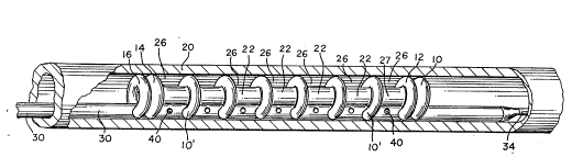
Referring to FIGURES 1, 2a, 2b, 3a and 3b, a preferred
of the present invention includes a baUoon assembly comprising a
single piece of baUoon tubing laced in a manner that wiU be described below to
form toroidal or donut-shaped baUoons 10, 12, 14, 16. When inflated the
baLloons 10, 12, 14 and 16 are intended to impinge upon or engage the vessel
waU 20 as explained in more detail below. The figures depict the baUoons 10,
12, 14, 16 in their inflated form.
In an exemplary ul.ll)od~ , these baUoons 10, 12, 14, 16 are
about 3 " (mm) in outside diameter in their inflated form. The
balloons, however, can have an outside diameter ranging from about 2 mm to
about 20 mm in their uninflated form, depending on the different vessels of the
human (or animal) body in which the baLloons wiU be used. The siz~ of the
baUoons may also vary for different procedures.
The most distal baUoon 10 and the next most distal baUoon 12
are preferably adjacent to each other as are the most proximal baUoon 16 and
the next most proximal baUoon 14. The distance between the next most
proximal baUoon 14 and the next most distal balloon 12 may be, for example,
about 20 mm.
- 12 -
2~7~304
- 13 -
Providing the balloons in pairs adv~ulL6~.Jly provides two
points of anchoring for each pair. This is IJ~iuulally useful at the ends of the
balloon assembly where the two point anchoring reduces the tendency of the
balloons to rotate ~ .l.. 1i. . '., to their axis.
The balloons are preferably made of a polyolefin. A suitable
polyolefin material is available from E.I. DuPont de Nemours and Co.
(Wilmington, Del.) under the tradename Sur~yn~ Ionomer.
The balloon assembly also imcludes a cylindrical sheath 22 which
connects the balloons 10, 12, 14 and 16. The diameter of the sheath 22 is less
than the diameter of the balloons. Therefore, when the balloons are inflated,
the sheath 22 is attached to the balloons at a point radially mward of the outer
diameter of the balloons in order to create a pocket. Preferably, the sheath 22
is disposed tbrough and cormected to the interior portion of the toroidal-shaped
balloons. In an exemplary ~ ' t, the sheath 22 is typically 2~i mm from
end to end lon~ ly and is preferably about 0.001 inches tbick.
The sheath 22 is situated coaxially to the vessel wall 20 and is
open at each end, thereby formmg a l~ IY or blood lumen 24 for the
blood to flow through when the balloons are inflated. Thus, the sheath 22
creates a barrier for separation of the drug medium and the blood. The sheath
22 is supported or held open by the tUlUi~ h~r~ balloons 10, 12, 14, 16
and has the capability of having a relatively large internal diameter. For
example, the internal diameters may be about 0.060 inches providing a very
large volume of blood flow. This internal blood flow lumen 24 formed by the
- 13 -
2Q7~3~
- 14 -
sheath 22 has the capability of being si~";l~,Lly larger than the shaft tubing
of the device.
The sheath 22 may be made of Surlyn0 Ionomer. More
preferably, the sheath 22 may be made of a polyester copolymer such as a
random copo]ymer. The random copolymer used to make the sheath of the
present invention may be prepared according to standard procedures from
ethylene glycol, and a mixture of dimethyl t~ ' and dimethyl
phfhl~' As used in the random copolymer, the ratio of terephthalate to
~50pl th~ fP im the random copolymer may be varied over the range of between
99:1 to 80:20. Suitable cu~l~ are . ~ Jly available and are sold
under the tradename SelaP Pr, such as SelaP X257, available from E.I.
Dupont de Nemours and Company (Wilmington, Delaware).
In this C..,IJ- '- t, the blood flow lumen 24 is created by the
inflation of the balloons 10, 12, 14, 16 and can be ' i '.y collapsed upon
deflation of the balloons 10, 12, 14, 16 (FIGURE 4 illustrates the balloons in a
deflated state). The dimensions of the deflated device will, of course, vary
depending on the specific use, ,' ~, but suitable sizes range from
0.035 inches to 0.1 inches.
As illustrated best in Figures 1 and 5, when the balloons are
inflated, a ,~ pocket or region 26 is defined between the sheath 22
and balloons 12, 14 and the vessel wall 20. When the balloons 12, 14 are
inflated they in effect form a seal between the vessel wall 20 and the
balloons 12, 14 and thus the balloons 12, 14 define the outer boundary of the
- 14 -
-15- 2a74304
pocket 26. The sheath 22, which is bonded to the balloons 12,
14, defines the rest of the ~ pocket 26. This . pocket 26
provides region in the vessel which is isolated or separate from the fluid
flowing through the flow lumen 24. Therefore, the drug medium may be
contained in this, pocket 26 in the desired ~ for a
A period of time without entering the blood stream for that period
of time.
The backbone of this; ' - ' is a catheter (or shaft) formed
by an elongated tubular shaft 30 into which the balloon tubing is laced in a
manner that will be described in detail below to form the toroidal or donut-
shaped balloons 10, 12, 14, and 16. The specific size and c.. ~ of the
shaft tubing 30 will, of course, vary depending on the specific application for
which it is intended. In one exemplary: ' - ' t, the shaft tubing is about
135 ~ (cm) long, about 0.040 inches in distal outside diameter and
about 0.050 inches in proximal outside diameter, and has a wall thickness of
from about 0.003 inches to about 0.004 inches. The diameter of the tubing 30
can range from about 0.030 inches to about 0.090 inches depending on the
. .' ' use. These exemplary ~' are suitable for a catheter
intended for use in the coronary arteries. As will be recognized, the
dimensions for catheters intended for use m the prostate, urethra, etc. may
have other suitable ~' This shaft tubing 30 is preferably made of a
flexible material such as a high density ~I~ ,ne OEIDPE!J.
- 15 -
- 16- 2~743~'1
The shaft tubing 30 is advd~ ~u~ly necked at its distal end to
allow for variable stiffness or variable flexibility along the shaft. The necking
of the shaft tubing 30 may be , ' ' by several ~ " ,. ,~ For
example, a necked region, that is a region which is more flexible tban the rest
of the tubing, may be provided by reducing the outer diameter of the tubing,
by reducing the wall thickness of the tubing, or by providing a section of the
tubing made of a more flexible material than the rest of the tubing. The
necked portion may also be provided by any . ' of these i ~ ' .
FIGURE 3b best illustrates this necking in one preferred
JO~I,- t FIGURE 3b depicts a slice through the plane perr~n~ r to
the plane of FIGURF 3a and for purpose of clarity does not depict the
structure of the device which is not in this plane. The shaft tubing 30 is ?
bonded to a necked section 30a of proximal shaft tubing. The bonding-152
commercially may be accomplished with an adhesive, such as BiPax~ BA-
2135D Tra-Bond available from Tra-Con (Medord, r~- ~ ). The
necked section 30a of proximal shaft tubing may have, for example, an outside
diameter of about 0.040 inches and a wall thickness of about 0.003 ir~ches.
Proximal to the necked section 30a of proximal shaft tubing, there is a
transition section 31 followed by the unnecked section 30b of proximal shaft
tubing 30. The uMecked section 30b may have an outside diameter of about
0.050 inches and a wall thickness of about 0.004 inches.
By way of example, the shafting tubing 30 may be about 135 cm
long, the necked section 30a of proximal shaft tubing may be about 25 cm
- 16 -
- 17- 2~7~30~
long, and the unnecked section 30b of proximal shafting tubing may be about
110 cm long. Preferably, the shaft tubing 30 has a tapered section 33 at its
distal end to prevent damage to the vessel walls as it is being inserted.
A necking may be r- L~ ' ' by pulling the tubing through a
heated die to elongate it and therefore change its inside and/or outside
diameters.
An inner shaft tubing 32 runs coaxially through the shaft tubing
30 in order to ~ ~ ' a PTCA guide wire 34. This type of guide wire
is known in the a~t and therefore no further discussion is given here. A
suitable (~nnfi~-~tinn for the inner shaft tubing 32 is a flexible tube made of
HDPE which is about 135 cm long, and has a wall thickness of about 0.003
inches.
This inner shafting tubing 32 may also be necked to provide
added flexibility to the catheter. As illustrated in FIGURE 3b, in one .
exemplary . lm' the inner shafting tubing 32 has a transition region and ~J
an unnecked proximal portion 32b. The distal, necked portion of the inner
shaft tubing 32 may be about 0.021 inches in outside diameter and about 30 cm
long. The unmecked proximal portion 32b may be about 0.024 inches in
outside diameter and about IOS cm long.
The necked or more flexible portion of the catheter may be
provided by necking one or both of the shaft tubing 30 and the inner shaft
tubing 32.
- 17 -
3o~
As illustrated there are basically three different lumens through
the proximal portion of the shaft tubing 30, one being the inner lumen for the
guide wire 34 formed by the inner shaft tubing 32, another being an inflation
lumen 36 for the balloon inflation medium which is continuous with the donut-
shaped balloons 10, 12, 14, 16 (described in greater detail below), and the
final one being the drug delivery lumen 38 formed in the space between the
inner shaft tubing 32 and the shaft tubing 30.
In one exemplary f-mhoflimf-n~, the inflation lumen is made at its
distal end (where the balloons 10, 12, 14, 16 are formed), from Surlyn~, and
has an inside diameter of 0.006 inches and an outside diameter of 0.011
inches.
In a presently preferred ~ the inf~ation lumen is
formed of three sections: 1) the section attached to the balloons described
above; 2) a middle inflation lumen section 164; and 3) the proximal inflation
lumen section 166 which eventually is attached to the manifold (not shown).
Both the middle and proximal inflation lumen sections 164, 166 are preferably
made of a polyimide material. The bond 170 of the balloon tubing 36 to the
middle inflation lumen section 164 and the bond of 172 of the middle inflation
lumen tubing 164 to the proximal inflation lumen tubing 166 is ~f~ ,rA
with a suitable adhesive, such as Loctite~ 405 available from Loctite Corp.
- 18 -
207~30~
- 19 -
(Newington, ('onm~rtitll~), The middle inflation lumen section 164 may have
an inside diameter of about 0.012 mches, a wall thickness of about 0.001
inches, and may be aboue 25 cm long. The proximal inflation lumen section
may have an inside diameter of 0.016 inches, a wall thickness of 0.001 inches,
and may be about 100 cm long. It will be recognized that these dimensions
are given by way of example and other suitable sizes may be employed.
The shaft tubing 30 has a number of apertures 40 in order to
ac~o...lllo ' the drug flow out of these apertures 40 into the,
pocket 26 formed between the two inner balloons 12, 14, the sheath 22, and
the vessel wall 24. The apertures 40 are placed k~n~ih~rlir~lly along the shaft
hubing 30 between the inner most balloons 12, 14. The apertures 40 are on
both sides of the shaft tubing 30 and the area in which they are placed does not
come into contact with the vessel wall 20. Preferably, the apertures 40
generally imcrease in diameter from proximal to distal end so that uniform flow
out of each aperture 40 is achieved. The apertures are preferably sr~ed such
that the drug medium is not pressure injected out of the apertures 40, but
rather the vessel tissue is "bathed" with the drug medium. The size of the
apertures will, of course, depend on the pressure at which the drug medium is
being provided at the proximal end by the drug medium source (not shown).
In an exemplary . :l " t, the more proximal apertures 40 are about 0.003
inches in diameter and the more distal apertures are about 0.005 inches in
diameter. These apertures 40 are placed about 2 to 3 mm apart from each
other.
- 19 -
2~ 7~30~
- 20 -
This ~ B~ul . ,. .,l is ~ .r~ ulcid by drilling the apertures into
the shaft tubiltg 30 and then laciltg non-blown balloon tubing 36 through these
apertures in such a way that the tubing is loored around a temporary fornting
mandrel (not shown) to form the section in which the sheath will be placed.
~Jpon completion of this lacing, the tubmg is ~ . 1 and dipped into a hot
water bath, preferably at 80 C, for expansion of the Ih. .. ~ balloon
tubing 36 to form the outer, .'( ~h~ balloons 10, 12, 14, 16.
After this process, another tnin-walled cylindrical balloon is
inserted into the interior of the toroidal or donut-shaped balloons 10, 12, 14,
16 to form the sheath 22 which separates the drug and blood mediums. The
sheath 22 is then bonded to aD of the ~ ~h~reA balloons 10, 12, 14, 16 and
the ends are cut off flush to the most proximal and distal donut-shaped balloon
10, 16. The bonding may be ~. ~o ~ A by the use of a suitable a&esive,
such as an epoxy resin with an aliphatic amine hardener. The rO"I, I.lLhJ~ for
a suitable epoxy is given below in Table 1.
TABLE I (EPOXY ADHESIVE)
Epoxide
Wt. Equi~alcnt
(in ~elams~ ~t. DescriDtion
Araldite GY 508 148.8 (g) 400~55 Bisphenal A based cpoxy
tciba Geigy] blendod with polyglycol di-
epoxidc
Arald~lte GY 506 59.0 (g) 17~-185 Bisphcnal A based epoxy
[Ciba Geigy]
Epon 828 9~6 (g) 187.5 Bisphcnal A based cpoxy
- 20 -
~ -21- 2~7~3~4
(Shell)
2~Me~byl-1,5- 32.~ (g)
pel~tadiamme
A from Dupol~t)
Ihe inner shaft tubing 32 is then put through the shaft tubing 30
in order to ~ - ' a guide wire 34. Tttis inner shaft tubing 34 is
fastened to the manifold on the proximal end of the catheter with an adhesive
bond, such as a urethane adhesive bond available from the 3M Company
(M- ~ -'- Mirtnesota) under the tradename ~ - Scotch Weld0
Brand 3549 B/A Urethane Adhesive Kit. The inner shaft tubing 32 is fastened
to the outer shaft 30 on the distal end of the catheter using the same urethane
adhesive. This bonding material on the distal end may also be used to form
the tapered section 33.
As is illustrated in Figure,~, the toroidal-shaped balloons 11 12,
14, 16 are formed from one continuous piece of tubing since they are laced
through the shaft tubing 30 and therefore the balloons lO, 12, 14, 16 are all in
fluid c, with each other.
T}te proximal end of this catheter comprises a manifold (not
shown) known in the art. A suitable manifold is a ~ ~. ' three-port
manifold. A vacuum source is placed on the balloon inflation port of the
manifold to evacuate air from the balloons. Ttte drug irtfusion port of the
manifold is then filled with a sterile liquid to evacuate air.
- 21 -
.
-22- 2~7~3~4
Thus, the blood flow lumen 24 is not an integlal part of the
shaft of the device. Rather the lumen is created by inflation of the balloons
10, 12, 14, 16 of the device. Since the blood flow lumen 24 is not an integral
part of the shaft, the ultimate diameter of the blood flow lumen 24 is not
limited by the diameter of the shaft. When the balloons 10, 12, 14, 16 are
deflated, the device is collapsed with essentially no blood flow lumen and
therefore small enough in diameter to easily maneuver through the patient's
vascular system. As discussed above, in some prior art devices, the lumen
was created by the shaft tubing itself thereby limiting the size of the blood flow
lumen. Unlike prior alt devices, when the balloon member is inflated, the
cross-sectional area of the blood flow lumen 24 is a significant percentage of
the cross-sectional area of the blood vessel. It is presently believed that with
the present invention, the blood flow through the device will be ahout 60% and
may be as much as 80% of the blood flow through a healthy vessel without the
device in place.
nifi~ ly, in this ' ' the balloon inflation medium,
which may be any suitable inflation medium such as saline and contrast dye, is
separate from the drug medium. This separation is imporlant because it
provides flexibility as to the amount of drug delivered, the time frame over
which it is delivered as well as flexibility as far as d~lu r of the device.
Also, as will be recognr~ed by those skilled irl the art, this, ~ '
minimizes the problem of tissue damage caused by a very high jet stream of
the drug being ejected from the catheter. As mentioned above, preferably the
2~3~
- 23 -
apertures 40 are not directed toward the vessel waU 20 and the apertures 40
are d~yluyli~'vly sizeo such that the drug emerges from the apertures to the
wall in a slow gentle stream instead of emerging from the apertures 40 in a
pressurized stream. Thus, the ~ region 26 created between the shaft
tubing 30 and the vessel waU 20 is used to "bathe" the vessel waU 20.
Other r~ O~ of the present invention are illustrated in
FIGURE 4, FIGURE~ 5, FIGI~RE 6, and FlGURE 7 with elements similar to
the previûus, ' :' numbered similarly. FIGURES 4 and 5 iUustrate this
device in the blood vessel 20 both deflated (E'IGURE 4) and inflated
(FIGURE 5). As iUustrated in these figures the baUoons in the baUoon
assembly may be spaced differently and additional baUoons may be placed at
closer intervals. The additional baUoons will aid in keeping the perfusion
lumen open. The t nnfif~ ti~-n and plj~;Lio~ g of the toroidal-shaped balloons
have multiple l~.J~ More baUoons may be formed between the
previously-described toroidal-shaped baUoons 10; and the baUoons 12 and 16
may even be eliminated. (FIGURE 7). For aU of these Pmho~" the
distance from the most proximal baUoon 16 to the most distal baUoon 10 can
range from about 20-30 mm and the inside baUoons 10' may be disposed about
2-3 mm apart.
The number of baUoons and the spacing between baUoons is
important in g, the dyy~ol blood flow through the vessel being
treated. It is possible that the flow of blood through the sheath 22 may be cut
off by any one of the foUowing: (I) the lesion may deform the sheath; (2) the
-23 -
207~04
- 24 -
device may be piaced at a bend and the sheath could kink; and/or (3) the
pressure of the drug could force the blood lumen shut. Therefore, the radial
support needed for the sheath 22 wiU vary depending on the specific conditions
of the treatment site and the particular treatment being ad~ ,u.,t~ d. The
radial support for the sheath 22 needed to maintain the blood flow lumen 24
through the center of the sheath 22 is provided by the baUoons. The different
~r,. ,l ic,. . ,.~ .C iUustrated may be used to provide more or less radial support as
needed. Increasing the number of balloons in the balloon assembly increases
the ability of the balloon assembly to maintain the perfusion lumen open.
Referring now to FIGURES 8, 8a, and 8b, another .
of the inventions is shown. In this embodiment the balloon assembly is com-
prised of an outer cylindrical sheath 50 and an inner cylindrical sheath 52
sealed to each other at the ends of the sheaths. The cylindrical sheaths 50 and
52 are also '~, sealed to one another at sections 54. An inflation
region or pouch 56 is defined between the two sheaths 52 and 54. These seals
54 run along the ~,u. U,llr~ of the cylindrical sheaths 50, 52 except that
they are not complete in that spaces are lert at certain points to aUow the
inflation medium to migrate from one pouch formed between the cylindrical
sheaths 50, 52 to another similar pouch. The method for forming these seals
is discussed in greater detail below.
Cutouts 58 are provided in the proximal cone section 60 of the
sheaths to allow blood to flow through the center of these sheaths 50, 52. At
the proximal portion of the cone, the outer sheath 50 and the inner sheath 52
- 24 -
2Q7~3~4
- 25 -
come to an outer balloon waste 100 and an inner balloon waist 102. The outer
balloon waist 100 is bonded with an adhesive, such as Tracon~, to an outer -
shaft 104 and the inner balloon waist 101 is bonded with a similar adhesive to
an inner shaft 106. The outer and irmer shafts are made in a similar fasbion to
the ~ ..,l,~.l - '~ previously described. The inner shaft 106 def~nes a lumen
for a guide wire 108. The space between the ~uter and inner shafts 104-
defines the d~ /illlL.iion medium lumen.
The double sheath/balloon assembly may be formed for example
from . ~ ;~lly available PTCA catheter balloons. The outer sheath 50 and
an inner sheath 52 are, however, may also be made from a polyester
copolymer such as that described above.
In an exemplary P.mh~' the outer sheath 50 and inner
sheath 52 are about 2.9 mm and about 3.0 mm respectively in outside
diameter. Depending on the use, however, these diameters may range from
about 1.9 mm and about 2.0 mm to about 5.5 mm and about 6.0 mm,
respectively. The sheaths 50, 52 are preferably about 20 mm in length, but
may vary from about 10 mm to about 30 mm. The length of the total device
is d~ / 135 cm, but may range from about 75 to about 150 cm. Tbe
seals 54 may be between about 0.005 inches and 0.01 inches wide and about 2
mm away from each other, though other ~ I~..~c,..,~;l,..~ may be employed.
Iu tbis ' ' t, the inflation medium and drug medium are
one and the same. When the balloon assembly is inflated, as shown in Figures
8-8b, the outer sheath 50 contacts the wall of the vessel 64 at the areas
- 25 -
2~7~3a4
- 26 -
designated by reference numeral 63. The contact area 63 is defined by the
section of the sheath 50 which is not bonded to the other sheath 52. The area
54 where the two sheaths 50, 52 are bonded, however, does not contact the
vessel wall 64. Therefore, a, pocket or region 11 for the drug is
defined in the space between two adjacent contact areas 63. The outer sheath
has apertures or holes 62 in order to deliver the drug to the vessel wall 64 in
the c pocket 11. These apertures 62 allow for ~ y of the
inflation medium (which contains the drug) out to the vessel wall 64.
Preferably, these apertures 62 are about 0.003 inches in diameter and spaced
radially at 90 for each pouch. EIere again other ~I, r~ .. may be
suitable as well. For example, both the number and pattern of spacings of the
apertures in each pocket defined by adjacent ribs may vary. The polymer that
the outer balloon is formed of can either have the apertures 62 as discussed
above or alt~ ly may be semi-permeable to the infiation/ drug solution.
This ~ ho~ also provides very good separation of drug
from the blood lumen and excellent blood flow because the blood flow lumen
66 is created by the inflation and expansion of the double sheath 50, 52 balloon
assembly. The sheaths 50, 52 are inser~ed into the patient in a deflated form
(not shown) and when they are positioned properly, they are inflated until the
outer balloon 50 comes into contact with the vessel waUs 64. The details for
using this ' . are similar to those described below for the; ' "
of FIGURES I, 2a, 2b and 3.
- 26 -
`` 2~7g304
- 27 -
Similar to the other Pmho limPnt~ discussed above, the position
and number of inflation pouches 56 may vary for different uses. To
r- '` this, the seals between the two cylindrical sheaths 50, 52 can have
different ~I-..i;L .~ depending on what type of lifting and expansion force
would be needed.
This: ' ~ " of I~IGURES 8, 8a, and 8b, is preferably
made by blowing two different sheaths 50, 52, one slightly smaller than the
other 52. The second smaller inner sheath 52 is inserted coaxially inside the
outer sheath 50. These are then completely sealed distally, creating an
occlusive seal 70 between the two sheaths 50, 52. These two sheaths 50, 52
may have seals through the body of the balloon assembly similar to
what an inflatable air or water mattress would have; these seals are mcomplete
in places, aUowing the inflation medium/drug to flow throughout the device
In an exemplary i ' ' t, the seals are 2-3 mm apart with a 0.01 inch
wide bond On the proximal end in the cone area 60 of the sheaths 50, 52,
there are sealed cutaway portions 58 for blood flow~ This sealing is around
the cutaway portions 58 and allows the blood to flow while still ~
inflation space 72 in parts of the cone to the body of the cylindrical sheaths.
The sealing can be done in a number of different ways. One
preferred method is by creating a heat seal between the two sheaths. This heat
seal can be made by a laser weld, a radio frequency weld, or an ultrasonic
weld. Other equivalent methods can be ' J In each case, the polymer
- 27 -
` 207g~0~
- 28 -
of the two cylindrical sheaths would be heated very locally to form a heat bond
between the two pieces of the polymer.
Referring now to FIGURES 9, 9a, and 9b, another; ' - '
of the invention ls shown. FIGURE 9a shows a cross section of FIGURE 9
along line A - A' and FIGURE 9b shows a cross section of the ' ' of
FIGURE 9 along line B - B' This; ' ' is comprised of an outer
polymer cylindrical sheath 72 and an inner polymer cylindrical shedth 74
~1 sealed to one another at areas 76 to form a balloon assembly
with an inflation area 78 being defined between the two sheaths 72 and 74.
This l ..,ho~ is similar to the; ' ' of FIGURES 8, 8a, and 8b
except the seals in the balloon assembly are forming welds which
are similar to spot welds.
In an exemplary ~ ..,Io~l, .1 the outer sheath 72 and inner
sheath 74 are about 2.9 mm and about 3.0 mm ~ ,ly in outside diameter
and about 0.001 inches thick. In an exemplary - ~ , the balloon
assembly comprised of sheaths 72, 74 is about 20 mm long and the total device
is preferably about 135 cm long. The total device length, however, may range
from about 75 to about 150 cm long. The outer sheath 72 and an inner sheath
74 are preferably made from the dbu~ .n~ n~A Selal3 copolymer. The
seals 76 are d~ y 0.005 inches wide and d~ / 1-2 mm from
each other. The method for forming these seals is similar to the methods
discussed in greater detail above.
- 28 -
2~74304
- 29 -
- The inflation/drug medium is delivered into the balloon
assembly, which comprises the sheaths 72, 74, through shaft tubing 82 that
runs lr~n~ih- ' 'Iy along the device. Again, in this . ' ' t, the inflation
medium and drug medium are one and the sarne. There are apertures or holes
80 put into the outer sheath in order to deliver drug to the vessel wall. These ~'~
apertures 80 allow for 1~ y of the inflation medium (which contains the
drug) out to the vessel wall in a, pocket defined between adjacent
areas where the outer sheath contacts the vessel wall. A suitable diameter for
these apertures 80 is about 0.003 inches. Apertures 84 in the shaft tubing 82
allow the inflation medium/drug to pass from the shaft tubing 82 to the space
78 between the sheaths 72, 74. These apertures 84 are 0.01 inches in
diameter, 0.50 mm apart and run Ir)n~ifllrfin~1ly along the shaft tubing 82. The
shaft tubing 82 may also be necked to provide variable stiffness along the
length of the shaft.
In an exemplary r~ rl;. \ ..f this shaft tubing 82 is about 135
cm long, about 0.040 inches in outside diarneter, semi-rigid yet flexible, and
preferably made of HDPE.
An inner shaft tubing 120 runs coaxially through the shaft
tubing 82 in order to ? - ' a guide wire 122. The guide wire 22 may
be a PTCA guide wire as is known in the art. In one exemplary ~ L~ " t,
this inner shaft tubing 120 is also flexible, about 135 cm long, about 0.021
inches in distal outside diameter, about 0.024 inches in proxirnal outside
- 29 -
` 207430~
- 30 -
diarneter, has a wall thickness of about 0.003 inches, and is made of ~DPE.
This inner shaft tubing 120 may also be necked as described above.
Similar to the other ~ L ' described above, the position
and number of inflation pouches and/or seals can have multiple ~
depending on what type of lifting and expansion force would be needed.
This . ' - ' may be made by blowing two cylindrical
sheaths 72, 74, one slightly smaller than the other 74. The second smaller
inner sheath 74 is inserted inside the outer sheath 72. The sheaths 72, 74 are
then completely sealed proximaUy and distally at section 88, creating an
occlusive seal between the two sheaths 72, 74. These two sheaths 72, 74 may
have il.L~IIlliLhl.L seals 76 through the body of the balloon similar to what an
inflatable water mattress would have. The sealing can be formed in a number
of different ways as described above.
This; ' - ' also provides good separation of the drug from
the blood flow lumen and excellent blood flow because the blood flow lumen
86 is created by the inflation and expansion of the double sheath 72, 74
sleeve/assembly. The balloon assembly is inserted into the patient in a deflated
or collapsed form (not shown) and when positioned properly, it is inflated until
the unsealed sections of the outer balloon wall 72 come into contact with the
vessel wall. Again, details for the use of this; ' ' are similar to those
described below for other 1. L
With all of the, ' ' disclosed above, because the blood
flow lumen is created by the inflatable balloon assembly (which may consist of
- 30 -
207~3~
- 31 -
any or all of a balloons/sheatb~shaft/etc.) the overall device can be kept to a
minimal size. The blood flow lumen is only formed upon inflation of the
balloon assembly and the device is in effect collapsed in its uninflated form.
This physical attribute allows the catheter to be of a very small diameter when
it is inserted into the patient's body and i~vd to the desired position,
yet provides a relatively large blood flow lumen when the balloon member is
inflated. Unlike prior art devices the blood flow lumen is not formed by solid
tubing, but instead by an inflatable and collapsible means. Since blood is not
flowing through the shaft of the device like prior art devices, the competing
balance of v the size of the device and making the shaft large enough
to ArcommorlA~ the larger volume of blood flow is eliminated. In all of the
I,od~ .lL~ disclosed, it is presently believed that when the device is inflated
the flow will be at least 60% of what the flow would be without the device in
place. Additionally, the drug may be delivered to a pocket which is separate
or isolated from the blood flow. This permits the drug to be ~ ' ' at
higher ~IJ'.. . I.llA~ and locally at the selected treatment site in the blood
vessel.
A preferred use for this device is a method of treating the vessel
waD after PTCA and the following description of the use of the invention after
PrCA is given so as to provide a practical example of the procedure for using
the present invention. One skilled in the art, however, can envision many
other uses of the invention for delivering drugs to blood vessel walls or to
walls of other ducts or cavities. The following example is given with respect
- 32 - 2074304
to the r~ of Figure 1. It will be recognized that similar procedures
will be used with respect to other embodiments of the invention.
Before use, the drug delivery catheter of the present invention
must be prepared. First vacuum is pulled on the balloon inflation port of the
manifold to evacuate any air from the balloons. Tben the drug infusion port is
filled with a sterile liquid, again to evacuate air therefrom.
Following a typical well known PTCA procedure, the drug
delivery catheter of the present invention is exchanged over the existing guide
wire 34 used in the PTCA. The drug delivery catheter is slid over the guide
wire 34 and positioned at the same site as the balloon dilation was performed.
Tbe balloons are then inflated to between S and 10 ;~ Oa~ ta such that the
region is defined as explained above. Drug infusion iâ then
initiated through the apertures in the balloon assembly to provide the desired
drug to the ~ OIIIA;IIII.~ .,1 pocket or region and thus to the vessel wall. The drug
or agent is provided in a thprlrc-lltir~lly effective amount and c.~ for
preventing aestenosis. For example, 100 mcglrnl of Heparin may be used, as
disclosed in 'IEffect of Controlled Adventitial Heparin Delivery on Smooth
Muscle Cell Proliferation'l by Edelman et al., Proc. Natl. Acad. Sci. (USA)
Ig90; 87: 3773-3778. The drug is
provided at a pressure rangimg from a minimal value over zero to 50 pounds
per square inch (depending on the volume and ~ .,1 "~ of drug desired).
Other pressures are ~u- t~ ] for other uses as per the flexible nature of
this device. The blood in the vessel continues to flow through the center of the
- 32 -
~' ~
2~7~30~
- 33 -
flow lumen created through the center of the balloons and the sheath. Since
the flov. lumen crfated through the center of the balloons and sheath is
relatively large (compared to the size of the blood vessel), the illt~ U~)tiUIl of
blood flow through the blood vessel is minimized. Further, smce the blood
flow is isolated from the, pocket, the drug is only ~
locally and does not enter the blood stream until the balloons are deflated.
This allows for the drug to be provided to the vessel wall in high
~OI~ f ..~ without providing a high of the drug in the blood
stream. After the drug has been applied to the vessel wall for the desired
time, the device is removed. Because of the large volume of blood flow
accommodated by this invention, numerous a~' of the drug may be
effected without removing the drug delivery device for a relatively long period
of time.
Therefore, the present drug delivery catheter of the present
invention may be used to more safely deliver agents intended to eliminate the
occurrence of restenosis. Some exemplary agents for this purpose are heparin,
calcium channel blockers, ~ul~; converting en~yme inhibitors, fish oil,
and growth peptides, lh.u~bu.~ nf, synthetase inhibitor, serotonin :~nt~nict~
HMGCoA reductase inhibitors, platelet derived growth factors, n . y
cell factors, platelet ~l~iUII inhibitors, and thrombin inhibitors such as
hifudin or its analogs.
The catheter of the present invention may also be used to deliver
agents which are intended to dissolve existing stenosis as an altemative to or in
- 33 -
2~7~3Q~
- 34 -
with other procedures such as ~.6iu,ul~aLy or dlh~ u~
procedures. The agents delivered may dissolve thrombus at the lesion site.
Suitable drug agents for the purpose are eisSue 1 ' O activator,
~L~ and urokinase.
The foregoing description of the exemplary and preferred
' ~ ' of the present invention has been presented for purposes of
illustration and fl~.~rrir~inn They are not intended to be exhaustive or to limit
the invention to the precise forms disclosed, and obviously many rn~lifin~tinnc
and variations are possible in light of the above teachings. The ....I.u.l; ,~
were chosen and described in order to best explain the principle of the
invention and its practical applications and to thereby enable others skilled in
the art to best utili7e the invention in various, ' ~ ' and with various
rnr~iifir:l~jnnc as are suited to the particular use A 1~ It is intended
that the scope of the invention be deflned by the following claims, including all
eo~uivalents.
- 34 -