Note: Descriptions are shown in the official language in which they were submitted.
.
EXTERNAL LAPAROSCOPIC SEAL NG MEANS
I
BACKGROUND ~ THE INVENTION
1. Field of the Invention
The present invention relates to surgical
instruments for performing laparoscopic and endoscopic
surgical procedures, and more particularly to devices ~or
preventing leakage o~ gases around the exterior of a cannula
particularly after extensive instrument manipulation~
2. Discussion of -the Prior Art
In recent years, laparoscopic and endoscopic
surgical procedures have become increasingly popular for
performing major surgical operations. In such a procedure,
a small incision or puncture is made in the patient's body
to provide access for a tube or a cannula device. The -tube
or cannula device is inserted into the patient's body to
allow for viewing the surgical site and for the insertion of
instruments used in performing the surgical procedure.
Typically, a trocar device including, for example, an
obturator and a cannula is employed. A pointed obturator
may assist in penetrating the trocar beyond the body wall of
the surgical site. Similarly, an incision may be made using
a scalpel or similar device before inserting a blunt
obturator. When either obturator is removed, the cannula
` 25 remains in place to maintain access to the surgical site
Several incisions may be made to provide numerous access
ports to the surgical objective. Once the cannulas are in
place, various surgical instruments such as scissors,
dissectors, retractors or the like, may be inserted by a
surgeon to perform the surgery. Typically, a scope device
is used to view the area directly, or a miniature camera is
,, , ,, , . : '' ', ,:
2 ~17~f~
used to display the surgical site on a video monitor in the
operating room.
The primary benefit o~ such minimally invasive
surgical techniques is the reduction of scarring, and
consequently, minimizing damage to surrounding tissue and
organs. As a result, recovery time is greatly reduced for
the patient.
- During a laparoscopic surgical procedure, gas is
int oduced into the body cavity by means of a
` lO pneumoperitoneum needle. The gas inflates the cavity to
provide greater access to the surgical area and minimize
obstruction during surgery. The trocar assembly is then
inserted nto the body cavity, usually the abdomen, to a
point adjacent the tissue or organ which is the surgical
; 15 objective. After the obturator is removed from the trocar
assembly, the cannula remains in place in the patient's
body. Due to the gas insufflation, it is necessary to
maintain a desired gas seal at each of the cannulas in
position in the body. Ordinarily, a flapper valve in a
housing at the proximal end of the cannula prevents the
insufflation gas from escaping through the cannula after the
` obturator is removed. However, gas leakage ~nay also occur
from around the incision where the cannula has been
positioned. Further, axial, lateral and/or angular movement
of the cannulas may result in gas leakage from around the
incision, and thereby, negatively affect the surgical
procedure.
In order to maintain adequate integrity of the gas
seal between a cannula and a body cavity it has been known
to provide various mechanisms and devices. Typical devices
include a medical device for control of enemata disclosed in
, :, ~ ;, i,
3 ~7~2~3
I U.S. Patent No. 3,459,175 to Miller, which provides a hollow
pipe adapted for insertion by its forward end through the
anal canal into the lower region o~ a patient's bowel. The
device includes an inflatable annular element o~ resilient
flexible material surrounding the pipe and a U-shaped
abutment element including legs and a base. The annular
element and the abutment element work in concert to prevent
flow of ~luid from the bowel.
U.S. Patent No. 4,077,412 to Moossun, as well as
10 U~So Patent No. 4,627,838 to Cross et al., disclose devices
to prevent the inadvertent removal or backing off o~ the
` cannula during the surgical prccedure and provide some
sealing properties, and include an in~latable diaphragm
member which is inflated once the cannula is positioned in
the body cavity to prevent inadvertent removal of the
cannula from the incision until the diaphragm is deflated.
U.S. Patent No. 5,002,557 to Hasson, discloses a
device which provides a laparoscopic cannula including a
truncated cone-shaped collar surrounding the cannula. An
expandable ~ember at the device's distal end is used in
concert with the collar to capture tissue therebetween.
U.S. Patent NG. 3, 817,251 to Hasson discloses a conical
- sleeve which may be adjusted to various positions on the
cannula to limit insertion of the cannula to specific
depths.
The novel sealing device for use with a cannula ox
trocar assembly of the present invention obviates the
disadvantages encountered in the prior art and provides a
simple device for attaching to the cannula or trocar
assembly which provides desired sealing between the cannula
and the body cavity during a surgical procedure.
, ' - ' ' ` ' i ' '. '
' ' . . j~ ;; ,
4 2 ~
Alternatively, the device may be constructed integrally with
a cannula, or as part of a tissue gripping device attached
to a cannula. Moreover, the sealing device provides a non-
invasive sealing means that will minimize trauma to the
tissue surrounding a cannula. The device of the present
invention thus provides the desired gas sealing necessary to
perform endoscopic or laparoscopic surgical procedures.
'' ,
; SUMMARY OF THE INVENTION
The present invention provides a novel sealing
; device for use with a cannula or similar device. The device
maintains a gas seal between the body cavity and cannula
after the body cavity has been insufflated during endoscopic
or laparoscopic surgical procedures. The sealing device of
the present invention may be used with various cannulas, or
may be constructed integral with a cannula that is part of a
trocar assembly.
The sealing device of the present invention
comprises a substantially cylindrical member positioned
about a cannula and includes a flange at a distal end which
is positionable against a patient's body. Also, preferably,
the sealing device i5 longitudinally adjustable.
In use, the device of the present invention is
attached to a cannula. The cannula is inserted into a
patient, so that the sealing mem`oer of the present invention
is resiliently engaged against the patient's body. The
sealing device helps avoid desufflation of the body cavity
by retarding escaping gases at the incision.
It is further contemplated that the d~vice of the
present invention be constructed as part of a trocar
, ~ ,,
~7~2~
1 assembly which includes a cannula, a tissue gripping member,
and an obturator.
BRIEF DESCRIPTION OF THE DRAWINGS
Preferring embodiments of the invention are
described hereinbelow with reference to the drawings
wherein:
Fig. l illustrates a perspective view of the
sealing device according to an embodiment of the invention;
Fig. 2 illustrates a perspective view of the
sealing device used with a cannula;
Fi~. 3 illustrates a cross-sectional side view of
the sealing device used with the cannula shown in Fig. 2;
Fig. 4 illustrates a perspective view of the
sealing device used with a trocar assembly;
Fig. 5 illustrates an exploded view of the sealing
device used with the trocar assembly shown in Fig. 4;
Fig. 6 illustrates a perspective view of the
sealing device used with the trocar assembly shown in Figs.
4 and 5 having a tissue gripping member in an engaged
position;
Fig. 7 illustrates an exploded view of the sealing
device used with another trocar assembly;
Fig. 8 illustrates a perspective view of the
sealing device used with still another trocar assembly; and
Fig. 9 illustrates a perspective view of the
sealing device according to another embodiment of the
inventlon .
. ... 1 ' ~ .' ' ' .
, , '; ' ,, '~,' ; ,~; ' . : .
6 2 ~ 7 ~
I DETAILED DESCRI TION OF THE PREFE~RE~ FMBODIMENTS
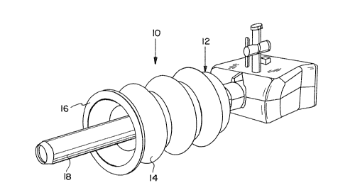
A sealing device 10 is provided for use with a
tubular member such as a cannula or similar device to deter
the escape of gases from the body cavity when, for example,
a surgeon is engaging in endoscopic or laparoscopic
procedures requiring insufflation of the body cavity.
A first embodiment of the sealing device 10 is
shown in Fig. l. The illustrated sealing device comprises a
cylindrical member 12. The cylindrical member 12 is shaped
; 10 similar to bellows in which a plurality of bulbous portions
14 are integrally molded to each other. The cylindrical
member 12 includes a flange 16 at its distal end that is
positionable again6t a patient's body.
It is also contemplated that the sealing device's
bulbous portions 14 be sealingly attached to each other to
allow varying of the number o~ bulbous portions 14 employed.
The sealing device 10 will thereby be longitudinally
extended or retracted by varying the number of bulbous
portions 14 employed.
Referring to Figs. 2 and 3, the sealing device 10
in use with or as part of a cannula is shown. As described
above, the sealing device 10 includes a cylindrical member
12 having a flange 16 at its distal end that is positionable
against a patient's body. However, in Fig. 2 the sealing
device 10 is shown positioned about the cannula 18.
As shown in Fig. 3, the inner diameter of the
sealing device 10 at its distal end is greater than the
diameter of the cannula to provide room for the cannula to
change its angular juxtaposition with respect to the
patient's body. HoweYer, the illustrated sealing device 10
comprises a ridge 20 around the inner circumference of the
. I , '. j ~, , -
; 1 sealing device's proximal end which preferably cooperates
with a receiving indentation or groove 22 at the proximal
end of the cannula. It is also contemplated that the inner
diameter of the sealing device at its proximal end may be
defined by a neck dimensioned to fit securely about the
cannula. The sealing device 10, as shown in Figs. 1, 2 and
3, is longitudinally adjustable and resiliently responds
when compressedO
In use, the cannula 13 may be part of a trocar
assembly 24, as described below. The trocar assembly 24
enables the sealiny device 10 of the present invention to be
used in concert with a tissue gripping device 26 which may
include, for example, mal~cot wings, an inflatable balloon,
or an expandable gripping device.
- 15 The sealing device 10 in use with or as part of a
trocar assembly 24 is shown in Figs. 4, 5 and 6. The
sealing device 10 includes a flange 16 at its distal end as
in the previous embodiment shown in Figs. 1, 2 and 3.
However, in the embodiment shown in Figs. 4, 5 and 6, the
sealing device 10 is part o~ a trocar assembly 24 which
preferably includes a cannula 18, a tissue gripping device
26, and an obturator 28. Further, the tissue g~ipping
device 26 includes a housing mel~er 29 fixedly secured to
the cannula 18.
The housing member 29 is concentrically secured at
a distal end of the cannula 18 by conventional means, such
as adhesives, heat staking, sonic welding, set screws, or
the like. The tissue gripping device 26 is provided with a
plurality of expansion members 30 which surround the cannula
and terminate in tissue contact faces 32. The housing
member 29 preferably has a truncated conical end, and has a
j . . .
.. . . . . . .
; , . -,. .. ., ~ ,, , . . .
$
diameter at a proximal end which is greater than an outside
diameter of the cannula 18 for accepting the expansion
members 30 of the tissue gripping device 26. The expansion
members 30 act in concert with slots 34 in the housing
member 29 to flex the expansion members 30 outwardly and
thereby engage tissue to hold the cannula 18 in place.
Further, the housing member 29 has a generally
cylindrical shape which extends from a proximal gripping
flange 36, which remains outside the body during the
surgical procedure. Adjacent to the gripping flange 36 is a
circumferential groova or indentation 38 designed to mate
with a ridge 20 at the proximal end of the sealing device.
The groo~e 38 and ridge 20 mate to form a desired seal that
allows the sealing device to pro~ide the desired gas seal
between the cannula 18 and the body cavity when the flange
16 at the distal end of the sealing device 10 is against a
patient's body.
The sealing device 10 may be, for example,
constructed of an elastomeric material, -~hich is preferably
an elastomer commercially available under the trademark
"SANTOPRENE", manufactured by MonsantoO
In operation, the sealing device as shown in Figs.
4-6 is used with a trocar assembly 24, which includes an
obturator 28 and a cannula body with 18 the tissue gripping
device 26 positioned thereon so that the cannula 18 passes
through the tissue gripping device 26 and extends through
the distal opening 40 of a housing member 29. Referring to
Fig. 6, after the cannula 18 is inserted into the body, the
tissue gripping member 26 is urged towards the body into the
housing member 29. Camming surfaces 31 on each of the
finger-like expansion members 30 engage the distal end wall
,~ . .
"
: ' . ,. . .,.; ~ :
9 2~7~2~
I of the slots 34 which flexes the expansion members 30
outwardly into engagement with the tissue at the incision.
The cannula 18 is thereby secured in the incision ~y the
outwardly deflected expansion members 30. The sealing
device 10 is urged towards the patient's body in concert
with the extension of the expansion members 30. The flange
16 contacts the patient's sXin providing a desired gas seal
for maintaining insufflation pressure within the body cavity
in the event there is leakage about the cannula 18 at the
incision. The sealing device 10 is designed to remain in
substantial contact with a patient's body while
accommodating the cannula 18 in varying angular positions
with respect to a patient's body. More specifically, the
sealing device 10 includes an inside diameter which allows
for angular juxtapositioning o~ the cannula with respect to
a patientls body while maintaining the relationship between
the flange 16 and a patient's body.
To remove the cannula 18, the tissue gripping
member expansion members 30 is withdrawn from the housing
member 29 so that the flexible nature of the expansion
members 30 draws the expansion member 30 back through the
slots. The sealing device 10 is withdrawn in concert with
the expansion members 30, and as a result, the flange 16 at
the distal end of the sealing device 10 is disengaged from
the patient's skin. Then, the entire tissue gripping device
26 and cannula 18 may be withdrawn from the incision.
The sealing device 10 in use with or as part of
-~ another trocar assembly 42 is shown in Fig. 7 which includes
a flange 16 at its distal end as in the previous embodiment
shown in Figs. 4-6. However, in the embodiment shown in
Fig. 7, the sealing device 10 is part of a trocar assembly
t ;~
1 42 where the tissue gripping member 30 is an integral part
of a cannula 18, or is fixedly secured thereto by, for
example, a set screw, adhesives, etc. Moreover, a pointed
obturator 44 is used to create an incision into the body
cavity. As discussed above, the tissue gripping device 26
is provided with a plurality of expansion members 30 which
surround the cannula 18 and terminate in tissue contact
faces 32. A separate housing member 29 is provided which is
similar to that described in reference to Figs. 4-6 above.
After the trocar assembly 42 is placed in the patient's
body, the obturator 44 is removed and the cannula 18 and the
sealing device 26 is secured in place as described in
reference to Figs. 4-6 above.
The sealing device lO in use with or as part of
still another trocar assembly 46 is shown in Fig. 8. The
sealing device 10 includes a flange 16 at its distal end as
in the previous embodiments shown in Figs. 4-7. However, in
the embodiment shown in Fig. 8, the sealing device lO is
part of a trocar assembly 46 where the cannula 18 itself has
a diameter sufficient to accept the obturator 44 and the
tissue gripping device 26 which has elongated flexible
finger-like expansion members 30. The cannula 18 itself,
preferably, includes a plurality of slots 48 through which
the expansion members 30 would extend. The slots 48
preferably are positioned about the circumference of the
cylindrical cannula 18. The cannula 18 is inserted into the
body using a pointed obturator 44 to penetrate the patient's
skin. Preferably, the cannula 18 tapers at its distal end
to facilitate penetration into the body such that the inner
diameter at the distal end would be slightly greater than
the outer diameter of the obturator 44 which passes
.
2~ 792 ~f~
1 therethrough. The tissue gripping device 26 is
substantially identical to that described in the previolls
embodiments, where a plurality of expansion members 30
extend into the cannula 18 terminating in camming surfaces
31 that engage the distal end walls of the slots to fle~ the
tissue contactiny surfaces 30 outwardly securing the cannula
18 in the tissue at the incision. The obturator 44 is then
removed so that the cannula 18 can be usPd for endoscopic or
laparoscopic activities. To remove the tissue gripping
device 26, the expansion members 30 are slid rearwardly away
from the body to withdraw the expansion members 30 back into
the cannula 18 so that the cannula 18 may be removed. As
described above, the sealing device 10 acts in concert with
the tissue gripping device 26 and thereby, the ~lange 16 at
the distal end of the sealing device 10 is disengaged from
the patient t s skin.
Another embodiment of the sealing device S0 is
shown in Fig. 9. The sealing device includes a flange 16 at
its distal end as in the previous embodiment shown in Figs.
1-8. However, in the embodiment shown in Fig. 9, the
sealing device 50 includes a generally frustoconical member
52. The generally frustoconical member 32 has a smaller
diameter at its proximal end than at its distal end. A
; flange 16 is located at the distal end of the generally
frustoconical member 52 that is positionable against a
patient's body in the same manner as the embodiment shown in
~igs. 1-8. It is contemplated that the generally
frustoconical member 52 may be used with or as part of a
cannula 18 or trocar assembly 24, 42, 46 similar to the
embodiment shown in Figs. 1-8. Thus, the generally
frustoconical member 52 may include an inner diameter at its
'" "'
12 ~7~
I proximal end defined by a neck dimensioned to fit securely
about a sannula. Further, the generally frustoconical
member 52 may include an inner circumference at its proximal
end defined by a ridge which cooperates with a rPceiviny
indentation or groove in the proximal end of a cannula.
Moreover, the generally frustoconical member 52 may be used
with or as part of a trocar assembly.
While the invention has been particularly shown
and described with reference to the preferred embodiments,
it will be understood by those skilled in the art that
various modifications and changes in form and detail may be
made therein without departing from the scope and spirit of
the invention. Accordingly, modifications such as those
suggested above, but not limited thereto, are to be
considered within the scope of the invention.
.. ~.0
- ~ .
~j . .. .