Note: Descriptions are shown in the official language in which they were submitted.
2~
BACRGRO~ND OF T8E INVEN'rION
2 1. FIELD OF THE INVENTION: The present invention is
3 directed to a medical device, and, more particularly, to a trocar
4 assembly which can be inserted a short distance in the abdominal
cavity during laparoscopic surgery and expanded to prevent the
6 device from sliding in and out of a surgical incision.
7 2. BRIEF DESCRIPTION OF THE_PRIOR A~: Laparoscopic
8 surgical procedures gain access to the interior of an anatomical
9 cavity by first using an implement, such as a trocar ~pike, cannula
or a needle having a sharpened point, to pierce or puncture the
11
12
13
14
16
17
18
19
21
22
2~ r~ `r~j~
1 bodily tissues, muscles, membranes, or the like, which may form a
2 portion of or surround the cavity wall.
3 Similarly, in many laparoscopic procedures, a small incision
4 may be made in the skin of the patient along the abdomen, for
example, and the sharp point of a larger penetrating implement,
6 such as a trocar spike of suitable length and diameter, may be
7 inserted into the incision, and pushed until the point punctures
8 the cavity wall. A Sleeve accompanies the implement into the
9 puncture wound to serve as a lining for preserving the shape of the
passageway created by the implement and for insertion of an
11 endoscope~ laparoscope, or the like, to view and operate upon
12 organs within the cavity.
13 In many such applications, a trocar is used which incorporates
14 a sleeve which may have a tendency to slide in and out of the
incision in the abdominal wall, particularly when the surgeon is
16 trying to move the laparoscopic instrument through the interior of
17 the trocar sleeve into or out of the abdominal cavity.
18 In some endoscopic procedures, it has been found necessary to
19 incorporate an electrosurgical procedure which uses a device which
is electrically activated and incorporates blades, needles, and the
21 like which are electrically activated by an electrical circuit
22 which generates a radio fre~uency current with different wave form
--3--
64405\BILL~'
2~
1 shapes to optimize tissue cutting, hemostasis, and the like. A
2 hand piece is configured to receive a working electrode by which
3 the radio frequency currents can be applied to the patient's tissue
4 for cutting or hemostasis. Such electrosurgical devices have been
utilized in laparoscopy in which the handpiece i~ carefully
6 inserted through the trocar sleeve into the abdominal cavity. In
7 such instances, when the handpiece is within the trocar sleeve,
8 electric "arcing" can occur between the handpiece and the metallic
9 components of the trocar sleeve causing electrical shocks to the
patient and/or surgeon.
11 Typical of such electrosurgical apparatuses is that as
12 disclosed in U.S. Patent No. 4,754,754, entitled "Electrosurgical
13 Handpiece for 81ades and Needles", issued July 5, 1988.
14 The present invention addresses the deficiencies of the prior
art, as set forth above.
16 8UMMARY OF IH~ INV~NTION
17 The present invention provides a surgical trocar assembly for
18 insertion of a electrically actuated laparoscopic instrument
19 through an inner abdominal wall. The trocar assembly compri6es a
trocar sleeve having a first end extending into the abdominal
21 cavity through a laparoscopic incision in the abdominal wall, with
22 the first end of the sleeve having a first external dimension for
6~s~n~
2~ 3~.
1 passage through the incision. Means are provided for expanding a
2 portion of the first end of the sleeve within the abdomen so that
3 the external dimension of the first end of the 61eeve within the
4 abdomen is expanded to a larger, second external dimension. The
first end of the sleeve abuts the abdominal wall about the incision
6 when expanded to the second external dimension to resist withdrawal
7 of the trocar assembly from the abdominal cavity.
8 A passageway is defined ~hat through the assembly and interior
9 of the trocar sleeve for introduction and removal of the
10 laparoscopic instrument relative to the abdomen. Insulation means
11 are provided to electrically insulate the patient from the
12 laparoscopic instrument while the instrument is within the
13 assembly. The insulation means may comprise an insulation sheath
14 which may ~e disposed within the sleeve and the passageway.
15 Alternatively, the trocar sleeve may be formed of a conductive
16 plastic having an electrical conductance of less than about 100
17 ohms per square inch. The sleeve may include an insulating
18 elastomeric coating which is disposed thereon to electrically
19 insulate the trocar assembly from the patient while the
20 instrument is within the assembly.
21 In an alternative preferred embodiment, the trocar assembly is
22 additionally provided with first and second interengageable gear
6..0s;~
2~
1 members with the second gear member carried on the housing and the
2 gear members being manually manipulatable to move the second end of
3 a sleeve member between retracted and enlarged diameters.
4 The invention includes a laparoscopic surgical method in which
an incision is made within the abdomen after which the trocar
6 sleeve is inserted therein and manipulated to expand one end
7 thereof to an enlarged diameter from a first, smaller diameter,
8 such that, upon enlargement of the diameter of such end of the
9 trocar sleeve within the abdomen, resistance is afforded to
retractive movements of the trocar assembly relative to the inner
11 abdominal wall.
12 After expansion of the end of the ~leeve diameter to the
13 enlarged position, the electrosurgical instrument is inserted
14 through a passageway through the trocar assembly and the insu}ation
provided between the exterior of the electrosurgical apparatus and
16 the sleeve of the trocar assembly buffers the trocar assembly
17 against "arcing" of electric current thereacross, thereby avoiding
18 electric shock to the surgeon or the operator of the trocar
19 assembly, or to the patient, possibly resulting in burning of
tissue and the like during surgery, and/or establishing a field of
21 static electricity immediate the area of ~urgery.
6~5~B~
2~
1 After use of the electrosurgical laparoscopic device inserted
2 through the passageway within the trocar assembly, 6uch device may
3 be removed and the trocar sleeve subsequently manipulated, such
4 that the diameter of the end of the sleeve within the abdominal
wall is reduced from the expanded position to the retracted,
6 initial position, and the trocar assembly removed from within the
7 abdominal wall.
8 BRIEF DE~CRIPTION OF T~F DRA~INGB
9 Fig. 1 is a cross-sectional view of a trocar sleeve forming a
first embodiment of the invention prior to expansion.
11 Fig. 2 is a cross-sectional view of a trocar sleeve of Fig. 1
12 after expansion within the abdomen and interior of an abdominal
13 wall.
14 Fig. 3 is a side view of a trocar used with a trocar sleeve.
Fig. 4 is a perspective view of a seal within the trocar
16 sleeve.
17 Fig. 5 is an illustrative view of a first modification of the
18 trocar sleeve where an expanding sponge is used.
19 Fig. 6 is an illustrative view of another modification of the
trocar sleeve where an inflatable balloon i~ used.
21 Fig. 7 is a view in cross-section of a trocar sleeve forming
22 a second embodiment of the present invention.
~UD5\BIIll;
2~
1 Fig. 8 is an exploded cross-sectional view of the trocar
2 sleeve of Fig. 7.
3 Fig. 9. is an exploded perspective view of an alternative stop
4 locking mechanism.
Fig. lo which appears with Fig. 7, is a cross-sectional
6 view of another alternative stop locking mechanism.
7 Fig. 11 is a view in cross-section of a modified trocar
8 sleeve.
9 Fig. 12 is a sectional schematic illustration of an
alternative embodiment of the present invention in position prior
11 to insertion into an abdominal cavity, with the cylindrical members
12 being in normally retracted position relative to one another~
13 Fig. 13 is a view similar to that of Fig. 12, but illustrating
14 the position of one of the cylindrical members relative to the
other when the one cylindrical member's outer diameter is expanded,
16 such as subsequent to insertion through the abdominal wall into the
17 cavity, during surgery.
18 Fig. 14 is a perspective illustration of one of the
19 cylindrical members and sleeve and an inter-engaging gear means.
Fig. 15 is a view similar to that of Fig. 14, illustrating the
21 position of the respective components during activation such that
22 the outer cylindrical sleeve is in expanded position.
-8-
2~ ?..~.5~
1 Fig. 16 is an outer prospective view of the alternative
2 embocliment of the present invention illustrating, in particular,
3 the manual manipulating means through the housing and the seal
4 assembly for insertion of an electrically activated laparoscopic
instrument.
6 Fig. 17 is a view similar to that of Fig. 16, showing the
7 insertion of the electrically activated laparoscopic instrument.
8 Fig. 18 is a view similar to Fig. 12 illustrating a device
9 with insulation provided by an elastomeric coating.
Fig. 19 is a view similar to Fig. 13 ~howing the embodiment of
11 Fig. 18 in expanded position.
12 DE8CRIPTION OF T~E PREF~RRED EMBODIM~NT~
13 With reference now to FIGURES 1-3, a trocar sleeve 10 forming
14 a first embodiment of the present invention is illustrated. With
the assistance of a trocar 12, seen in FIGURE 3, a first end 14 of
16 the sleeve 10 is inserted through an incision 16 in the abdominal
17 wall 18 of a patient. The trocar 12 is inserted through passage 26
18 in the trocar ~leeve so that only the 6barp pointed end 13 extends
19 from the first end 14 of sleeve 10. The pointed end 13 of trocar
12 then is pushed through the abdominal wall along with first end
21 14. The 61eeve 10 i6 secured therein by expansion of a mushroom
22 hinge 20 which abuts the inside surface 22 of the abdominal wall
6~5~D~
2~
1 about the incision 16 to prevent premature withdrawal of the trocar
2 sleeve. After removal of trocar 12, laparoscopic instruments and
3 the like can be inserted into the ab~ominal cavity 24 through
4 passage 26 in the trocar sleeve 10.
With reference to FIGURES 1 and 2, the trocar sleeve 10 can be
6 seen to include an inner cylindrical metal sleeve 28 and a
7 concentric outer cylindrical plastic sleeve 30 preferably of
8 polypropylene or other suitable material. The sleeves 28 and 30
g are secured indirectly one to another through an insulating ~leeve
member 28A at the first end 14. Near the first end 14, the outer
11 plastic sleeve 30 defines a mushroom hinge 20 with a series of
12 hinge members 32 about the circumference of the sleeve 30 separated
13 by elongated slits 34. An outer elastic coating or sleeve 36,
14 preferably of latex, overlies the hinge member 32 and slits 34 to
cover the mushroom hinge 20. Coating or sleeve 36 insures that no
16 body tissue is trapped between portions of the mushroom hinge,
17 particularly when contracting the hinge for removal of the sleeve
18 10 from the patient.
19 With continued reference to Figs. 1 and 2, interior of the
metal sleeve 28 is disposed an additional concentric cylindrical
21 sleeve member 28A extending all or part of the length of the member
22 28 to electrically insulate the trocar assembly fro~ arcing caused
--10--
~05\BIIl~;
2<~ ~J~
1 by insertion therethrough of an electrically activated laparo~copic
2 instrument while such instrument is within the trocar assembly.
3 The inner sleeve 28A preferably ifi ~ade of teflon or other chemical
4 material known to those skilled in the art to insulate the
electrically activated laparoscopic instrument from the metal tube
6 28. In lieu of providing a teflon sleeve 28A, any plastic or other
7 material may be utilized to make the sleeve 28A provided that it is
8 non-conductive.
9Alternatively, the assembly as shown in Fig. 1 and Fig. 2, may
provide insulation means in a form other than a sleeve 28A. For
11 example, the outer sleeve 30 may be made of a conductive plastic
12 with a conductance of less than about 100 ohms per square
13 inch. In such configurations, such a plastic sheath will not
14 act like a capacitor, thereby shielding the surgeon and patient
against shock as a result of arcing attributable while the
16 electrically activated laparoscopic instrument is within the
17 trocar 10.
18An additional alternative means of providing insulation ~eans
19to the trocar assembly is by means of providing an elastomer sheath
20312A over the outside of the plastic ~heath 312, as shown in Figs.
21 18 and 19. Such an elastomeric sheath may be made of silicone,
22 latex or a natural rubber.
--11--
6~40s~;
2~
1At the end 38 of the trocar sleeve opposite the first end, the
2insulating sleeve 28A and sleeve 28 have a series of openings 40.
3A plastic end member 42 is secured to the ~leeve 28, insulating
4sleeve 28A and outer sleeve 30 at end 38 by latches 44 received in
5the openings 40. A plastic seal retaining cap 46 is, in turn,
6secured to the end member 42 by latches 48 on the cap. On O.D.
7seal 50, a split seal 52, and a pack ring 54 are confined between
8the end member 42 and cap 46 to prevent the pressurized gas within
9the abdominal cavity from escaping through the passage 26 through
10the metal sleeve 28 as will be described hereinafter. Seals 50 and
ll52 are preferably of silicon rubber.
12A handle 58 is secured to the outer plastic sleeve 30 at end
1338. A coil spring 60 acts between the end member 42 and the handle
1458 to urge the handle 58 toward the first end of the trocar sleeve.
15In the configuration illustrated in FIGURE 1, the coil spring
1660 is held in a compressed state between the end member 42 and the
17handle 58 as resilient latches 62 on the end member 42 are in
18contact with an end surface 64 of the handle.
l9A latch release 66 is received about the outer plastic sleeve
2030 for movement relative the sleeve 30. The latch release 66 can
21be seen to have a conical ca~ming surface 68 which can be moved
22into engagement with the ends of latches 62 to deflect the latches
6~405~1~1N
2~
1 inward toward the axis 70 of the trocar sleeve 20. The latch
2 release 66 can be activated by the ~urgeon directly, or, as an
3 alternative, by providing a mechanism whereby withdrawal of the
4 trocar after inserting the sleeve through incision 16 activates
the latch release. The latches 62 will be deflected inward enough
6 to release the bandle 58 from the end member 42, which permits the
7 spring 60 to expand to the position shown in FIGURE 2. As the
8 spring expands, the spring forces the handle 58, and attached outer
9 plastic sleeve 30, toward the first end relative to the end member
1042 and inner metal sleeve 28 and insulating ~leeve 28A. This
11 causes the portions of the plastic sleeve 30 t the living hinges
12 to bend and expand the mushroom hinge 20, as seen in FIGURE 2. It
13 can be readily understood that the expanded mushroom hinge abuts
14 the inside surface 22 of the abdominal wall 18 to resist removal of
the trocar sleeve.
16A dense rubber foam stop 72 can be frictionally engaged with
17 the outer plastic sleeve 30. When the mushroom hinge has been
18 expanded, the stop 72 can be slid downward along the plastic 61eeve
19 30 toward the first end *o contact the outer surface 74 of the
abdominal wall 18 to resist movement of the trocar sleeve into the
21 abdominal cavity.
~405\BIIl~'
;~t ;~J~..)t'J~L~
1 It can be readily understood that the trocar ~leeve 10
2 provides a ~table platform for insertion of an electrosurgery or
3 other laparoscopic instrument through the ~leeve 10 into the
4 abdominal cavity through passage 26 without concern for electric
shocks. The expanded mushroom hinge 20 also forms a good seal with
6 the patient's abdominal wall 18 to prevent C02 loss. Any tendency
7 for the trocar sleeve 10 to move relative the abdominal wall as the
8 instrument is being inserted or removed will be greatly reduced by
9 the expanded mushroom hinge 20 and stop 72.
~hen the trocar sleeve 10 is to be removed from the patient,
11 surface 46A is pressed towards the handle 58 at surface 58A in a
12 manner similar to a syringe to collapse the mushroom hinge 20 and
13 compress the spring 60 until the latches 62 again latch against
14 surface 64 of the handle 58 to allow removal of the trocar sleeve.
The m~shroom hinge 20 can be collapsed and removed without having
16 to move stop 72.
17 With reference now to FIGURES 1, 2 and 4, the mechanism for
18 preventing gas from escaping the abdomen through the sleeve 10 will
19 be described. When no electrosurgery or other laparoscopic
instrument is inserted through the trocar sleeve 10, a split seal
21 52 prevents the escape of gas. The split fiaal 52 i5 formed of a
22 resilient material which has a concave curvature facing the first
-14-
~SUI~'
2 ~
1 end 14 of the trocar sleeve. A slit 76 is formed in the split seal
2 which forms a first ~eal lip 78 and a 6econd seal lip 80 on
3 opposite sides of the slit 76. With the concave ~hape, the
4 pressurized gas within the abdominal cavity acts to force the lips
78 and 80 together to form a tight seal to prevent the gas escape.
6 When an instrument is inserted in end 38 of the trocar ~leeve 10,
7 the resilient lips 78 and 80 simply deflect away from the
8 instrument, permitting the instrument to pass through the passage
9 and into the abdominal cavity.
The O.D. seal S0 forms a seal against the outer cylindrical
11 surface of an instrument as the instrument is inserted into the
12 passage 26 of the trocar sleeve 10. With the combination of the
13 O.D. seal 50 and split seal 52, very little gas is lost as
14 instruments are inserted and removed from the trocar sleeve 10.
With reference to FIGURE 5, a first modification of the trocar
16 sleeve 10 is illustrated. In the first modification, an expanding
17 sponge 90 replaced the mushroom hinge 20 at the first end of the
18 trocar sleeve 10. The sponge 90 can be expanded in a manner
19 similar to the mushroom hinge 20 to hold the trocar sleeve within
the abdominal cavity.
21 With reference to FIGURE 6, a second modification of trocar
22 sleeve 10 is illustrated. In the second modification, an
-15-
6~5~8D~;
2f`~
1 inflatable balloon 92 is mounted at the first end of the trocar
2 sleeve which can be inflated to an expanded condition to secure the
3 trocar ~leeve within the abdominal cavity.
4 The sleeve 10 of the present învention reduces the frictional
forces encountered by laparoscopic instruments being inserted into
6 or removed from the sleeve as compared to prior designs. One
7 reason for this advantage is that the entire sleeve 10 can be made
8 shorter than past designs which required a longer length to insert
9 through the abdominal wall to resist accidental removal of the
sleeve from the patient. Another reason for the friction reduction
11 is the use of efficient seals 50 and 52.
12 The trocar 12 can be used to automatically expand mushroom
13 hinge 20 as the trocar is removed from ~leeve 10. One possible
14 mechanism for this is illustrated in FIGURES 2 and 3. A lever 100
can be pivoted to trocar 12 about hinge 106 and urged outwardly by
16 a spring 60. The lever will be retracted into the trocar 12 as the
17 trocar is inserted into the sleeve 10 to place the sleeve 10
18 through incision 16. When the trocar i6 removed from ~leeve 10,
19 the lever extends outward in the direction of arrow 108 through a
slot 102 in the sleeve 60 that the lever 100 contacts latch release
21 66. Further movement of trocar 12 would cause the release 66 to
22 move upward and deploy the mushroom hinge. The lever 100 can be
-16-
~UQ5\BIIl':
2~ 'r~
1 mounted to retract back into trocar 12 in the direction of arrow
2 110 after sufficient force has been exerted on trocar 12 to move
3 release 66 to allow the trocar to be removed from ~leeve 10.
4 In one possible construction of a sleeve in accordance with
the teachings of the present invention, the inner diameter of the
6 sleeve could be about S to 6 mm. with the diameter of the expanded
7 hinge about 0.845 inches. The stroke of the sleeve 30 to activate
8 the hinge could be 3/8 inches and ~leeve 36 could be lO mil6 thick.
9 With reference now to FIGURES 7 and 8, a trocar 100 forming a
second embodiment of the present invention is illustrated. The
11 trocar 100 includes an outer cylindrical sleeve 102 and a
12 concentric inner cylindrical electrically insulative sleeve 104.
13 The sleeves 102 and 104 are secured together at a first end 106 of
14 the trocar. The attachment is made by interlocking inwardly
protruding lug 108 on sleeve 102 which fits within similarly shaped
16 openings 110 in the inner cylindrical sleeve 104. The outer
17 cylindrical sleeve 102 has formed thereon a mushroom hinge ~12 much
18 as hinge 20. As with trocar 12, relative movement between the
19 outer cylindrical sleeve 102 and the inner cylindrical electrically
insulative sleeve 104 will expand the mushroom hinge 112 to secure
21 the trocar lO0 in place, or to retract the mushroom hinge for
22 insertion or removal of the trocar.
-17-
64~05~BD~;
2~ J~'~
1 The end 116 of the outer cylindrical sleeve 102 opposite the
2 first: end 106 flares outwardly to form a cylindrical portion 118.
3 the c:ylindrical portion 118 includes a cylindrical wall 120 with a
4 series of apertures 122 through the wall in a first plane 124
perpendicular to the axis 114 and a similar series of apertures 126
6 in a second plane 128 perpendicular the first axis. A series of
7 helically directed guide slots 130 are also formed in the wall 120
8 which extend from onè end in the first plane to an opposite end in
9 the second plane. The cylindrical portion 118 also has a radially
extending flange 132 which assists a surgeon to hold the
11 cylindrical sleeve.
12 The end 134 of inner cylindrical sleeve 104 is electrically
13 insulated by sleeve 102. A member 138 is slid over the inner
14 sleeve 104 so that the portion 136 of the inner cylindrical sleeve
is received within an annular recess 140 in the member 138. The
16 engagement between the portion 138 and recess 140 is sufficiently
17 close to be essentially airtight, yet permit the member 138 to
18 rotate about the axis 114 relative to the inner cylindrical sleeve
19 104. The member 138 also has a series of resilient arms 142 which
extend into the cylindrical portion 118, with each arm 142 ending
21 in a lug 144. Every other lug 144 is received in one of the guide
22 slots 130. If the member 138 is rotated about axis 114 relative to
-18-
~405~LL';
2~ ~
1 the cylindrical portion 118, the interaction between the lugs and
2 the guide slots 130 will cause the member 138, and 61eeve 104, to
3 move along axis 114. When the member 138 i~ turned 60 ~hat lugs
4 144 abut a first end 146 of the guide slots, the mushroom hinge 112
is retracted and a trocar can be inserted or removed from the
6 patient. When the member 138 is rotated so that lugs 144 are
7 guided along slots 130 to the second end 148 of the ~lots, the
8 member 138 and inner cylindrical sleeve 104 are translated along
9 the axis 114 to cause the mushroom hinge 112 to expand to lock the
trocar within the patient. The intermediate lugs 144, i.e., those
11 lugs 144 not received in one of the guide slots 130, act as detents
12 in the retracted or extended positions, with the intermediate lugs
13 144 engaging apertures 126 when the mushroom hinge 112 is retracted
14 and the lugs 144 engaging the apertures 126 when the mushroom hinge
is deployed.
16 An end member 150 is snap fit to member 138 with a plurality
17 of resilient arms 152. An 0-ring 153 seal6 between end member 150
18 and member 138. The sealing effect of the O-ring i6 enhanced by a
19 conical seal surface 155 on member 150 which urges the o-ring into
compression between members 138 and 150 to form an effective seal.
21 The end member 150 mounts the O.D. seal 54, split 6eal 52 and
22 packing ring 50 as used in trocar 100.
--19--
6~5\HLU~
2~ ~;?,,~
1 A significant advantage of the trocar 100 is the deployment
2 and retraction of the mushroom hinge by rotating one part of the
3 trocar relative to the other about its elongated axis. Since the
4 forces necessary to insert the trocar in the patient and remove the
trocar from the patient are generally along the axis 114, there is
6 little opportunity in trocar 100 for an inadvertent deployment or
7 retraction of the mushroom hinge during insertion or removal of the
8 trocar. Further, as the laparoscopic instruments are also inserted
9 or removed by exerting forces along the axis 114, these procedures
will likewise entail little risX of inadvertent actuation of the
11 trocar.
12 The dense rubber foam stop 154 is seen to have ridges 156 in
13 contact with the outer surface of the outer cylindrical sleeve 102.
14 These ridges are oriented to make the movement of the stop along
the sleeve 102 toward the patient relatively easy so that the stop
16 can be guickly pushed against the outer surface of the patient's
17 skin. However, the ridges resist movement along the sleeve in the
18 opposite direction to prevent inadvertent loosening of the trocar
19 100 from the patient.
As shown in Fig. 8, insulation means may be provided by a
21 teflon or other insulation-formed inner insulating member number
22 104A extending concentrically within the inner 61eeve member 104,
-20-
zr~
1 such that the inner sleeve may be made of metal. As with the
2 embodiment shown in Figs. 1 and 2, such insulation means may also
3 be provided in lieu of provision of sleeve 104A by means of a
4 plastic sheath made of a non-conductive plastic for the outer
S sleeve 102 or by insulating the outer sleeve 102 with an ela~tomer
6 sheath over the outside of the plastic of the outer sleeve 102.
7 With reference to FIGURE 9, the trocar 100 is shown with a
8 modified outer sleevè 158 and 8top 160. The outer sleeve 158 is
9 formed with a discontinuous thread 162 which defines two unthreaded
grooves 164 along axis 114. The stop 160 has a similar
11 noncontinuous thread 166 with grooves 168. By turning the stop 160
12 to one orientation relative to the sleeve 158, the threads of one
13 part are in the grooves of the other part, which permits the stop
14 to be moved freely along axis 114 relative to the outer sleeve.
When the stop is properly positioned relative to the patient, the
16 stop need only be rotated 90 degrees to engage the threads of the
17 stop and sleeve to lock the stop in the desired position.
18 With reference now to FIGURE 10, yet another modification of
19 trocar 100 is illustrated which includes an outer cylindrical
sleeve 170 and a stop 172 with electrically insulative sleeve
21 member 170-A. The outer cylindrical sleeve 170 has a continuous
22 thread 174 along its length. The stop has resilient rachet teeth
-21-
1 176 to engaqe the threads 174, which allows the stop to be easily
2 moved in a direction along the axis 114 to move into engagement
3 with the patient by the teeth 176 deflecting over the thread~ 174,
4 but locks against threads 174 to resist removal. The threads 174
can be part of a continuous thread so that the stop can be backed
6 off from the patient by rotating the stop to thread the stop bacX
7 away from the patient.
8 FIGURE 11 illustrates a trocar 200 which is a modification of
9 trocar lOo. Trocar 200 uses mating threads 202 on cylindrical
portion 118 and threads 204 on member 118 to move outer sleeve 102
11 and inner insulative sleeve 104 relative each other to activate the
12 mushroom hinge. The threads have a pitch selected 80 that the
13 desired axial movement between sleeves 102 and 104 can be achieved
14 by rotating the threads less than one-half turn.
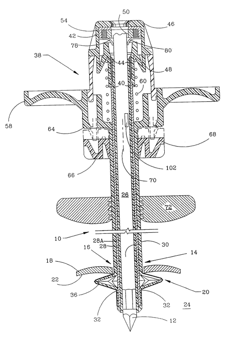
Now with reference to Fig. 12, there is shown a trocar
16 assembly 310 having a housing 311 with an upper opening 332 defined
17 therein for receipt of the outwardmost end of a hand-manipulatable
18 control assembly 333. The housing 310 has a distal end with an
19 abutment skirt 311A thereon terminating in a receptacle 311B for
receipt of first and second concentrically di~posed cylindrical
21 members 312 and 313 therethrough and into the housing 311. Placed
22 between members 312 and 313 is an insulative sleeve 313A.
6~-05\BLLr;
1 A passageway 316 extends completely though the assembly 310
2 and 1:he innermost of the cylindrical members 313 to an opening 315.
3 An end 314 is defined on the distal end of the first elongate
4 cylindrical member 312, which is inserted into the abdominal
cavity. As shown in Fig. 12, the first elongate cylindrical member
6 312 has a retracted outer diameter 317.
7 Now referring to Fig. 13, the first and second elongate
8 cylindrical members 312 and 313 and the insulative sleeve 313A are
9 secured, one to another by means of plastic securements 330
radially spaced there-across at the distal end.
11 The first elongate cylindrical member 312 carries thereon a
12 portion within the housing 311 defining a gear member 319 having
13 inter-engaging teeth at 320 thereon and, preferably, in a circular
14 spaced configuration, 322. A cylindrical extension 326 has a no-go
shoulder 327 (Fig. 14) thereon which contacts the end 319A of the
16 gear member 319 when the cylindrical members 312, 313A are moved to
17 the normally retracted position, as shown in Fig. 12, to thereby
18 limit travel of the moving cylindrical memher in one direction,
19 i.e., when the cylindrical members 312, 313A are moved by the
control 333 from the expanded position, Fig. 13, to the retracted
21 position, Fig. 12.
6~5~
1 Companion inter-engaging teeth members 321 are provided
2 circularly around a portion of a ring component 321A of the control
3 333 for inter-engagement with the teeth 320 on the gear members 319
4 of the first elongate cylindrical member 312.
The extension member 326 has a 6eries of outwardly protruding
6 wing members, 328A, 328B which are inserted in companion slots 29A,
7 329B on the housing 311 for ~ecurement thereto. An outer seal
8 member 331A and companion lip seal 331B, as shown in Fig. 16,
9 prevent transmission of gasses, and other fluids, from within the
passageway 316, when the assembly 310 is within the abdominal
11 cavity, during surgery.
12 An auxiliary instrument I, such as an electrically activated
13 cutting device, may be inserted through the seal member 331B and
14 into the passageway 316, subsequent to insertion of the assembly
310 within the abdominal cavity and manipulation of the first
16 elongate cylindrical member 312 to the expanded outer diameter 318,
17 as shown in Fig. 13.
18 Now referring to Fig.'s 14 and 15, a sleeve portion 325 is
19 defined immediate the distal end of the fir~t elongate cylindrical
member 312 and has a series of radially extending serrated flexing
21 members 323 thereon with openings 324 inter--defined there between.
22 A serration 323A is cut on each of the flexing members 323 to
6~5\8Dl~;
2~ 3~1.
permi.t flexing movement to the expanding outer diameter position
2 318.
3 As shown in Fig's. 12 and 13, the embodiment shown therein
4 contains an insulating 61eeve 313A made of teflon or other
insulating material, the sleeve 313A being concentrically
6 securingly disposed around the outer diameter of the inner sleeve
7 member 313.
8 As with the other embodiments shown in the previous Figs., the
9 embodiments shown in Figs. 12 and 13 may provide insulation means
10 other than by positioning of a concentrically disposed inner teflon
11 or other insulated sleeve member 313A, by means of making the outer
12 member 312 of a plastic non-conductive material having a
13 conductance of less than about 100 ohms per square inch.
14 Additionally, and alternatively, the outer sleeve 312 may be
15 covered with an insulated elastomer such as silicone, latex or
16 natural rubber, or the outer sleeve 312 may be coated with a
17 conductive material having a conductance of less than about 100
18 ohms per square inch to provide an electrically resistant
19 coating 312A.
20 abdominal wall through an incision, or the like, during surgery,
21 the assembly 310 is inserted therein by the 6urgeon ~imply grasping
22 the housing 311 and insexting the assembly 310 through such
--25--
1 incision, or opening. Thereafter, prior to introduction of the
2 electrically activated auxiliary instrument I through the assembly
3 310, the control 333 is contacted by the finger or thumb of the
4 surgeon and moved from the position 6hown in Fig. 14, backwardly,
to the position shown in Fig. 15. Accordingly, as the control 333
6 is manipulated, the teeth 321 in the ring 321A will travel across
7 the companion inter-engaging teeth 320 in the configuration 322 of
8 the gear member 319, such that the first elongate cylindrical
9 member 312 moves away from the no-go ~houlder 327 on the
cylindrical extension 326.
11 Because the first and second cylindrical members 312, 313 and
12 313A are attached one to another by means of the securements 330 at
13 the distal end of the assembly 310, movement of the first elongate
14 cylindrical member 312 relative to the second member, 313, will
cause the sleeve portion 325 flexing members 323 to be urged
16 regularly outwardly from the cylindrical member 313 from the
17 retracted outer diameter of position 317 (Fig. 12) to the expanded
18 outer diameter of 318 (Fig. 13). In the position ~hown in Fig. 13,
19 the trocar assembly 310 thus resists removal from the abdominal
wall W in the cavity, during 6urgery.
21 The apparatus may be moved from the position shown in Fig. 13
22 to the position shown in Fig. 12 by rever6ing the procedure
-26-
6.-05~
2~
1 described above, and the assembly 310 completely withdrawn from the
2 abdominal wall w~
3 Accordingly, as the electrically activated auxiliary
4 instrument I is introduced within the as~embly 310, arcing between
the metallic housing of such instrument I and the metal sleeve 313
6 is prevented by provision of the insert sleeve member 313A which
7 provides insulation means. If ~uch sleeve 313A i5 not provided,
8 but the outer sleeve 312 is made of a conductive plastic, as
9 described herein, shocking of the patient will be eliminated
because such plastic sheath will not act as a compacitor and
11 therefore dissipate any electrical shock to the patient during
12 surgery.
13 Although the invention has been described in terms of
14 specified embodiments which are set forth in detail, it should be
understood that this is by illustration only and that the invention
16 is not necessarily limited thereto, since alternative embodiments
17 and operating techniques will become apparent to those skilled in
18 the art and view of the disclosure. Accordingly, modifications are
19 contemplated which can be made without departing from the spirit of
the described invention.
-27-
~S\B~;