Note: Descriptions are shown in the official language in which they were submitted.
W O 92/09633 PCT/GB91/02077
~96953
IMMUNOGLOBULIN-3I~DING PROTEINS AND RECOM9INANT DNA ~OL_CUL_S
CODING TEEREFOR
This invention relates to immunoglobul~n-binding proteins
ana recombinant DNA molecules coding therefor.
Protein-A (SpA) is a cell w~ll component of Sto~nyiococcus
o~reus which binds to the Fc region of immunoglobulins from a variety
of sources (Langone, 1982). For example, it can bind to human IgG
sub-classes 1, 2 and 4 but not in general to IgG3. It can also
efficiently bind IgG from rabbit and pig, but it binds horse and cow
IgG with lower affinity, and binds rat IgG only very weakly (Boyle and
Reis, 1987).
This specific interaction with IgG molecules makes SpA a
very useful immunolosical tool. It has been used in immur.ologlobulin
purification when immobilised into chromatography columns, and as an
antibody probe in enzyme-linked immunosorbent assay (FTISA) systemsi
together thece uses have been exploited in the screer~ng and
purification of monoclonal antibodies. Recently SpA has also found
use in chemotherapy to remove immune complexes from serum (see Palmer
et al., 1989 and references therein), and in biotechnology wnere it
has been incorporated nto cloning vectors in wn~c~ the c oned g_ne
can be expressed as a fusion with SpA (Nilsson et al, 1985).
The isolation of SpA from S. sureus cells is not
s~-aightforward and also no entirely sat~sfactory techniques are
available for anchoring SpA to solid supports. The gene for SpA has
been cloned and sequenced (Lofdahl et al. 1983, Uhlen e~ al. 1984,
W O 92/09633 PCT/GB9l/02077
-- 2 --
2~9~9a3
Shuttleworth e~ al, 1387) and encodes a 4~ kDa proteln consisting of
homologous IgG binding domains termed E,D,A,B and C, and a C-terminal
cell wall spanning and membrane anchoring region. resion X. In
addition an N-terminal signal sequence is thought to target the
protein out of the cell. The crystal structure of a single IgG
binding domain-fragment ~ (Sp ~) bound to human Fc nas been resolved
at the 2.8 A level (Deiser~ofer, 1981) and recent NMR studies show
that SpAB contains 2 c-helices, the residues of which for~s most of
the contact points of Fc. The Fc binding Q-helices of successive SpA
domains are apparently separated by flexible polypeptide spacer
regions.
Site directed mutagenesis is a powerful tool which could be
used to probe the SpA-Fc interaction. However, mismatch primer
mutagenesis is very difficult since the repeated na~ure of the gene
means that the primer could anneal to multiple sites. Other workers
(Nilsson ee al, 1987: Saito et al, 1989) have reported the production
of IgG binding proteins, based upon the B domain of SpA, from
synthetic genes. Their studies highlighted the difficulties often
encountered when expressing small foreisn proteins ln ~. co~i eg.
proteolysis of the product by host enzymes or difficulties in the
purification of the expressed protein. Accordingly it has hitherto
not been practical to prepare mutated proteins, derlved from SpA,
which have properties adapted to specific purposes. Specifically, the
production of modified forms of SpA which avoid the above- mentioned
difficulties has consequently proved to be problematical.
W O 92J09633 PCT/GB91/02077
~D~ 69~'3
The present invention has solved these problems by designing
a synthetic Fc-binding domain which is highly amenable to site
directed mutagenesis. Expression of polypeptides comprising this
synthetic Fc-binding domain has enabled the production of immuno-
globulin-binding proteins having distinct advantages compared to SpA,
rendering them particulsrly useful in preparative and diagnostic
techniques and in therapy.
According to one aspect thereof, the present invention
provides a polypeptide capable of forming a complex with an
immunoglobulin, said polypeptide being characterised by having at
least 2, but not more than 4 binding domains, each capable of binding
to the Fc region of an immunoglobulin of the IgG class.
Preferably the polypeptide is characterised by having 2, but
nct more than 2 of said binding domains.
In one e~bodiment of the invention, the binding domains
possess a high degree of sequence homology with the binding domains of
Stophylococcus o~reus Protein-A (SpA). Thus preferably esch of said
binding domains has at least 75% sequence homology, preferably at
least 90% sequence homology, with at least one of the binding domains
designated A, B, C, D and E of StophyIococcus oureus Protein-A.
Most preferably each of said binding domains has at least
75% sequence homology, preferably at least 90Z sequence homology, with
the binding domain designated B of StophyIococcus oureus P-otein-A.
It is not necessary for the bind_ng domains of the
palypeptide to match precisely the size of the binding domains of SpA,
but preferably each of said binding domains consists of from 40 to 55
smino ac d residues.
W O 92/09633 PCT/GB91/02077
-- 4 --
2 0 9 ~ 9 .5 ~.
The following are especially prefer-ed sequences for the
binding domains of polypeptides according to the invention:
(1) the sequence
Ala Pro Lys Ala Asp Asn Lys Phe Asn Lys
Glu Gln Gln Asn Ala Phe Tyr Glu Ile Leu
His Leu Pro Asn Leu Asn Glu Glu Gln Arg
Asn Ala Phe Ile Gln Ser Leu Lys Asp Asp
Pro Ser Gln Ser Ala Asn Leu Leu Ala Glu
Ala
(2) sequences consisting of at least 40 amino acid residues
and derived from sequence (1) by
(a) deleting up to 11, preferably not more than 8 and
most preferably not more than 3 amino acid residues
of sequence (1) and/or
(b) substituting up to 11, preferably not more than 8
and most preferably not more than 3 amino acid
residues of sequence (1) by other amino acid
residues and/or
(c) inserting up to 11, preferably not more than 8
and most preferably not more than 3 amino acid
residues into sequence (1) .
W O 92/09633 PCT/GB91/02077
2 ~ .5 ~
?olypeptides according to the invention having a~ least one
binding domain as specified in alternative (2) above may be produced
by site-directed mutagenesis, using as a start~ns point recombinant
DNA molecules containing DNA sequences coding for sequence (1) above.
It is particularly preferred according to the invention for
the derived sequences 2(a), 2(b) and 2(c) to confer on the
polypeptides according to the invention a bindins capacity which has a
different pH dependance compared to that of protein A itself. This
may be achieved for example by replacing a non-ionisable amino acid
residue in sequence (1) by an ionisable amino acid residue.
Alternatively an ionisable residue may be replaced by a non-ionisable
residue. As a further alternative, an ionisable residue may be
replaced by another ionisable residue having a different p~a or p~b.
Examples of ionisable amino acid residues include
His, Arg, Lys, Glu, Asp, Cys and Tyr.
Of these residues, Glu, Asp, Tyr and Cys ionise when the pH is raised;
whereas His, Arg and Lys ionise as the pH is lowered.
Thus according to a preferred aspect of the invention Tyr 8 (the Tyr
residue which occurs in the partial sequence Ala Phe Tyr Glu) may be
replaced by a residue selected from:
His, Arg, lys, Glu. Asp and Cys.
W O 92/09633 PCT/GB91/02077
-- 6 --
2a96~s3
Thus, for example, specific examples of derived sequences
(b) include sequences wherein the or at least one of the partial
sequences
Ala Phe Tyr Glu
is replaced by one of the following sequences:
Ala Phe Glu Glu
Ala Phe Phe Glu
Ala Tyr Tyr Glu
Ala Phe His Glu
Ala Phe Lys Glu
Ala Phe Cys Glu
Further examples of derived sequences (b) for the binding domains of
polypeptides according to the invention are sequences having the
following general sequence:
Ala Pro Lys Ala Asp Asn Lys Phe Asn Lys
Glu Gln Gln Asn Ala Phe X Glu Ile Leu
His Leu Pro Asn Leu Asn Glu Glu Gln Arg
Pro Ser Gln Ser Ala Asn Leu Leu Ala Glu
Ala
wherein X can be phenylalanine, glutamic acid. histidine. cysteine or
lysine.
Sequences for the binding domains of polypeptides according
to the invention such as those above are preferably mutated by
cassette mutagenesis.
Preferably each of the binding doma~ns of the polypeptide of
the ~nvention has the same amino acid sequence.
W O 92/09633 PCT/GB91/02077
2 ~ 9 ~
Thus for example it is preferred that where a polypeptide
according to the invention comprises two or more derived sequences as
defined in paragraph (2) above. each of said derived sequences is
identical, i.e. the derived sequences contain the same amino acid
substitution(s) at the same position(s). As indicated. preferred
polypeptides according to the invention having derived binding domain
sequences as described above may exhibit modified, pH-dependent
binding affinities. Particularly preferred polypeptides according to
the invention are provided at their C-terminal ends with an amino acid
residue having a functional group allowing the polypeptide to be bound
covalently to a solid support. Thus preferably the polypeptides
according to the invention are provided with a cysteine residue at the
C-terminal end.
One such preferred polypeptide has the following C-terminal
sequence
Ala Pro Lys Ala Asp Asn Lys Phe Asn Lys
Glu Gln Gln Asn Ala Phe Tyr Glu Ile Leu
His Leu Pro Asn Leu Asn Glu Glu Gln Arg
Asn Ala Phe Ile Gln Ser Leu Lys Asp Asp
Pro Ser Gln Ser Ala Asn Leu Leu Ala Glu
Ala Lys Lys Leu Asn Glu Ser Gln Ala Pro
Lys Ala Asp Asn Lys Phe Asn Lys Glu Gln
Gln Asn Ala Phe Tyr Glu Ile Leu His Leu
Pro Asn Leu Asn Glu Glu Gln Arg Asn Ala
Phe Ile Gln Ser Leu Lys Asp Asp Pro Ser
Gln Ser Ala Asn Leu Leu Ala Glu Ala Cys
W O 92/09633 PCT/GB91/02077
2~.9~9~3 - 8 -
This polypeptide has two binding domains of formula (~)
separated by a 7 amino acid linker (Lys Lys Leu Asn Glu Ser Gln) based
upon the sequence linkins adjacent IgG binding domains in native SpA.
The above polypeptide is further provided with a Cysteine residue at
the C-te~minus.
Preferred polypeptides according to the invention are
produced in the form of fusion proteins, espec ally fusion proteins
having a molecular weight in the range 18 - 30 kDa. It is further
preferred that the fusion proteins of the invention comprise a
polypeptide according to the invention. fused to an amino acid
sequence capable of acting as a nucleus for protein folding events.
An example of such a sequence is the first 50 to 85 amino acids of
DNasel.
In one preferred embodiment of the invention, said fusion
protein is one having an N-terminal amino acid sequence comprising the
sequence of the first 50 eo 85 amino acids of DNasel, preferably the
sequence of the first 81 amino acids of DNasel. Said fusion proteins
in accordance with the invention may further be in the form of
inclusion bodies.
According to a further aspect of the invention there is
provided a recombinant DNA molecule having insert coding for the amino
acid sequence
Ala Pro Lys Ala Asp Asn Lys Phe Asn Lys
Glu Gln Gln Asn Ala Phe Tyr Glu Ile Leu
His Leu Pro Asn Leu Asn Glu Glu Gln Arg
Asn Ala Phe Ile Gln Ser Leu Lys Asp Asp
Pro Ser Gln Ser Ala Asn Leu Leu Ala Glu
Ala
W O 92/09633 PCT/GB91/02077
_ g _
~Y6.~5~
and characterised by the presence of at least one unique restrictiOn
site, preferably at least two unique restriction sites. Preferred
recombinant DNA molecules according to ehe invention are characterised
by the presence of at least three. preferably four unique rest~iction
sites, particularly restriction sites selected from DdeI, MluI, BglII
and MaeII~.
One such preferred DNA insert has the sequence
gcg cct aag gct gat aac aaa ttc aac aaa
gaa cag cag aac gcg ttc tac gag atc tta
cat ctg ccg aac ctg aac gaa gaa cag cgt
aac gct ttc att cag tct ctg aaa gac gac
ccg agc cag tct gct aac ctg ctg gct gaa
gct
and a second has the sequence
atg gcg cct aag gct ga~ aac aaa ttc aac
aaa gaa cag cag aac gcg ttc tac gag atc
tta cat ctg ccg aac ctg aac gaa gaa cag
cgt aac gct ttc att cag tct ctg aaa gac
gac ccg agc cag ~ct gct aac ctg ctg gct
gaa gct tgc
In the above sequences, one or more codon may be replaced by
a degenerative codon (i.e. one coding for the same amino acid).
Thus the initial codon gct in the first sequence (and the
corresponding second codon in the second sequence) may be replaced by
gcg, which is the codon present in the corresponding position in the
sequence coding for the natural binding domain of SpAB. ~he
subst tut on gcg <--> gct has no effec~ on the restric~ion mar, of ~he
overall sequence.
W O 92/09633 PCT/GB91/02077
-- 10 --
209~53
The e~pression, purification and activity of novel
Fc-binding proteins according to the invention designated 81-Sp ~ *-2
and 53-Sp ~ *-2 consisting of two such synthetic domains fused to
parc of the bovine DNAasel gene will now be described by way of
example, with particular reference to the following drawings of
which:
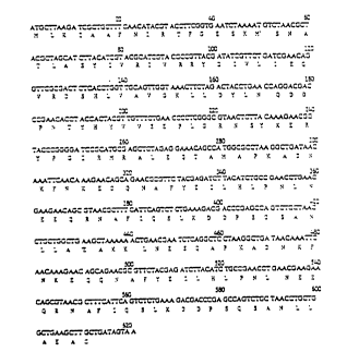
Figure 1 shows the complete nucleotide sequence and the
encoded amino acid sequence of the synthetic
SpA * gene
Figure 2 shows the formation of a gene encoding two SpAB*
douains.
Figure 3a shows the construction of gene fusion plasnid
p81-Sp ~ *-2.
Figure 3b shows the construction of gene fusion plasmids
p81-Sp~ *-2 and p53-Sp ~ *-2
Figure 4 shows the compl0te DNA and amino acid sequence of
fusion protein 81-SpAB~-2.
Figure 5 shows an SDS-PAGE gel illustrating the tire course
of induction of fusion protein 81-Sp ~ *-2.
Figure 6 shows an SDS-PAGE gel illustrating inclusion body
purification.
Figure 7 shows an enzyme linked immunosorbant assay for IgG
binding.
Figure 8 shows the formation of 81-Sp ~ *-1 or
81-Sp~ *-2-IgG complexes by light scattering.
Figure 9 shows helic~l wheel represen~ation of residues
13-'1 and 29-40 from Sp ~ *.
W O 92/09633 ~ PCT/GB91/02077
, 2~6Q~'~
Figure 10 sAows the affinities of 81-_pA~-2 and SpA for IgG
at different pHs.
Figure ll shows the relationship between binding of IgG by
mutated 81-SpAB~-2 proteins and pH.
In the following Example the production of I~G-binding
proteins (81-SpAB*-2 and 53-SpAB*-2) by total gene synthesis is
described. Unique restriction sites have been placed alons the genes
to facilitate the production of variant proteins. 81-SpAB~-2 is the
product of the fusion of part of the gene for bovine DNAasel and a
gene coding for the two B domains (SpAB) of Protein A from
S~phy Zococc~Ls c~ureus .
The fusion product is expressed in high yields in
~scherichia coIi JMl03 as an inclusion body which can be purified by
centrifugation and washing with aqueous denaturants such as Triton and
urea. The protein may be extracted into Z.5M urea and IgG-binding
activity is restored on removal of the urea by dialysis.
The protein has a single cysteine residue placed at the
carboxyl terminal of the protein which facilitates either
immobilisation of the protein to an insoluble matrix or the labelling
of the protein by radioactive or fluorescent reagents and has the same
affinity and specificity for IgG from various sources as Protein A.
The protein can be precipitated from solution by adjusting
the p~ to 6.0 and is very heat stable and loses no activity by heating
at 85 C for 30 min.
W O 92/09633 PCT/GB91/02077
2~69 3 - 12 -
Variations of 81-SpAB~-2 have been ~roduced by amino acid
subs~itutions, and some of these mutated proteins show changes in IgG
binding ac~ivity.
In this Example, a gene encoding a s_ngle synthetic IgG
binding domain was contructed by automated DNA synthesis. This
synthetic domain, termed Sp~ ~ was based upon one of the five IgG
binding domains of Protein A; domain B (SpAB) which has an amino
acid sequence closest to the consensus sequence of the five domains.
Further i; is strongest binding of all isolated single domains. The
amino acid numbering system used to refer to residues in the synthetic
binding domains throughout this description is based upon that devised
by Uhlen et al (1984) and is shown in Figure 1 for ease of reference.
Bacterial strains, clonin~ vectors and cell growth
E.co~i JM103 (Messing et al, 1981) was used as a bacterial
host. Plasmid and phage vectors used were pUC19 (Yanisch-Perron et
al, 1985) pkk223-3 (Brosius and Holy, 1984) and phage M13mpl9
(Yanisch-Perron et al, 1985). Bacteria were routinely grown in
L-broth (1,. bactotryptone, 0.5Z yeast extract, 0.5,. NaCl) supplemented
where appropriate with 50 ~g/ml ampicillin (Sigma).
DNA techniques
Restric ion enzymes (purchased from Boehringer Mannheim,
Northumbria Biolosicals Ltd) were used according to the supplier's
recommendations, as were the enzy~es T4 DNA ligase, T4 polynucleotide
kinase and calf intestinal alkaline phosphatase (Boehringer Mannheim).
DNA sequencing was performed using 'Sequenase', modified phage T/ DNA
polymerase (Tabor and Richardson, 198l; 'Sequenase' kit purchased from
W O 92/09633 - 13 - PCT/GB91/02077
~9~353
United Sta~es Biochemical Corporation). .~ll sequencing proeocols
including template preparation, were performed according to the
supplier's recommendations. Oligonucleotides were synthesized on a
fully automated Applied ~iosynthesis 380A DNA synchesiser wnich
empioys the phosphoramidite method of solid phase synthesis (At~inson
and Smith, 1984). De-protected oligonucleotides were pur .ied by
electrophoresis on a 7M Urea 12% polyacrylamide gel from which the
band corresponding to the full length DNA sequence was excised and
eluted (Maniatis et al, 1983).
DNA const-uctions
A synthetic gene, termed SpAB~ was constructed. based on
the B domain of SpA.
The DNA sequence was modified to maximise where possible the
codon usase for translation in E.coZi (Guoy and Gautier, 1982;
Grosjean and Fiers, 1982). Oligonucleotide cassette based site
directed mutagenesis is facilitated by the introduction of a series of
unique restriction sites at intervals in the DNA sequence.
Specifically, SpAB* was constructed as a series OI six
oligonucleotides of length 58-66 bp (see Fig l); adjacent
oligonucleotide pairs had a 7 bp cohesive overlap with the
neighbouring pair. The internal 5' ends were phosphorylated
separately, then complementary oligonucleotide pairs were annealed
together by heating separately, to 85 C followed by slow cooling to
room temperature. The three pairs were ligated together and cloned
into Bam~I/Pstl cut M13mpl9. DNA sequencing was performed to check
the construction, then the resultant BamHI/Pst' SpA3* _nser~ was
W O 92/09633 PCT/GB91/02077
- 14 -
9 ~ ~
subcloned into pUC19 to create plasmid pSpA3*. Two a-helices within
the encoded domain are predicted to be largely involved in IgG binding
and these are represented in Figure 1 by boxes over the amino acid
sequence. Residues which are predicted to make close contacts with
the ~c molecule have been underlined. The amino acid sequence of
Sp ~ * remains identical to that of SpAB except for the
substitution of an alanine residue for glycine-29, and the
introduction of a C-eerminal cysteine.
The gly --> ala replacement occurs at a non-essenti~l
position in the 2nd a-helix (i.e. away from the face that interacts
with Fc) and is not believed to affect Fc bindins (Nilssen et al,
1987). This substitution was done to remove the single Asn-Gly
peptide bond, making the domain resistant to hydroxylamine treatment.
This will permit the inclusion of such a bond at the junction between
the DNAasel and SpA3 moieties of the fusion protein so that the two
may be split by hydroxylamine and separately purified.
~ he additional Cys is introduced at the C-terminus which is
away from the Fc-binding region and provides a reactive site for
possible fluorescent labelling or immobilisation onto Sepharose to
give an IgG purification column.
A gene encoding two Fc-bindins domains (SpAB*-SpA3*) was
constructed by linking 2 of the SpAB~ genes together by the
methodology shown in Figure 2. The synthetic linker DNA encodes those
amino acids which separate adjacent Fc-binding domains in native
Protein A. This technique also ensures that the cysteine residue and
stop codons are removed from domain 1, giving an in-frame protein with
W O 92/09633 PCT/GB91/02077
- 15 -
2~3~J3
a single .erminal cyste: residue. This construction was also
cloned into pUC19 to give plasmid pSp~ ~-2.
The technique used for linking genes for 2 Sp~ * domains
is shown in Figure 2. pSpAB* was digested with Ba~I/HindI~I and the
185 bp fragment was purified away from vector DNA to give the
'upstream' domain 1. In a parallel procedure pSpAB* was digested
with Ddel/Pstl and the 166 bp fragment corresponding to the
'downstream' domain 2 was purified. These 2 molecules were then
ligated together using a short synthetic linker sequence (see Table 1)
containing the appropriate HindIII/Ddel restriction site cohesive
ends. The resultant SpAB* - SpAB* gene was cloned into Ml3mpl9,
sequenced, then sub-cloned into pUC19 to create pSp~ *-2.
Although SpAB* was designed with its own Shine-Dalgarno
ribosome binding site (Figure 1), high level expression was not
achieved following sub-cloning into expression vector pkk223-3, so a
gene fusion approach was used to increase expression.
The Sp~*-SpAB* sequence was linked to synthetically
constructed genes encoding mutated and inactive bovine DNAasel protein
(Worrall and Connolly, 1990) to create two genes, the first encoding
the first 81 amino acids of DNAasel followed by a 12 amino acid spacer
and then the 111 amino acids of SpAB*-SP~ and the second
encoding the first 53 amino acids of DNasel followed by a 12 amino
acid spacer and then the 111 amino acids of SrA_~-SpA3*. The
resulting plasmids containing the fused const- - s cloned into the
polylinker of pkk223-3 were termed p81-Sp~ *-2 and p53-SpAB~-2 and
their construction is shown in Figures 3 and 3a. Figure 4 shows the
W O 92/09633 - 16 - PCT/GB91/02077
2~969~3
complete DNA and amino acid sequence of the fusion pro~ein
p81-Sp~*-2 whic~ consists of 204 amino acids and has a calculated
molecular weight of 27.0 kDa. On inductmon of E.coIi J~103
(p81-Sp~ *-2 ) with 2mM IPTG. fusion pro~ein 81-Sp~*-2
accumulates within the cell (Figure 5) such that it becomes the major
cell protein. equivaient to at least 15,. of totPl cell protein as
estimated by gel scanning of a Coomassie Blue stained SDS-P.~GE gel
whereas the fusion protein 53-Sp~ *-2 was expressed at a lower
level. This is almost certainly an underestimate since only the
protein moiety derived from the DNAase 1 gene is stained well by this
reagent.
Construc~ion of gene fusion plasmids
Gene fusion plasmids were constructed by purifying fragments
of the SpA8*-2 gene from pSp~ *-2 and inserting these into
appropriately restricted pAW2 (WorrPll and Connolly. 1991). Figure 3b
summarises the construction of the plasmids encoding two IgG binding
domains and 81 or 53 residues from the N-terminus of the DNasel.
p81-Sp~ *-2 was created by ligation of the Kpn I-?st I fragment of
pSpAB*-2 into Kpn I-Pst I cut pAW2. p53-Sp~*-2 was constructed
in the same way, using the Xbal-PstI res~riction sites. The plasmids
encoding fusion proteins with single Sp~* domains were constructed
by digesting each respectivè plasmid with Bsl II, removing the
released fragment and religating the shortened. linearisea plasmid.
Restriction analysis and DNA sequencins were performed to confirm the
generation of recombinant DNA molecules. ,he complete
nucleotide/amino acid sequence of the encoaed fusion protein
W O 92/09633 PCT/GB91/02077
- 17 -
~0969~3
81-SpAB*-2 is shown in Figure 4 and its amino acid composit:on is
shown in Table 1.
Table 1. Amino acid composition of 81-SpAB*-2 and its component
parts.
DNase I 'INTER'SpA~*-SpA~* TOTAL
Ala 5 2 14 21
Cys - - 1 1
Asp 5 6 11
Glu 4 2 11 17
Phe 2 - 6 8
Gly 3
His 2 - 2 4
Ile 6 1 4 11
Lys 4 - 10 14
Leu 8 1 13 22
Met 2 1 1 4
Asn 5 ~ 15 20
Pro 2 1 6 9
Gln 2 _ 11 13
Arg 7 2 2 11
Ser 6 - 7 13
Thr 3 1 - 4
Val 8 - - 8
Trp
Tyr 7 - 2 9
81 12 ~11 204
Protein induction and inclusion body isolatlon
The expression of recombinant fusion proteins was induced by
the addition of ImM IPTG (Northumbria Biologicals Ltd) to cultures of
E.co~i JM103 containing the appropriate plasmid which had reached an
optical density of A600 0 7 ~ 0.9. Cell growth was at 37 C in a 251
batch fermentation vessel. Cells were harvested 4 hours post
induction and were stored frozen at -20 C. Aliquots equivalent to lOg
wet cell paste were thawed and resuspended ~n 30 ml 10 mM Trls-HCL
W O 92/09633 PCT/GB91/02077
- 18 -
2~969~3
pH.8.5, then treated with lysozyme at 0.1 mg/mi. stir-ing at 4 C for
10 minutes. Cells were disrupted by sonication (4 x 20 second bursts
at medium amplitude, MSE Soniprep 150) and the sonicate was treated
with 10 ~g/ml DNAasel for 30 minutes on ice.
Further induction an~lysis (Figure 6~ lndicated that
81-Sp~ *-2 was produced as an inclusion body within the cell, a
theory which was confirmed by microscopy (data not shown). When whole
cells of an induced culture are disrupted by sonication and then
subjected to low speed centrifugation, 81-Sp~*-~ is found
exclusively in the pelleted insoluble fraction (as shown in Figure 6,
lane 3). This enabled a purification protocol to be developed
involving repeated sonication and washes in detergent to solubilise as
much contaminating protein as possible away from the inclusion bodies.
Urea is then used to solubilise 81-Sp~ ~-2, which is found to regain
IgG binding activity on removal of urea by dialysis (see Materials and
Methods Section for deta ls). It was found that a sinilar pattern of
results was obtained for the fusion proteins 81-Sp~*-l,
53-Sp~*-l which were synthenised with one Sp~* dom~n and
53-Sp~*-2 and the inclusion bodies containing these proteins could
be recovered by the procedure described above. Inclusion bodies were
pelleted by centrifugation at /,500 g for 15 minutes and were
resuspended in 10 mM Tris-HCL pH 8.5, 1,. v/v Triton X-100 detergent.
The sonication process was repeated to ensure that all cells had been
disrupted, the sonicate was stirred at 4 C for 15 minutes then the
inclusion bodies were pelleted by centrifugation as before. Two
washes were performed in this way, then the pellet was washed x 2 -n
W O 92/09633 PCT/GB91/02077
2 ~
10 mM Tris-~:CL p~ 8.5 containing 1 M urea. SO1UDi1 isation of the
fusion protein was achieved by extraction in 10 mM Tris-HCL pH 8.5
containing 2.5 ~ urea at 4 C for 1 hour. The solution was spun at
27,000 g for 20 minutes and the supernatant was retained. A further
extraction in 10 ~M Tris-HCL pH 8.5 containing 4 M urea was performed
on the pellets and again the supernatant was retained. The urea was
dialysed away against 2 x 4 litres Z0 ~M XP buffer pH 8Ø and
aliquots of the resulting protein solùtion were freeze dried.
The observation that the level of expression of the fusion
proteins is dependent upon the number of residues from the D~ase 1
included in the fusion protein is significant and possibly explained
by close examination of the 3 dimentional structure of DNasel (Suck et
al, 1984). The amino terminal 80-85 amino acid residues appear to
exist as a domain distinct from the remainder of the protein. This
domain includes several secondary structural features. Two a-helices
(I and II, see Suck et Al, 1984) consisting of residues 18-29 and
42-54 respectively are present, and six 3-strands (A,B,C,D,E and F)
four of which (A,C,E and F) form a B-pleated sheet and the other two
form a parallel 3-pleated sheet with each other. Thus it is possible
that the N-terminal 81 residues of 81-SpAB*-1 or 2 may fold into the
same stable tertiary structure as in DNase 1. The first 53 residues
of DNase 1 (used in 53-SpAB*-1 or 2) also contain helices I and II
but contain only four 3-strands (A,B,C and D). This truncated
sequence may still fold into its 'native' structure but will be less
stable having lost possible hydrogen bonds between strands C and F.
W O 92/09633 PCT/GB91/0207
- 20 -
2~969~
The existence of the complete and thus presumably more
stable ~-terminal domain of DNase 1 in 81-SpA~*-1 or 2 may act as a
nucleus for protein folding events and hence maylead to a compact
fusion protein, better protected from proteolysis.
Protein Analysis
Protein concentration was estimated by the absorption of
light at 280nm, (E28o=10,800), An alternative Dethod for protein
concentration estimation is the bicinchoninic acid protein assay of
Smith et al (1985)(Sigma).
Determination of I~G-bindin~ activity
The interaction of 81-SpAB*-2 and 81-SpA3*-1 with Swine
anti-sheep IgG coupled to Horse radish peroxidase was determined by
ELISA experiments. In the standard method 81-Sp~ *-2 was shown to
have the same affinity for this IgG species as whole Protein A from
Stophy~ococcus oureus (figure 7). It was found. however. that
protein 81-SpAB*-l interacts approximately 100 fold more weakly than
either SpA or 81-Sp ~ *-2. Both IgG binding domains of 81-SpAB*-2
are functional since immunoprecipitates are formed when 81-SpAB*-2
is mixed with IgG. These precipitates can be detected either by
observation of an increase in light scattering or by using OuchterlonY
plates. Light scattering experiments used to monitor the interaction
between IgG bindlng proteins and IgG were performed using a Perkin
Elmer 650S Spectrofluorimeter with an incident and emission wavelength
of 320nm.
W ~ 92/09633 PCT/GB91/02077
- 21 -
2 5~ 9 ~ 9 ~ 3
IgG binding activity was quantified us ng an Enzyme Linked
Immunoabsorbent Assay (ELISA) technique modifiec from Hudson and Hay
(1980). Serial dilutions of protein in 50mM sodium carbonate buffer.
about pH 9.0 (coating buffer) were used to coat ~he wells of a
microtitre plate at 37 C for at least two hours. Wells were then
washed three times in 0.1,. Tween 20 in phosphate buffered saline. pH
8.2, (PBS-Tween)) before in-ubation at room temperature with lOOul
Swine anti-sheep-horse radish peroxidase conjuga~e (Serotec. UX)
diluted to lug per ml in the same buffer. After 30 min the wells were
washed three times again with PBS-Tween, then 200ul of substrate was
added to each well (0.35 ms/ml 0-phenylene diamine. and 0.1,. v/v
H202 in O.lM sodium citrate-phosphate buffer pH 5.0). The
reaction was stopped after 15 min by the addition of 50ul per well of
12.5Z H2S04 and the absorbance read at 495nm.
Com~etitive ELISA ExPeriments
IgG from other species was also shown to be bound by
81-SpAB*-2 by using competitive ELISA techniques. For competitive
ELISA all wells were coated with 200ns of 81-SpA3~-2 in sodium
carbonate buffer pH 9.6 as above. Seri~l dilut ons of the test
antibody in PBS-Tween were then made and allowed to bind to the
81-SpAB*-2 coating the wells for about lOmin. ,Oul of Swine
anti-sheep-horse radish peroxidase conjugate was then added (2~g per
ml in PBS-Tween) and the rest of the ELISA techn~que carried out as
described above.
W O 92/09633 PCT/CB91/0207
- 22 -
2~n~953
Exmeriment 1
The results of experiment 1, sl~marised in Table 2
demonstrate that 81-SpAB*-2 binds IgG with a similar species
specificity as whole Protein A. The affinity for the IgG decreases in
the following order:
Guinea Pig/Human/Pig/Mouse/Rabbit/Cow/Horse/Rat/Chicken/and Goat.
Table 2. Inhibition of 81-SpAB*-2 binding to peroxidase
conjugated swine-antl-sheep IgG from different species.
The figures given represent the amount (ng) of
competing antibody required to inhibit binding of the
test antibody by 50%, according to the conditions of
the ELISA described (see Materials and ~ethods).
Amount of competing
antibody required to
Source of I~G give 50~ inhibition
n5
Guinea pig < 20
Human 40
Pig 5
Mouse 5
Rabbit 60
Cow 320
Rat 3200
Horse 4
Chicken > 5000
Goat > 5000
A further comparison was carried out as follows.
Experiment 2
Experiment 2 used an identical procedure to Experiment 1,
except that the serial dilutions of the test antibody in P3S-Tween
were allowed to bind to the 81-Sp~ *-2 coating the wells of Che
microtitre plate for 15 minutes, instead of about 10 minutes. The
results of this experiment are shown in Table 3.
W O 92/09633 PCT/GB91/02077
- 23 -
2096~
Table 3
ng of immunoglobulin giving 50~ inhibi~ion
Source of I~GSDAS`DAB*-~
Goat 2,500 6.000
Rat 2,250 5,600
Cow 900 400
Mouse 150 70
Pig 20 65
Guinea Pig 40 50
Human 10 25
Rabbit 10 22
The af~inity for the IgG decreases in the following order
from this experiment as follows:
Rabbit/Human/Guineapig/Pig/Mouse/Cow/Rat/Goat.
Horse and Chicken IgG were not tested in this experiment.
This experiment also demonstrates that 81-SpAB*-2 binds
Ig5 with a similar species specificity as whole Protein A.
AB* - I~G complexes
Precipitation of SpAB*-IgG complexes can only occur if
both participants have two or more functional sites for the other.
Nephelomet.y measurements were made in order to determine whether both
IgG bindin~ domains of our construct 81-SpAB*-2 were able to bind
W O 92/09633 PCT/GB9l/02077
- 24 -
2~969~3
IgG. Figure 8 illustrates the changes in the light scatterlng pattern
from a cell containing 81-SpAB*-2 on addition of isolated Fc
fragments or whole IgG (human or chicken). A large precipitation
indicated by an increase in light (320nm) scattering at 90 occurs if
81-Sp~ *-2 is mixed with 5 fold excess of Fc or human IgG wheareas
only slight precipitation occurs when human IgG is mix~d with
81-Sp~ *-1. No precipitation occurs if chicken IgG is added to
81-Sp~*-2 human fc is added to 81-SpA3*-1 or any immunoglobulin
or Fc is added to a solution of DNAasel. Thus it appears that both
IgG binding sites in 81-SpA9*-2 are functional in the presence of
human Fc or IgG but that there is either no affinity for IgG from
chicken or that the complexes formed remain soluble. Evidence from
competitive ELISA experiments described above suggest that the former
is the case. The lack of formation of any precipitate with DNase 1
and IgG removes the possibility that the DNase 1 part of the fusion is
involved in any binding phenomenon to either Fab or Fc. The slight
increase in light scattering observed when 81-SpA3*-1 is mixed with
human IgG must arise from possible interactions involving the Fab (but
not antigen binding site) of the IgG since no such event occurred when
Fc or chicken IgG was used. The possibility of a recognition site for
SpA on the Faba section of IgG has been noted before (Langone~ 1982,
Lindmark, 1983). Our studies suggest that such recognition is to part
of the SpAB domain.
SDS-PAGE
SDS-PAGE was performed according to the method of Laemmli
(1970).
W O 92/09633 PCT/GB91/02077
- 25 -
2~5~3
_lectro~horetic Transfer of Protein f~om SDS-PAGE Gel to PVDF membrane
The electrophoretic transfer of proteins from SDS-PAGE gels
to membranes was performed essenti~lly as described by Towbin et al
(1979). However, a PVDF membrane was used in preference to
nitrocellulose, and CAPS buffer was used as the transfer buffer
(Matsudaira, 1987). Electroelution was carried out for 2 hours at a
constant 45 volts. For total protein staining the membrane was
immersed in Coomassie Blue stain for 2-5 min, then destained as for
SDS-PAGE gels. For the qualitative detection of IgG-binding activity
of protein bands electro-blotted onto PVDF membrane, the membrane was
blocked with 3% gelatine in P9S for 5-10 min, then the IgG-HRP
conjugate was added (diluted 1:5,000 in 1% gelatine). The solution
was agitated at intervals for 20-30 min, then the solution was
discarded and the filter was washed 5 times in 0.1X v/v Tween 20 (in
phosphate buffered saline (PBS)-Tween) and once in PBS.
Chloronaphthol solution was added to cover the filter along with
H202 (to 0.1~ v/v) and the solution was agitated. A purple/blue
colour is indicative of IgG-binding activity.
Heat denaturation studies on 81-SpAB*-2 have shown that
the protein is extremely resilient, and any denaturation caused by
heating up to 85 C for periods up to 30 min was found to be comPletelY
reversible with no loss of activity being detected in by ELISA carried
out at room temperature or 37 C after recooling the heat treated
81-SpA3*-2. However, the protein is extremely susceptible to
ienaturation and precipitation from solution if subjected to very mild
W O 92/09633 PCT/GB91/02077
2~969~3 - 26 -
acidic conditions. The protein precipitates from solution when the pH
is 6.2 or lower. This provides a simple method for removing the
protein from solutlon under extremely mild conditions. The
precipitated 8l-spAB~-2 can be refolded to produce active protein by
dissolving the precipitate in 4M urea and removing the urea by
dialysis.
PRODUCTION OF MUTANTS
Mutants were produced by site-directed mutagenesis in which the amino
acid residues designated l7Phe and l8Tyr (see below) were replaced
as follows:
Phe --> 17Tyr
Tyr - > l8Glu
Tyr - > l8Phe
Tyr - > l8His
8Tyr--> 18Lys
8Tyr--> 18Trp
Tyr - > l2Cys
Ala Pro Lys Ala Asp Asn Lys Phe Asn Lys
Glu Gln Gln Asn Ala Phe Tyr Glu Ile Leu
12 17 18
His Leu Pro Asn Leu Asn Glu Glu Gln Arg
Asn Ala Phe Ile Gln Ser Leu Lys Asp Asp
32
Pro Ser Gln Ser Ala Asn Leu Leu Ala Glu
A12a
W O 92/09633 PCT/GB91/02077
27 ~ ~ a 9~) a ~ 3
Figure 9 is a helical wheel representation of the amino acid
residues in the SpAB domain shown by X-ray crystallographic studies
of Diesenhofer (1981) to fall into two helical secondary s~,uctural
motifs. Residue 18 in the helix closest to the ~ino terminus of the
protein has been implicated to be essential for binding to the Fc of
IgG and possibly to participate in a hydrogen bond (ring hydroxyl
group) with the carbonyl group of the peptide bond for~ed between
residue 432 and 433 of the IgG. We have examined the requirement for
this tyrosine residue and the effect of the disruption of the proposed
hydrogen bond. Mutations described herein have therefore all been
made at this position numbering 111 and 169 in the fusion protein with
two IgG binding domains. Three mutations are described in
81-SpAa*-2, both domains having the same residue altered. Firstly,
Tyr 111, 169 to Phe 111, 169 replacements, the most conservative of
all, remove the possibility of the intermolecular hydrogen bonds to
two IgG molecules. Secondly, the Tyr was replaced in each domain by
Trp to examine the possibility of accomodating a bulkier side chain.
Finally, the Tyr was replaced by Glu to assess the effect of charge at
this position in each domain. These mutants were termed
81-Sp ~ *-2(YlllF, Y169F), 81-SpAB*-2(YlllW, Y169W) and
81-Sp ~ *-2(YlllE, Y169E) and the interactions between them and
porcine IgG-HRP conjugate were ~etermined by the modified T T ISA
technique described above.
Figure 7 shows the level of porcine IgG-HRP activity
retained per well containing various amounts of each IgG binding
protein. The daea demonstrate clearly that the residue Tyr lli (and
169) may be replaced by both Phe 111 (and 169) or Trp 111 (and 169)
W O 92/09633 PCT/GB91/02077
205653 8 -
without severe disruption of the binding interactions. Analysis of
such binding data shows that replacement of Ylll, Yl69 with Flll, ~169
causes a three fold decrease in affinity for the IgG, this being
equivalent to a loss of 0.6kcal per mol of bindins energy due to the
loss of the inte~colecular hydrogen bond per domain. The replacement
by the bulkier amino acid tryptophan decreases the affinity of the
protein for IgG by a factor of two suggesting that the loss of the
hydrogen bond is compensated by an increase in other favourable
interactions, presumably hydrophobic. In contrast, the presence of a
negative charge at this position, accomplished by the replacement of
the Tyr residue in each domain by Glu, appears to almost completely
destroy the interactions between the two proteins. This data suggests
that the loss of binding of Sp ~ to IgG after nitration of the Tyr
residue is not due solely to the loss of the hydrogen bond or to
steric effects (Sjoholm et al, 1973) since the Phe and Trp mutants
described above still possess reasonable binding activity. D~ase l
alone does not give any positive signal in ELISA tests.
In a further experiment, mutants with Tyrl8 replaced by
other amino acids were made by digestion of the plasmid 8l-spAB*-2
with MlaI and Bgl II to release a short fragment encoding residues
'7-19 inclusive. This wa replaced by short synthetic
oligonucleotides bearing sing~e amino acid substitutions in position
'8, coding for Phe, Glu, His or Lys (see Table 4) but having the same
cohesive ends. The manipulations necessary to generate a gene
encoding the same fusion protein but bearing identical mutations in
the two IgG binding domains have been aescribed elsewhere (Popplewell,
W O 92/09633 PCT/GB91/02077
2~95~3
1991). In order to demonstrate that the IgG binding domains in the
fusion protein behave as those in native Protein A both were comPared
for IgG binding ability by the modified ELISA protocol described
above. The results in Fig. 10 demonstrate that the non-mutated
protein 81-Sp ~ -2 has a very similar affinity for I~G as P-otein A
in the pH ranse 6.0 to 8.0 although it has a three fold lower affinity
at pH 5Ø This latter difference arises due to an instabil~ty of the
fusion protein at this low pH.
The data displayed in Fig. 11 shows the amoun~ of
porcine IgG-HRP conjugate bound to various amounts of fusion proteins
in the wells of a standard microtitre plate. The mutant YlllH,Y169K
shows less binding, approximately 8X of that of the native protein and
the mutant YlllE,Y169E shows virtually no IgG binding under these
conditions. However, the interaction of All three proteins with
Porcine IgG-HRP was found to be very sensitive to pH unlike the native
protein. At pH values where the replaced residue would be expected to
have a charge, the binding is less strong than under conditions where
the equilibrium between charged and uncharged species lies towards the
uncharged side. The mutant YlllE,Y169E therefore shows maximal
binding at pH 4 and minimal binding at pH 6 or above i.e. at pH values
where the carboxyl group of the side chain has a negative charge. In
contrast, both the mutants YlllH,Y169H and YlllK,Y169K show the
ir.creased binding of IgG-HRP a. ~igher pH where both side chains
become less protonated. Significantly the apparent pK (6.~) of the
binding curve shown by YlllH,Y169H is lower than that shown by
YlllK,Y169K (7.4), as would be expected. Table 4 gives the percen~age
WO 92/09633 PCr/GB91/02077
- 30 -
~ V~b~53
of IgC binding shown by each mutant compared with the non-~u~ated
construct at two pH values where the binding is minimal or maximum.
It can be seen that the Glu mutant has a maximum binding of only 10,.
of the 'native' protein at pH 4.0 whereas much higher relative binding
is obtained for the Lys (20%) or His (50%) mutants at pH 9Ø
SUMMARY
From the foregoing it can be seen that the present invention has
successfully overcome the problems of the prior art. Recombinant DNA
techniques are provided for the production of polypeptides having
between 2 and 4 modified IgG binding domains which allow high levels
of expression in E co ti without incurring proteolysis by host
enzymes or difficulties in purification. Said binding domains are
highly amenable to site-directed mutagenesis and therefore enable the
production of immunoglobulin-binding proteins having distinct
advantages compared to Protein A. Examples of such mutated proteins
are given and their properties investigated.
W O 92/09633 PCT/GB91/02077
- 31 - ~ ~ ~6953
.able
f~u~an~~
consr uc~ Q~ ~eia~ive 3i~di~g ~a~i eJ
Y111,169 4.0 lOG~
(na~ive)
~ . O iOO%
Ylll~., n69~ 4.0 15
9.0 iO~
n llK, n 69K 4.0 <5%
9.0 23
YlllE, Y169E 4.0 iO~
9.0 <0.5~ ,