Note: Descriptions are shown in the official language in which they were submitted.
2103.1 ~3
- 1 -
ATRAOlIATIC END08COPIC APPARATUS
Technical Field
The field of art to which this invention relates is
surgical instrumentation, in particular endoscopic
surgical instruments.
Background of the Invention
The use of endoscopic surgical procedures has become
increasingly common throughout the surgical community.
There are many advantages associated with the use of
endoscopic surgical techniques including decreased trauma,
improved post-operative recuperation, decreased avenues
for infection, and decreased post-operative hospital
stays. The term endoscopic as used herein is meant to
encompass all minimally invasive surgical techniques
utilizing a scope including endoscopic, laparoscopic,
thoracoscopic and arthroscopic.
In many endoscopic surgical techniques, it is necessary to
enter a body cavity to obtain access to the target
surgical site. This is conventionally done by using a
trocar. A trocar typically consists of a trocar obturator
having a sharp piercing point and a trocar cannula. The
trocar obturator is concentrically housed within the
trocar cannula during insertion through the musculature
and fascia surrounding the body cavity. The trocar
obturator is then removed from the trocar cannula after
the trocar has been maneuvered into the body cavity,
SEN-110
21031?3
- 2 -
leaving the trocar cannula as a pathway into the body
cavity, e.g., the abdomen.
Numerous surgical instruments have been developed and
adapted for endoscopic surgical procedures. For example,
there are stapling apparatuses, suture and cannula
assemblies, electrocautery devices, tissue manipulating
devices, tissue cutting devices, tissue ligating devices,
and the like.
In most conventional, open surgical procedures, organs
must typically be manually displaced by the surgeon to
access a target surgical site. This must be done with
minimal trauma to the organs. This task is facilitated
during an open surgical procedure by the fact that the
surgeon has sufficient tactile sensory input through a
latex surgical glove to effectively prevent undue stress
upon the organs when they are being displaced. In an
endoscopic procedure it is also necessary for the surgeon
to manipulate or move tissue including blood vessels
internal organs such as the liver, the spleen, and the
gall bladder in order to access a target surgical site.
This is typically done with a variety of endoscopic tissue
manipulators which have been specially developed for this
task. However, there are certain deficiencies associated
with these tissue manipulating devices. The tactile
sensory input available in an open procedure is not
available to the surgeon during a conventional endoscopic
procedure since the endoscopic surgeon is manipulating
organs and tissue with instruments. The surgeon, when
manipulating organs with a manipulating instrument, has
a loss of tactile sensory input. An additional
complicating factor is that conventional endoscopes,
having video output to video monitors, do not provide the
sEN-110
210~~.'~~3
- 3 -
surgeon with depth of field as the surgeon attempts to
maneuver within the body cavity: Therefore, if the
appropriate care is not taken by the surgeon, it is
possible that organs and tissue, especially the capsular
organs, will be traumatized or damaged by the endoscopic
manipulating instruments as the surgeon attempts to
maneuver in a three-dimensional space with a two-
dimensional visualization system.
There is a need in this art for atraumatic endoscopic
surgical manipulating instruments. Such instruments when
used in a endoscopic surgical procedure would eliminate or
minimize trauma to tissue and organs, in particular,
capsular organs such as the liver, spleen and lungs.
Therefore, it is an object of the present invention to
provide an atraumatic endoscopic surgical instrument for
grasping and manipulating tissue and/or organs.
Disclosure of the Invention
Accordingly, an atraumatic endoscopic apparatus for
engaging mammalian tissue is disclosed. The endoscopic
apparatus comprises a frame having a proximal end and a
distal end. The frame has a passage therethrough. The
endoscopic apparatus has a handle means at the proximal
end of the frame for holding the apparatus. Jaw means are
attached to the distal end of the frame for engaging or
holding tissue or organs. The atraumatic endoscopic
apparatus has actuating means for moving the jaw means
between fully extended and fully closed positions.
Atraumatic means are mounted to the jaw means effective to
make the endoscopic apparatus substantially atraumatic
when the jaw means are actuated by the actuating means.
SEN-110
2IO~~.v3
- 4 -
The atraumatic means consists of a member having at least
one tissue contact surface, preferably a substantially
first planar tissue contact surface and a second proximal
tissue contact surface angulated with respect to the first
surface.
Another aspect of the present invention is a method of
manipulating tissue or organs in an endoscopic procedure
in a manner to effectively prevent or minimize trauma to
the tissue or organs by using the above-described
atraumatic endoscopic apparatus.
Yet another aspect of the present invention is an
atraumatic means for mounting in an endoscopic apparatus
for preventing or minimizing damage to tissue and organs.
Other features and advantages of the invention will become
more apparent from the following description and
accompanying drawings.
Brief Description of the DrawincLs
FIG. 1 is perspective view of the atraumatic endoscogic
apparatus of the present invention shown in an open
position.
FIG. 2 is a partial perspective view of the distal end of
the atraumatic endoscopic apparatus of the present
invention shown in a closed position.
FIG. 3 is a partial cross-sectional view of the handle
portion of the instrument as taken along View Line 3-3 of
FIG, 1.
SEN-110
- - 21~3~ ~
- 5 -
FIG. 4 is a partial cross-sectional view of the atraumatic
endoscopic apparatus as taken along View Line 4-4 of FIG.
1 with the jaws in the open position.
FIG. 5 is an enlarged cross-sectional view of the
atraumatic endoscopic apparatus as taken along View Line
5-5 of FIG. 2 with the jaws in a closed position.
FIG 6. is an exploded perspective view of the linkages
used to articulate the jaws.
FIG. 7 is an exploded perspective view of the atraumatic
means.
FIG. 8 is a perspective view of the atraumatic endoscopic
apparatus of the present invention inserted through a
trocar cannula into a mammalian body cavity.
FIG. 9 is a perspective view of an alternate embodiment of
the atraumatic means shown with the jaw members in the
open position.
FIG. 10 is a perspective view of the embodiment of FIG.
9 shown in a clamped position.
FIG. 11 is a perspective view of yet another embodiment of
the atraumatic means which may be used with the atraumatic
endoscopic apparatus of the present invention.
Hest Mode for Carryj~g~ Out the Invention
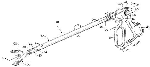
As can be seen in Figs. 1, 2, and 3, the atraumatic
endoscopic apparatus to of the present invention has
longitudinal frame 20. Longitudinal frame 20 has
SEN-110
~~~J~ rd c!
- 6 -
longitudinal passage 23 extending therethrough. The
proximal end 22 of frame 20 is mounted to handle 30 which
has finger grip 35 extending downwardly from the handle
30. Preferably, frame 2o is rotatably mounted to handle
30. Knob 28, mounted to proximal end 22 of frame 20, is
used to rotate frame 20. Handle 30 has a pair of opposed
mounting tabs 25 for mounting actuating lever 40.
Mounting tabs 25 have holes 27 therethrough for receiving
actuating lever pivot pin 50. Actuating lever 40 has pivot
l0 hole 49 for receiving pivot pin 50.
Actuating lever 40 is a substantially elongated member
having thumb ring 45 at one end and head 42 at the
opposite end. Head 42 has cavity 46 therein for receiving
the proximal end 72 of actuating rod 70. Spherical cavity
41 is also contained in head 42 for mounting ball member
48. Pin hole 43 extends through ball member 48 for
receiving actuator rod pivot pin 71. Ball member 48 has
slot 48A for receiving distal end 72 and eye 75 of
actuating rod 70. Although it is preferred that
proximal end 72 be rotatably mounted in head 42 so that
rod 70 can rotate when frame 20 is rotated, proximal end
72 may simply be pivotally mounted therein. Extending
from the top of head 42 is optional thumb grip 47. Thumb
grip 47 consists of a series of substantially parallel
members disposed substantially perpendicular to the
longitudinal axis of the apparatus lo. However, any
conventional gripping means such as knurling and the like
may be used. Actuating lever 40 is mounted to handle 30
at mounting tabs 25 by actuating lever pivot pin 50 which
extends through pivot hole 49 and holes 27.
Aa can be seen with reference to FIGS. 3, 4, 5 and 6,
actuating rod 70 is an elongated, substantially
SEN-110
2~6~~ r~
_ 7 _
longitudinal member which is slideably mounted within
frame 20 in passage 23. The actuating rod 70 has proximal
end 72 and distal end 74. Proximal end 72 has circular
eye 75 for receiving actuating rod pin 71. The proximal
end 72 of actuating rod 70 is pivotally mounted in slot
48A of ball member 48 (which in turn is mounted in
spherical cavity 41 of head 42) by pivot pin 71, which
pivotally engages eye 75 and pin hole 43. Ball member 48
and spherical cavity 41 function as a ball and socket
joint allowing frame 20 and rod 70 to rotate with respect
to handle 30, and additionally allowing end 72 to pivot.
Preferably, end 72 is rotatably mounted in the cavity 46
to allow rotation with frame 20. If rotation of frame 20
and rod 70 is not desired, then eye 75 is simply pivotally
pinned in cavity 46 of head 42. The distal end 74 of
actuating rod 70 has eye 77 which is pivotally mounted to
connecting members 90.
As best seen in FIG. 6, mounted to the distal end 24 of
frame 20 are the jaw mounting members 80. The jaw
mounting members are seen to have a semi-cylindrical shape
and are disposed substantially opposite to each other and
are separated by mounting slot 82. The jaw mounting
members 80 have upper and lower pivot holes 84 disposed
on either side of slot 86 for receiving jaw pivot pins 88.
The connecting members 90 are substantially flat elongated
members and have blunt, rounded ends. Centrally disposed
in each end of the members 90 are the pivot pin holes 95.
The jaws 100 are elongated members having a proximal end
105 and a distal end 110. Extending from the proximal end
105 of each jaw member 100 are the angulated lever members
120. Each angulated lever member 120 has slot 122 therein
for receiving a connecting member 90. It can also be
SEN-110
i~ :: ~., : ,, )
:'~, (.mi ti
- g -
seen that at the proximal end 105 of each angulated lever
member 120, there are pivot mounting holes 130 to receive
pins 135. At the proximal end 105 of each jaw member 100
there are pivot mounting holes 140 for receiving jaw
mounting pins 88. The jaws 100 are pivotally mounted to
jaw mounting members 80 by pins 88 which are inserted
through pivot holes 84 and through pivot mounting holes
140.
Members 90 are pivotally mounted on one end to jaws loo in
slots 122 by pins 135 which are inserted through pivot
holes 130 and 95. Members 90 are mounted at their other
end to distal end 74 of rod 70 by pin 93 through pivot
holes 95 and eye 77. The above-described pins are secured
using conventional techniques such as swaging, welding,
screw threads, bonding with adhesives, brazing, soldering,
mechanical fasteners and the like.
As can be seen from FIGS. 1, 2, 6 and 7, jaws 100 have a
substantially rectangular cross-section at the proximal
end 105 which tapers down to a substantially reduced
cross-section at the distal end 110 of each jaw member
100. However, other equivalent cross-sections can be used
including circular, elliptical, polyhedral and the like.
Proximal to the distal end 110 of each jaw member 100 are
the atraumatic mounting means 170, as seen in FIG. 6. The
atraumatic mounting means 170 are seen to be curved,
members extending from the reduced, distal section 110 of
the jaws. Each mounting means 170 has a blunt distal end
175 and optionally at least one or more parallel slots 172
extending therethrough. The gap 101 is seen to be
contained between the jaws 100. It is particularly
preferred that the jaw members 100 have a curved
configuration as seen in FIG. 5 to optimize the size and
SEN-110
el ~ r~
g
configuration of the gap 100. It is believed that the
presence of the gap 101 contributes to the manipulating
ability of the apparatus 10.
Mounted in each jaw mounting means 170 are the atraumatic
means 190. The atraumatic means 190 in one embodiment as
seen in FIGS 9, 10 and 11 consists of semi-cylindrical
pads 290 having the curved side 291 affixed to the
mounting means 270 and the flat side 292 projecting
inwardly to act as a tissue contact surface. The mounting
means 270 are seen to have a configuration which
substantially conforms to the semi-cylindrical shape of
the pads 290. As seen in the embodiment shown in FIG. 11,
the curved side 291 has optional projections 295 for
mounting in optional holes 275 contained in mounting means
270. The pads 270 are also seen to have proximal tissue
contact surface 299.
In a preferred embodiment as seen in FIG. 7, the
atraumatic means 190 consist of a wedge shaped pad 195
mounted in each mounting means 170. The pads 195 have a
substantially flat, lower surface 191 having a series of
optional projections 192 extending outwardly therefrom.
the surface 191 may also be curved. The projections may
have a variety of shapes including semi-spherical,
conical, cylindrical and the like. The pads 195 will be
mounted in mounting means 170 so that the flat surfaces
191 and projections 192 will contact tissue or organs.
The pad 195 is seen to have a rounded blunt tip 198 and a
curved upper surface 199 conforming to the shape of the
mounting means 170. Upper surface 199 has a series of
optional projections 200 which fit into slots 172 and are
preferably frictionally engaged therein. The atraumatic
pad 195 is also seen to have proximal surface 196.
SEN-110
210~~.~1~
- 10 -
Proximal surface 196 is seen to be substantially
perpendicular to surface 191, however, surface 196 may be
angulated with respect to the longitudinal axis of surface
191 at an obtuse or acute angle ranging from 45 degrees to
165 degrees. Proximal surface 196 may be flat or planar.
Tissue or organs grasped between jaws 100 in gap lol will
typically contact at least part of proximal surface 196.
The atraumatic means 190, in particular, the pads 195,
l0 simulate grasping by a surgeons gloved finger tips. The
atraumatic means 190, such as pads 195, are characterized
as sufficiently soft, and compressible, such that the
pads are effective to be atraumatic to tissue and capsular
organs. The atraumatic means 190 will also be
sufficiently flexible, and conformal to be effectively
atraumatic. By atraumatic is meant the capability to
contact, grasp and maneuver tissue with minimal trauma or
damage to the tissue or organs. The atraumatic means 190
will preferably have a high durometer, and more preferably
will have a durometer similar to the range of durometers
for human fingers (this range is widely known in the art).
It is particularly preferred to manufacture the atraumatic
means 190, e.g., pads 195, from polymeric foam materials.
Such polymeric foam materials include such biocompatible
materials such as polyethylene, polypropylene,
polyurethane and the like. In addition to foam pads, the
atraumatic means may be include air filled plastic pads,
saline filled plastic pads, gel filled plastic pads, gauze
pads, cotton pads, silicone filled pads, combinations
thereof and the like.
As can be seen from Figs. 1, 2, 3, 4 and 5, a counter-
clockwise rotation of actuating lever 40 about the lever
pin 50 causes a distal longitudinal displacement of
SEN-110
2103~~1
- 11 -
actuating rod 70. Actuating rod ?0 displaces connecting
members 90 which in turn displace the jaws 100 by acting
on the angulated levers 120. This causes the jaws 100 to
open by pivoting about pivot pins 88. Similarly a
clockwise displacement of actuating lever 40 will in a
similar manner~cause the actuating rod ?0 to displace in
a longitudinal, proximal manner causing the jaws 100 to
rotate to a closed position.
l0 The atraumatic, endoscopic apparatus 10 of the present
invention is used in conventional endoscopic surgical
procedures, and equivalents thereof, to manipulate tissue
and body organs, in particular, capsular organs. In a
conventional endoscopic surgical procedure, the patient is
prepared using conventional surgical preparatory
techniques including, as required, depilation of the
epidermis, scrubbing, and application of aqueous iodine
solutions in the area where incisions are likely to be
made. Then, the patient is anesthetized using
conventional anesthesiology procedures with a conventional
anesthesia and the patient is connected to a ventilator
an/or anesthesia machine, as required. Next, the
patient's body cavity, e.g., abdominal cavity, is
typically insufflated with a sterile gas such as carbon
dioxide, although it is possible to operate without using
insufflation. Then, using conventional endosurgical
techniques, several trocar cannulas are inserted into the
patient's abdominal cavity to act as pathways to and from
the body cavity. Next, an endoscope is inserted through
one of the trocar cannulas and the other trocar cannulas
are used for the insertion of various conventional
endoscopic surgical instruments including staplers,
electrocautery instruments, cannulas, ligating clip
appliers and the like. In order to access a particular
SEN-110
- 12 -
surgical site within the body cavity, the surgeon must
frequently manipulate internal organs such as the liver
out of the operating field. The movements must be made in
a delicate, gentle manner to minimize trauma to organs and
tissue, especially the organs and tissue described herein.
This can be accomplished by using the atraumatic
apparatus 10 of the present invention.
In order to use the apparatus 10, the surgeon grasps the
instrument by the surgeon by the handle 30 with the
actuating lever 40 rotated fully clockwise to a closed
position so that the jaws 100 are in a closed position.
Then the distal end of the apparatus 10 is inserted into
a trocar cannula and displaced into the patient's body
cavity. When the surgeon observes on the endoscope's
visual display, typically a video monitor, that the jaws
100 of the apparatus 10 are in the body cavity, the
surgeon maneuvers the jaws 100 proximate to. the organ
which must be moved in order to access the target surgical
site, as seen in FIG. 8. Then, the surgeon rotates the
actuating lever 40 in a counter clockwise manner using the
thumb ring 40, thereby actuating and opening up the jaws
100. The surgeon then manipulates the open jaws 100
around a section of the organ and once again engages the
actuating lever 40, this time rotating it in clockwise
manner to close the jaws 100 and engage the atraumatic
means 190 about the section of organ. At least one and
preferably both of the surfaces 191 and 196 contact the
organ and preferably tissue will be contained in gap 101.
This allows the surgeon to maneuver the organ in an
atraumatic manner. After displacing the organ, the
surgeon manipulates the actuating means 40 to open the
jaws 100 and release the organ from the atraumatic means
SEN-110
2~.~~1'~~
- 13 -
190. The apparatus 10 may then be withdrawn from the
trocar cannula.
The atraumatic, endoscopic apparatus 10 of the present
invention has many advantages for use in endoscopic
surgical procedures. In particular, it is now possible
for a surgeon to manipulate and move sensitive organs and
tissue, such as capsular organs including the liver,
spleen, and lungs, with minimal trauma, or possibly no
l0 trauma, caused to the tissue or organs. The use of the
apparatus l0 having compressible atraumatic means 190 also
provides the surgeon with tactile input to the hand which
is holding the apparatus 10. This tactile input is an
indicator of the force being applied to the organ or
tissue. This tactile input is not available with
conventional endoscopic instruments having hard, non
compressible surfaces. It is believed that the
combination of surface 191 and proximal surface 196
results in unexpectedly improved atraumatic tissue
grasping.
Although this invention has been shown and described with
respect to detailed embodiments thereof, it will be
understood by those skilled in the art that various
changes in form and detail thereof may be made without
departing from the spirit and scope of the claimed
invention.
SEN-110