Note: Descriptions are shown in the official language in which they were submitted.
RFSP File No. 7405-001
~~.~ ~~b~
- 1 -
Title: DENTAL IMPLANT
FIELD OF THE INVENTION
This invention relates to dental implants of the
type used in the mouth to stabilize dentures or support
dental crowns and bridges.
BACKGROUND OF THE INVENTION
Dental implants used to stabilize dentures or
support dental crowns and bridges have been known and have
been used fairly extensively in the recent past. Such
prior art devices are typically comprised of three
components, namely, an implant component for anchoring to
the bone, a transgingival component and a separate support
component. The support component usually attaches to the
transgingival component which, in turn attaches to the
anchoring component at about the level of the bone. An
artificial tooth or bridge may then be attached to this
separate support component. This support component is
sometimes referred to as an abutment portion, the
transgingival component is sometimes referred to as an
abutment connection or the transgingival collar or the
transepithelial connection and the implant is sometimes
referred to as a fixture.
An example of such a prior device may be found
in Canadian Patent No. 1,313,597. This patent describes
an implant for insertion into bone through an epithelial
and fibrous connective tissue layer to which a prosthesis
may be attached. This implant comprises a top portion for
supporting a mechanical component to which the prosthesis
may be connected and a body comprising an upper bone
attachment region which tapers to a lower bone engagement
region having a porous surface. The upper bone attachment
region comprises a substantially non-porous but
bioreactive surface and this patent teaches that this
results in an upper bone attachment region which is
claimed to be capable of enhancing bone attachment.
2~~'~2~2
However, several problems develop with an
implant of this type. In particular, the patent teaches
use of a collar 14 that is adapted to be coupled to the
implant 12. However the interface between the collar 14
and the implant 12 occurs at a level below the gingiva in
the installed position. Further, although the patent
teaches providing recesses 40 on the lower surface 42 of
the collar 14 to compliment projections 32 of the implant
12 to prevent rotation between the two components, in
practice this is not effective. The attachment between
the collar and the implant is accomplished by means of a
threaded screw identified as 46 in Figure 1. Such a screw
has a natural tendency to become loose during the vigorous
stresses to which an implant of this type is subjected.
To avoid problems associated with the loosening
of the threaded screw 46,.practitioners have resorted to
insertion of cement into the threaded portion to ensure a
locked and non-loosening joint between the implant
component and the support component. Unfortunately,
because the interface between the collar and the implant
is below the gum level, any excess cement will be squeezed
out at the interface and may not be noticed by the
practitioner since it is hidden from view. Such excess
accumulation of cement can create irritation of the gum
and the bone and can result in infection and/or implant
failure.
In addition, all implant systems, (fixture,
abutment connection, abutment) which have this type of
arrangement have a microgap between the fixture or implant
and the abutment connection or the transgingival collar at
the level of the bone. This microgap has been called an
"endotoxin generator" by some authorities because it is a
region for potential bacterial growth.
Other prior art devices include implants with
threaded exteriors which require extensive and complicated
methods for preparation of the gum and bone to accept the
insert. As a result, such implants are difficult and
~~~~~~7~
_ 3 _
expensive to insert and the surgery is most often done by
specialists. In any event they are not practical and
result in excess discomfort for the patient and
unnecessary difficulties for the dentist making the
installation. For example, some require incising the gum
to gain access to the bone; multiple drilling and reaming
steps; installation of the implant; reattachment, by
suturing or the like, of the gum over the implant site to
promote healing; a return visit several months later to
the dental office to have the gum again incised to allow
access to the implant; insertion and attachment of the
abutment portion; and final attachment of the prosthesis
to the abutment portion. The first incision into the gum
can promote scaring, making the second incision difficult.
SZJMMARY OF THE INVENTION
What is required is an implant which may be
installed in an easy one-step method and yet which is
secured to the bone and provides a firm anchor.
Preferably such an invention would also minimize the risk
of infection or irritation of the bone and the gingiva and
would provide for easy attachment with standard
components. Further, such an implant would not require an
excessive number of steps for installation, or excessive
patient discomfort and would be quickly and efficiently
installed in various types of installation conditions.
Therefore, according to the present invention
there is an implant far anchoring a prasthetic device in
bone, said implant comprising:
(a) a one piece body having:
(i) a root portion having a surface conducive to
bone ingrowth;
(ii) a tapered emergent portion having a smooth
biocompatible surface which is non-irritating to
living tissue, and
(iii) a coronal portion having a keyway; and
(b) an abutment portion for insertion into said keyway,
'-'
said abutment portion including a key at one end for
insertion into said keyway and a head for retention
of said prosthetic device at the other end.
In a further aspect of the present invention
there is provided a method of inserting a dental implant
comprising:
incising through a gingival layer;
drilling a pilot hole, at an appropriate position
into a bone located below said gingival layer;
drilling a recess, about said pilot hole, into the
bone with a second drill bit to form a tapered
recess of appropriate diameter;
positioning a one piece dental implant in the
tapered recess;
gently tapping said implant into surface engagement
with said bone wherein said step of tapping includes
wedging a smooth emergent portion against a hard
cortical bone portion and wedging a root portion
coated with a material conducive to bone ingrowth
into a cancellous region of the bone;
allowing said bone to grow into said root portion;
and
securing an abutment portion to said implant.
BRIEF DESCRIPTION OF THE DRAWINGS
Reference will now be made to preferred embodiments
of the invention by reference to the attached drawings
which are by way of example only and in which:
Figure 1 is a side view showing an implant according
to the present invention;
Figure 2 is a front view of the implant of Figure 1;
Figure 3 is a front view of an abutment portion for
an implant of Figure 1 adapted to act as an anchor
for overdenture retention;
Figure 4 is a front view of an abutment portion for
an implant of Figure 1 adapted to act as an anchor
for a false tooth cap;
Figure 5 is a cross-sectional view through the
2~~'~~~~
- 5 -
abutment portion along the lines 4-4 of Figure 4;
Figure 6 is a side view of a healing cap for use in
association with the implant of Figure 1;
Figure 7 is a view of the implant partially inserted;
Figure 8 is a view showing the implant of Figure 1 in
an installed position; and
Figure 9 is a view of a drill for inserting the
implant of Figure 1.
DETAILED DESCRIPTION OF THE PREFERRED EMBODIMENT
Figure 1 shows a one piece body or implant 10 having
a coronal portion 12 and an apical portion 14. Beginning
at the bottom of the one piece body 10 there is shown a
tapered section 16 above which is a cylindrical section
18. The tapered section 16 and the cylindrical section 18
are suitably coated with a material which is conducive to
bone ingrowth and which is identified as 20. These
sections 16, 18 together may be considered as a root
portion. The preferred form of bony ingrowth material is
of the type which creates a multitude of tiny passageways.
This can be accomplished, for example, by fine wire mesh
screens or the like, but the preferred form is to use
discrete particles of titanium alloy which are bonded to
the outside of the implant in a random fashion. The
preferred method of bonding is by sintering, as will be
known by those skilled in the art. The preferred size of
particles is between 45 to 3.50 microns although other
sizes may also be appropriate for bony ingrowth, as will
be known to those skilled in the art.
The one piece implant consisting of the coronal
portion 12, the body 10, with the apical portion 14, is
preferably made of titanium alloy, Ti6A14Va, and the
coating material is made of commercially pure titanium.
Above the cylindrical section 18 coated with bony
ingrowth material 20 is a tapered, smooth-walled portion
22. The taper of portion 22 may be referred to as the
"fourth taper". The smooth wall is an important aspect of
the present invention. To avoid gum and bone infection it
2~.~'~?62
-- 6 -
is important to have a relatively smooth nonporous surface
which is biocompatible at the emergent portion. In
particular, it is preferred if the emergent region is
machine polished rather than highly polished. Also it is
preferred if the emergent cylindrical portion 18 and the
tapered portion shown as A in Figure 1 is preferably 176
degrees. The preferred length of the smooth walled
portion 22 is .06 in. (1.50 m.m.).
The coronal portion 12 begins with a tapered section
24 increasing in diameter towards the end of the coronal
portion 12, and which ends at a generally horizontal top
portion 28. Between tapered section 24 and the top 28 are
located two further tapered sections, identified as 25 and
26 respectively. The preferred total length of the
coronal portion 12 is .16 in. (4.00 m.m.), of which the
axial length of tapered section 24 is .12 in. (3.00 m.m.)
and of each of sections 25, 26 is .02 in. (0.50 m.m.).
Located within the coronal portion is a keyway
identified as 30 which includes a tapered section 32 and
a part cylindrical section 34. The taper on section 32
may also be referred to as the "first taper". Located
between these two sections is a toroidal section 36 which
may be formed in a part circular shape with cross-section
having a .O1 in. (0.30 m.m.) diameter.
Located at the top of the coronal portion 12 is a
cross groove 38 which is shown more clearly in Figure 2.
The purpose of this groove is to provide an additional
keyway to resist rotation of the parts of the present
invention with respect to each other as will be
appreciated from the following description.
Turning to Figure 3, an abutment portion indicated
generally as 40 is shown. The abutment portion is formed
with a key 42 (also referred to as a keyway insertion
portion) for insertion into the keyway 30 of the implant
10 and a head 42 which is in the form of a common anchor
for overdenture retention. As can be seen in Figure 3, a
locking bar 46 is provided which mates with groove 38 on
the coronal portion of implant 10. Also, it can be seen
that a tapered section indicated at 48, also referred to
as a second taper, is provided on the key for the purpose
of locking the abutment portion 40 into the implant 10.
Figure 4 is a view of a second abutment portion 50 of
the type used to act as an anchor for a single false tooth
cap. The key portion 52 is identical to the key portion
42 of abutment 40 and includes a locking bar 56 which is
identical to locking bar 46. The head portion 44 is in
the form of a standard anchor for a single tooth cap.
Again, a tapered portion 58 is provided which is identical
to the tapered portion 48.
Figure 5 shows a cross-sectional view along line 4-4
of Figure 4. As will be appreciated this view is
identical for abutment portions 40 and 50, which according
to the present invention have the same key portion. As
can be seen, one face of the key portion 52, 42 is flat
and is indicated as 60. Also shown is the underside of
the head 44 which is shown as 62 as well as the locking
bar which is shown as 56. The main portion of the key is
shown as represented by the line 52 and the taper is
represented by the area shown as 58. It can now be
appreciated that the taper runs around the perimeter of
the key except for at the plane surface 60.
It can now be appreciated how the abutment portions
40, 50 may be securely retained in the implant 10. The
first aspect is that there is a non-rotational feature,
namely the locking bars 46, 56 and the groove 38. This
feature prevents the rotation of the abutment portion
relative to the insert, when the abutment portion is fully
inserted into the insert. It is preferred to use this
feature to achieve secure non-rotational attachment.
It can now be appreciated that two types of locking
are present between the abutment and the implant. The
taper of the portion shown as 48 and 58 is preferably less
than seven degrees and more than two degrees. Most
2~.0'~~~~
_8_
preferably this degree of taper is about four or five
degrees. This degree of taper is desired to take
advantage of a metallurgical phenomenon known as~cold
welding which occurs between relatively smooth metal
surfaces which have an interference fit and which are
closely angled to each other. It is a feature of the
present invention that a cold weld be formed between the
mating surfaces of the key and the keyway. The weld can
be effected by gently tapping the abutment portion into
the implant portion. As will be appreciated, to achieve
this weld the surfaces between which the weld is to be
formed need to be carefully and properly finished, such as
by sandblasting or the like.
The second manner of attaining secure attachment is
the use of standard dental cement. It will be understood
of course that this is an alternative to the cold welding
method outlined above. In this method the cement can be
inserted into the implant prior to the abutment portion
key 52, 42 being inserted. The cementable abutment
portions 40, 50 are about .001 in. (.035m.m.) smaller in
radius than the keyway 30 to allow for cement space. The
cement then forms a solid bond between the metal surfaces.
It is preferred in this approach to roughen the metal
surfaces by serrating or the like to ensure a good bond
with the cement. The toroidal section 36 can now be more
fully understoad. Its purpose is to provide a reservoir
into which excess cement may be driven upon insertion of
the key into the keyway. Suitable cements for the metal
to metal bonding are composite cements commonly used in
clinical dentistry. Even in the Qvent that there is
excess cement present, according to the present invention
it will be forced out at an interface between the
underside 62 of the head of the abutment portion and the
top of the coronal portion, shown as 37 above the gum line
and thus will be clearly visible to the practitioner and
readily removed while still soft.
Figure 6 shows a healing cap which may be used to
~~~~2u2
_ g _
protect the keyway 30 while the bone is healing and
growing into the implant prior to an abutment portion 50
or 40 being inserted. The healing cap is identified
generally at 70 and includes a coronal portion 72 which is
gently curved and a keyway portion 74 which is intended to
be press fit into the keyway 30. The keyway portion 74 is
comprised of two limbs 76 and 78 separated by a groove 80.
By means of the groove, the portion 76 and 78 may be
compressed inwardly into the keyway 30 and securely
retained in position.
It will be also noted that the healing cap 70 includes
an insert portion 82 which accommodates the upper part of
the coronal portion 12 of the one piece implant. The
surfaces 84, 86, 88, and 90 are intended to cover and
capture the coronal portion of the implant. By means of
the inward angle of the portions 84, the healing cap snaps
into place over top of the coronal portion of the one
piece implant 10. In this manner, the healing cap is
securely held in place in addition to the compression of
the keyway portions 76 and 78. Preferably the healing cap
70 is made of a polymer material for flexible retention in
the coronal portion:
With reference to Figures 7-9, it can now be
appreciated how the implant of the present invention may
be installed. The first step is to identify the site into
which installation is to occur. As will be understood by
skilled practitioners, the necessary ground work to
prepare for installation will involve the obtaining and
careful analysis of X-rays or other suitable imaging
techniques to enable a full understanding of the implant
site to be gained. Once the location has been identified
then it becomes necessary to identify the appropriate
insert. It will now be appreciated that by forming the
preferred implant with a cylindrical section 18 above the
tapered apical gortion 14 the implant length can be varied
according to site conditions. While the preferred axial
length of the tapered apical portion 14 is .12 in. (3.00
~~.0'~~62
- to -
m.m.), the cylindrical section 18 can be made .14, .26,
.37 or .49 in. (3.50, 6.50, 9.50 or 12.5 m.m.) This can
then provide to the practitioner a range of implant depths
to choose from again according to site specifics. In
general, providing there is adequate integral bone the
larger depths axe preferred.
Block et al. writing in J. Oral Maxillofac. Surg. 48:
174-178, 1990 and Stultz et al. writing in Compered.
Contin. Educ. Dent., Vol XTV, No.4, 478-486, and Walmsley
have shown that implant success is directly proportional
to implant length.
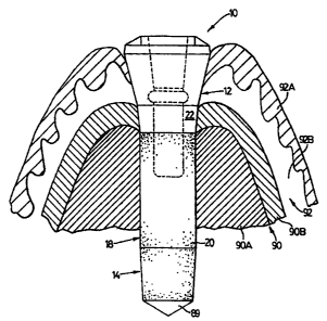
Once the proper implant length has been selected it is
then necessary to prepare the gingiva 92 and bone 90 for
the implant 10 (see Figure 7). This is accomplished by,
after adequate anaesthetic of the patient, drilling a
single recess 89 into the installation site. It is noted
that the bone 90 of the installation site is generally
composed of a soft cancellous portion 90A and a hard
cortical portion 90B, and the gingiva 92 is generally
composed of a gum 92A and a sub-gum or derma 92B.
According to the present invention only a single final
drill bit 93 need be used as shown in Figure 9. The drill
bit 93 has a tapered section 94 and a cylindrical section
95. It will be appreciated that the shape and dimension
of sections 94, 95 of the drill bit should closely
correspond to 'that of the selected implant 10. However,
it is preferable for the tapered section 94 to be just
slightly longer than the carresponding implant, for
example by .03in. (0.75 m.m.), for ease of fit.
The cylindrical section 94 has opposed cutting edges
96 (only one of which is shown). A tapered coronal
portion 97 has one or more discrete, protruding cutting
teeth 98 thereon. The taper on the coronal portion 97 may
be referred to as the "third taper". Reference numeral 99
indicates another set of cutting edges on the tapered
section 94 (which has three or four sets of such edges, as
desired). A smaller diameter upper portion 100 of the
2
- 11 -
drill 93 has a keyway adapter 101 at its top end for
engaging a drive mechanism of a dentist's drill (not
shown). A hollow stem or bore 102 extends through the
drill 93 along its length, as shown. A saline coolant is
delivered from the drill to an opening 103 through the
bore 102 to cool the drill bit and prevent heat build up,
which heat could damage the living cells being drilled
into.
The use of the single final drill bit 93 is preceded
by the use of a conventional smaller pilot drill (not
shown). The pilot drill also has a hollow stem or bore to
allow internal irrigation with normal saline solution.
Additionally, external irrigation maybe used (for both
drills) to cool the site being drilled to prevent damage
to the bone tissue during the site preparation.
The drill 93 of the present invention differs from
prior art systems which use a parallel sided final drill,
either with a hollow stem or with a solid stem (which
permits external irrigation only). The tapered portion of
the surgical site is prepared after, by a separate drill,
thus allowing no control over the length of the midsection
portion of the bone hole.
Once the recess 89 in the bone has been drilled, it
will have a tapered apical portion. Referring back to
Figure 7, an implant 10 according to the present invention
is shown being installed into the recess 89 in the bone 90
and the gingiva 92 after the bone has been drilled. As
can be seen, the lower section of the recess 89 tapers
slightly, preferably in an amount less than 5°, over the
depth of .12 in. (3.00 m.m.).
Figure 8 shows the implant in the fully installed
position. To attain this position it is necessary to
press the implant into the bone 90, by reason of the
interference fit between the bone 90 and the implant 10.
This is preferably accomplished by gently tapping the
coronal portion 12 of the implant to firmly seat the
implant into the bone. Thereafter the healing cap 70 is
2~.0'~~'~2
- 12 -
installed and the patient is allowed to leave. A suitable
period of a few months is allowed to elapse and then the
patient can return for installation of an appropriate
abutment portion. Those skilled in the art will
appreciate that the two styles of abutment portions
provided according to the present invention are
conventional above the gum line and thus will be readily
understood and used by practitioners.
At this point an important aspect of the invention can
be better appreciated, namely how the implant 10 of the
present invention and the bone 90 interact.' When the
implant 10 is tapped into the recess 89 as shown in Figure
8, the smooth-walled (i.e. emergent) portion 22 causes the
implant to initially be wedged into the hard cortical
portion 90B of the jaw bone 90 to stabilize the implant in
the recess 89. This wedging arises because of the taper
of emergent portion 22 and because the diameter of the
emergent portion 22 is slightly larger than the diameter
of the recess, and is not due to any surface effects
between the cortical bone 90B and the smooth biocompatible
surface on emergent portion 22. During the ensuing
months, the soft cancellous portion 90A of the jaw bone
gradually grows into the coating 20 and attaches to the
cylindrical section 18 of the implant. In the meantime,
the cortical portion 90B slowly subsides away from the
smooth biocompatible surface of the emergent portion 22.
Hence, the biocompatible surface of the emergent portion
22 only temporarily helps hold the implant 10 in place
while the cancellous portion 90A bonds to the cylindrical
section 18. It is believed that the stress induced into
(i.e. the displacement of) the cortical portion 90B by the
taper of the emergent portion 22 is insufficient to
prevent the cortical portion 90B from subsiding and
remoulding away from the implant.
It will be appreciated by those skilled in the
art that the foregoing description is in respect of
preferred embodiments and that various modifications can
- 13 -
be made to the invention without departing from the broad
scope of the appended claims. Some of these modifications
have been suggested above, and others will be apparent to
those skilled in the art. Fox example, the head portion
44, 54 may be angled relative to the key portion 42, 52,
respectively, to allow the location of the implant to be
varied based on site conditions without displacing the
overdenture or bridge. The angle by which the head
portion and the key portion may be angled is between 15° -
35°. Also, the precise dimensions may be modified without
changing the operation of the invention. And finally,
while the invention has been described in association with
overdentures and bridges, it will be appreciated that it
can be used to secure other cosmetic devices, such as
artificial ears or eyes.