Note: Descriptions are shown in the official language in which they were submitted.
WO 92/2129l PGT/US92/04392
2I~~937
A _ .~
_1_
APPARATUS AND METHOD FOR PERITONEAL RETRACTION
BACKGROUND OF THE INVENTION
The present invention relates to an apparatus
and method for mechanically lifting the abdominal
wall away from the underlying abdominal organs during
laparoscopic procedures. In its more specific
aspects, the invention is concerned With such an
apparatus and method wherein the abdominal wall is
lifted internally by a mechanical device which is
introduced blindly or laparoscopically and, once in
place, expanded to engage an extensive area of the
abdominal wall. The invention is also concerned with
an apparatus and method for draping the abdominal
organs and displacing a particular organ, such as the
gallbladder, for treatment.
Laparoscopy dates back to the turn of the 20th
Century. Early laparoscopic techniques were used
primarily for diagnostic purposes to view the
internal organs, Without the necessity of
conventional surgery. Since the 1930s, laparoscopy
has been used for sterilization and, more recently,
for the suturing of hernias. U.S. Patents 4,919,152
and 4,944,443 are concerned with techniques of the
latter type. Another very recent innovation is the
use of laparoscopic surgery for removal of the
gallbladder.
CA 02109937 1999-04-13
- 2 -
The concept of using mechanical retraction schemes to
lift the abdominal wall away from the underlying abdominal
organs during laparoscopic procedures is new to the present
invention. Procedures presently use carbon dioxide
insufflation to tent up the interior of the abdominal wall.
This requires gas seals to be present at all entry ports
through the abdominal wall; and because of the doming effect
of insufflation, the laparoscopic instruments (graspers,
scissors, electrocautery instruments, etc.) need long shafts
(on the order of 12 to 13 inches) to reach the treatment site.
Such instruments are difficult to control and result in
exaggerated movements during instrument application.
In United States Patent No. 3,774,596, Cook shows an
arrangement of flat, inflatable panels stacked in a collapsed
state on a base. The apices of the inflatable panels are
joined using longitudinal rods. After insertion into a body
cavity through an existing orifice, the inflatable panels are
inflated. This causes them to unfurl from the base and form a
hollow polygonal structure that permits inspection and
treatment of the body cavity. Windows can be formed in the
inflatable panels.
Cook's apparatus is unsuitable for lifting the abdominal
wall during laparoscopic surgery because its polygonal
arrangement of flat inflatable panels is incapable of
generating the force required to lift the abdominal wall
unless very high inflation pressures are used. This, in turn,
requires using a thick material for the flat, inflatable
66810-739
CA 02109937 1999-04-13
- 3 -
panels, which makes the device too bulky for the device to be
usable laparoscopically.
To retract the liver to gain access to treat the gall
bladder, published European Patent Application No. EP-A 0 246
086 shows a device with four fingers that extend through a
single laparoscopic puncture in the abdominal wall. After the
fingers have been inserted, a mechanism outside the abdominal
wall is operated to spread the fingers apart to expose the
gall bladder. This apparatus has the disadvantage that
additional incisions are required to insert the endoscope and
to insert instruments used for treating the gall bladder.
Moreover, the apparatus includes no provision for gripping the
gall bladder.
German Offenlegungeschrift No. 28 47 633 shows a balloon
catheter in which a balloon is carried on the distal end of a
hollow shaft. An additional lumen in the shaft provides a
passage for inflating the balloon once inside a body cavity.
Using the balloon catheter for extracting gall stones from the
bile duct is described. However, the structure shown is
incapable of piercing the gall bladder from outside.
66810-739
S
"~ r
SUMMARY OF THE INVENTION
The invention provides apparatus for lifting the
abdominal wall, said apparatus comprising: (a) a primary
balloon insertable through a small laparoscopic incision of
limited area in the abdominal wall, said balloon being
expansible from the exterior of the abdominal wall laterally
beyond the limited area of the laparoscopic incision to
interiorly engage an extensive area of the abdominal wall, and
being fabricated of a generally inelastic flexible material
shaped to expand laterally into engagement with an extended
area of the abdominal wall upon inflation; and (b) means to
selectively expand the balloon against the abdominal wall to
impart lifting force to the abdominal wall through the
balloon.
The various mechanical retraction schemes of the
present invention allow intraperitoneal placement via small
limited incisions or puncture sites. The abdomen does not
need to be sealed against gas leaks and doming up of the
abdominal wall is avoided. The abdominal wall is lifted by
means of either externally disposed posts or mechanical arms,
or by means of inflatable bags or balloons which are expanded
within the abdomen.
In practice, a small opening is formed in the
abdominal wall and the lifting device is inserted into the
abdomen through the opening in a contracted state. Once
within the abdomen, the device is extended to engage an
extensive area of the abdominal wall and the wall is lifted
with the device.
The apparatus additionally preferably comprises
lifting means for imparting lifting force to lift the
66810-739
CA 02109937 1999-04-13
- 4A -
abdominal wall through the abdominal wall engaging means.
In an apparatus according to the invention for
withdrawing the gall bladder from an abdominal cavity, the
apparatus comprises a compression balloon. The compression
balloon is laparoscopically insertable in a contracted state
into the abdominal cavity, and, upon inflation, compresses the
liver and displaces the gall bladder to provide access to the
gall bladder. The apparatus additionally comprises gripping
means, laparoscopically insertable into the abdominal cavity
externally of the compression balloon, for gripping the gall
bladder exposed by the compression balloon and for withdrawing
the gall bladder from the abdominal cavity.
The balloon may be transparent and viewing of the
procedure is provided by means of an endoscope disposed within
or passed through the balloon.
The invention also provides an apparatus for
laparoscopically gripping and removing the gall bladder from
the abdominal cavity. This apparatus comprises an elongate
tubular shaft carrying a balloon. The elongate tubular shaft
has a sharpened distal end for piercing the gall bladder. The
balloon is for insertion into the gall bladder and for
expansion into internal gripping engagement with the gall
bladder.
The apparatus may also include an opening in the shaft to
enable the contents of the gall bladder to be drawn into the
shaft.
66810-739
CA 02109937 1999-04-13
- 4B -
A principal object of the present invention is to provide
a peritoneal retraction system to lift the abdominal wall
without insufflation.
Another and related object of the invention is to provide
such a system which avoids the requirement of gas seals to be
present at a11 entry ports through the abdominal wall.
Still another object of the invention is to provide such
a system which avoids doming of the abdominal wall and the
requirement that the laparoscopic instruments be very long in
order to accommodate such doming.
Yet another object related to the latter object is to
enable laparoscopic surgery to be carried out with instruments
having relatively short shafts and to thus ease and increase
the control imparted to the instruments by the surgeon.
A further object of the invention is to provide a
peritoneal retraction system which drapes the
66810-739
WO 92/2129l PCT/US92/04392
-5-
abdominal organs and may serve to displace a
particular organ for treatment.
Another object of the invention is to provide a
peritoneal retraction system which is gentle and may
be controlled to effect the lifting of discreet areas
of the abdominal wall.
Yet another object of the invention is to
provide a peritoneal retraction system which employs
a balloon within or through which an endoscope may be
placed for viewing a laparoscopic operation to the
outside of the balloon.
Yet another and more specific object of the
invention is to provide a laparoscopic instrument
internally engagable with an organ to be treated to
distend and manipulate the organ or withdraw the
organ from the body.
Still another object related to the latter
object is to provide such an instrument which may be
used to withdraw the contents of the organ prior to
its removal.
Yet another object of the invention is to
provide a system of lifting the abdominal wall for
peritoneal retraction which avoids ~induly tensioning
body tissue.
Another general object of the invention is to
provide such a system which avoids gas leaks and the
need for trocar valves.
WO 92/21291 PCT/US92/04392
-6-
These and other objects will become more
apparent when viewed in light of the following
detailed description and accompanying drawings.
BRIEF DESCRIPTION OF THE DRAWINGS
Fig. 1 is a transverse cross-sectional
elevational view of a body, showing a first
embodiment of the invention in the process of lifting
the abdominal wall;
Fig. 2 is a transverse cross-sectional
elevational view of a body, showing a second
embodiment of the invention in the process of being
placed for lifting of the abdominal wall;
Fig. 3 is a transverse cross-sectional view
similar to Fig. 2, showing the second embodiment in
the process of lifting the abdominal wall;
Fig. 4 is a transverse cross-sectional
elevational view of a body, showing a third
embodiment of the invention in the process of being
placed for lifting of the abdominal wall;
Fig. 5 is a transverse cross-sectional view
similar to Fig. 4, showing the third embodiment in
the process of lifting the abdominal wall;
Fig. 6 is a transverse cross-sectional view of a
body, showing a fourth embodiment of the invention in
the process of being placed for lifting of the
abdominal wall;
WO 92/21291 PCT/US92/04392
2~Q993'~
_,_
Fig. 7 is a transverse cross-sectional view
similar to Fig. 6, showing the fourth embodiment in
the process of lifting the abdominal wall;
Fig. 8 is a perspective view of the lifting
device of a fifth embodiment of the invention, with
solid lines showing the device in contracted
condition and phantom lines showing the device in the
expanded condition;
Fig. 9 is a perspective view of a body in the
process of having the fifth embodiment lifting device
inserted into place in the contracted condition, with
the abdominal wall broken away for purposes of
illustration;
Fig. l0 is a perspective view similar to Fig. 9,
showing the lifting device of the fifth embodiment in
the process of being expanded;
Fig. il is a transverse cross-sectional
elevational view of a body showing the fifth
embodiment of the invention in the process of lifting
the abdominal wall;
Fig. 12 is a perspective view of the lifting
device of a sixth embodiment of the invention, with
the device shown in contracted condition and the
balloons therein deflated;
Fig. 13 is a perspective view of the lifting
device of the sixth embodiment, with the device shown
in expanded condition and the balloons inflated;
WO 92/21291 PCT/US92/04392
A0993'
_8_
Figs. 14 to 19 are perspective views
sequentially illustrating the steps of inserting the
fifth embodiment lifting device into place within the
abdominal cavity and expanding the device for
engagement with the abdominal wall, with part of the
abdominal wall broken away for purposes of
illustration;
Fig. 20 is a transverse cross-sectional
elevational view of a body, showing a modified
IO version of the fifth embodiment lifting device in the
process of lifting the abdominal wall, wherein the
device is shown illuminating the area beneath the
wall;
Fig. 21 is a perspective view of another
modified version of the fifth embodiment lifting
device wherein the distal ends of the expansible
elements are provided with balloons to shield them
against snagging on body tissue;
Fig. 22 is a transverse cross-sectional
elevational view of a body showing yet another
modified version of the fifth embodiment device
wherein the expansible legs of the device carry a
lifting balloon;
Fig. 23 is a transverse cross-sectional
elevational view of a body showing a seventh
embodiment of the invention in the process of lifting
the abdominal wall;
WO 92/2129l PCT/US92/04392
~1~J9~~1
_g_
Fig. 24 is a perspective view of the lifting
device of an eighth embodiment of the invention
wherein the device takes the form of a single
inflatable toroidal balloon;
Fig. 25 is a perspective view of the lifting
device of a ninth embodiment of the invention wherein
the device takes the form of three superimposed
inflatable toroidal balloons;
Fig. 26 is a transverse cross-sectional view of
a body showing the lifting device of the ninth
embodiment of the invention in the process of lifting
the abdominal wall;
Fig. 27 is a perspective view of the lifting
device of a tenth embodiment of the invention wherein
the device takes the form of a tubular rod having
balloons at its distal ends, with the balloons shown
in the contracted state;
Fig. 28 is a transverse cross-sectional view of
a body, showing the lifting device of tenth
embodiment in the process of lifting the abdominal
wall;
Fig. 29 is a perspective view of the lifting
device of an eleventh embodiment of the invention,
wherein the device takes the form of three
superimposed toroidal balloons with a draping
membrane extending across the lowermost balloon and a
WO 92/2129l PCT/US92/04392
'"
-10-
centrally disposed expansion balloon disposed within
the two uppermost toroidal balloons;
Figs. 30, 31 and 32 are transverse cross-
sectional elevational views of a body, sequentially
illustrating the eleventh embodiment lifting device
in the process of being inserted into the abdominal
cavity above the intestines and inflated to lift the
abdominal wall and drape the intestines;
Fig. 33 is a cross-sectional elevational view
similar to Fig. 32, illustrating a pair of
laparoscopic instruments extended through the center
balloon and draping membrane of the eleventh
embodiment lifting device to surgically treat the
intestines;
Fig. 34 is a perspective view of the lifting
device of the twelfth embodiment of the invention
wherein the device takes the form of a single U-
shaped balloon having a draping member secured
thereacross;
Fig. 35 is a perspective view of a modified
version of the lifting device for the twelfth
embodiment wherein the device takes the form of a
single U-shaped balloon having a draping member
secured thereacross, with the walls of the balloon
being tacked together at discreet locations;
Fig. 36 is a perspective view of the lifting
device of a thirteenth embodiment of the invention
WO 92/2129l PCT/US92/04392
210~~3'~
-11-
shown in place within the abdominal cavity of a body,
with parts broken away and shown in section to
illustrate the device in treating relationship to the
intestines;
Fig. 37 is a perspective view of a lifting
device of a fourteenth embodiment of the invention
shown in place within the abdominal cavity of a body,
with parts broken away and shown in section to
illustrate the device displacing the liver for
exposure of the gallbladder;
Fig 38 is a transverse cross-sectional
elevational view of a body showing a fifteenth
embodiment of the invention in the process of lifting
the abdominal wall and displacing the liirer for
exposure of the gallbladder, with an endoscope shown
in place within the lifting device;
Fig. 39 is a transverse cross-sectional view
similar to Fig. 38, illustrating a modified version
of the fifteenth embodiment wherein a secondary
balloon is positioned above the primary balloon to
shield the primary balloon from a trocar being
extended through the abdominal wall;
Fig. 40 is a perspective view of the lifting
device of the fifteenth embodiment shown in place
Within the abdominal cavity of a body, with parts
broken away for illustration and laparoscopic forceps
WO 92/2129I PCT/US92/04392
21U9937
-12-
extended into gripping engagement with the
gallbladder;
Figs. 41, 42 and 43 are perspective views of the
lifting device of the fifteenth embodiment of the
invention in place within the abdominal cavity of a
body, sequentially illustrating the steps of
inserting the gallbladder distension and manipulation
device of the invention laparoscopically into the
abdominal cavity and into gripping engagement with
the gallbladder;
Fig. 44 is a cross-sectional view showing the
gallbladder distension and manipulation device of
Figs. 41 to 43 in inflated condition within the
gallbladder;
Fig. 45 is a cross-sectional view showing a
modified version of the device of Fig. 44 wherein the
device has a single lumen for inflation of the
balloon and no lumen for withdrawal of the contents
of the gallbladder; and
Fig. 46 is a transverse cross-sectional view
similar to Fig. 38, showing a modified version of the
fifteenth embodiment wherein the endoscope extends
fully through the balloon of the lifting device.
DETAILED DESCRIPTION OF THE INVENTION
In those embodiments of the present invention
which employ balloons, the balloon material should be
WO 92/2129l PCT/US92/04392
214993'i
-13-
relatively inelastic and tough. Examples of such
material are Mylar, Polyethylene and Polyurethane.
The thickness of the balloon wall is typically from
.5 to 5 mils.
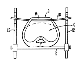
Referring now to Fig. 1, a body is designated
therein in its entirety by the letter "H" and is
shown having an abdominal cavity "C" with an upper
wall "W". The solid lines illustrate the wall in the
retracted elevated condition. The phantom lines
depict the position the wall would assume when
relaxed.
The lifting device of the first embodiment (Fig.
1) comprises a stiff transverse bar 10 passed through
a puncture site below the costal margin on one side
of the body and out another puncture site below the
costal margin on the other side. The puncture sites
are placed as far laterally as possible; close to the
anterior axiallary line on both sides. The bar is
then lifted and placed on slotted posts 12 secured to
both sides of the operating table 14. The placement
of the bar below the costal margin places the maximum
lift at the site of the gallbladder, for
cholecystectomy procedures. The bar may be placed
through puncture sites located more inferiorly for
other procedures. A second transverse bar may also
be used to define an entire plane of lift, as four
puncture sites are then made in the abdominal wall.
WO 92/21291 PCT/US92/04392
~~~993'7
-14-
Alternatively, instead of using a rigid bar, a cable
may be passed through the abdominal wall and variable
tension applied to the cable to yield different
degrees of retraction.
In the second embodiment (Fig. 2) a stiff bar 16
is passed into the abdominal cavity through one side.
A small puncture is then made at the mid-line of the
abdominal wall and the cable loop 18 is then passed
into the abdominal cavity. The bar 16 is passed
through the loop and the loop is then pulled up (Fig.
3) to achieve retraction. Directional control of the
bar is maintained by the portion of the bar that
remains outside the abdomen.
The lifting device of the third embodiment
(Figs. 4 and 5) comprises a rigid rod 22 having a
lateral offset 24 of a generally rectangular shape.
The rod 22 is threaded through entry and exit
puncture sites in the abdominal wall and then rotated
and clamped down to provide retraction, as shown in
Fig. 5. Slotted posts 12 support the proximal and
distal ends of the rod. A crank 26 is engaged with
one of the ends of the rod to rotate it to the
retraction position.
The fourth embodiment (Figs. 6 and 7) is
essentially a variation of the third embodiment. In
the fourth embodiment, the rod 28 is fabricated of a
shape-memory metal, such as NITINOL'~ which is
CA 02109937 1999-04-13
- 15 -
straight when cool and assumes a shape with a laterally offset
central portion 30 when heated. An electrode (not
illustrated) embedded into the rod is used to heat the rod at
the transition site, causing the rod to convert to a
rectangular shape for retraction, as seen in Fig. 7.
The rod 28 is passed through the abdomen at puncture
sites located at the costal margins, similarly to the Fig. 1
embodiment. Once in place, the proximal and distal ends of
the rod are engaged on slotted posts 12. A heater 32 (see
Fig. 7) is then activated to increase the rod temperature at
the transition site, resulting in retraction of the abdominal
wall.
The fifth embodiment shown in Figs. 8 to 11 comprises a
pair of angle-shaped rigid members 34 and 36 having
intermediate sections 38 and 40 extending in generally
parallel relationship to one another and rotatably received
within a sleeve 42. The proximal ends of the members 34 and
36 are formed with operating levers 44 and 46, respectively.
The distal ends of the members 34 and 36 are provided with
elongate arms 48 and 50.
In use of the fifth embodiment, an incision 52 is cut
into the abdominal wall and the arms 48 and 50 are extended
through the incision while in the contracted condition shown
in Figs. 8 and 9. The levers 44 and 46 are then moved toward
one another and held together to fan the arms outwardly
beneath the abdominal wall, as shown in Fig. 10. The
66810-739
CA 02109937 1999-04-13
- 16 -
abdominal wall may then be lifted as shown in Fig. 11 to
retract the abdomen.
The sixth embodiment shown in Figs. 12 and 13 differs
from the fifth embodiment only in that the arms, designated
48a and 50a carry elongate balloons 54 and 56, respectively,
and that the angle-shaped members are tubular to provide for
the conduit of inflation gas to these balloons. The elements
of the Figs. 12 and 13 embodiments corresponding to those of
the embodiments shown in Figs. 8 to 10 are designed by like
numerals, followed by the subscript "a" as follows:
intermediate sections 38a and 40a; sleeve 42a; and arms 44a
and 46a. Flexible conduits 56 and 58 are secured to the
levers 44a and 46a to provide for the conduit of gas thereto
to inflate the balloons 54 and 56.
The sixth embodiment is introduced into the abdominal
cavity and used for retraction in generally the same manner
depicted in Figs. 9 to 11, with the exception that after the
arms 48a and 50a are fanned out, the balloons 54 and 56 would
be inflated, as shown in Fig. 13. This expands and cushions
the area of contact between the arms and abdominal wall.
66810-739
WO 92/2129l PGT/US92/04392
-17-
Figs. 14 to 19 illustrate the preferred sequence
for forming the incision 52 and introducing the fifth
or sixth embodiment into the abdominal cavity. In
Fig. 14, a Veress needle with a thin plastic sheath
60 forms a puncture in the abdominal wall and enters
the abdominal cavity. The Veress needle is then
withdrawn, leaving the sheath 60 in place as shown in
Fig. 15 and the guidewire 62 is threaded through the
sheath and into the abdominal cavity. A small
incision 52 (.5 cm) is then made along the sheath
adjacent the guidewire and the sheath is removed,
leaving the guidewire in place as shown in Fig. 16.
A dilator 64 having a plastic guide sheath 66
thereover is then advanced over the guidewire and
into the abdomen as shown in Fig. 17 and then the
dilator and guidewire are removed, leaving the sheath
in place as shown in Fig. 18. The dilator may have a
fiberoptic scope to ensure that no bowel loops are
impacted by the sheath during its placement. The
lifting device or retractor is then introduced into
the abdominal cavity through the guide sheath, as
shown in Fig. 18, with the sheath protecting
abdominal organs from trauma during. insertion.
Thereafter, the arms 48 and 50 are fanned out to
provide expanded engagement with the inside of the
abdominal wall, as shown in Fig. 19. There it will
also be seen that a mechanical arm 67 is being
WO 92/2l291 PCT/US92/04392
21~ ~3'~ ~ ~~ ~~~
engaged With the levers 44 and 46 to impart lifting
force thereto and, in turn, retract the abdominal
wall. The arm 67 has a distal section 69a and a
proximal section 69b connected by a lockable swivel
5~ 7I. The proximal section is supported on a motorized
worm gear actuator 73 mounted on the side of the
operating table, designated 75.
The modified version of the fifth embodiment
shown in Fig. 20 corresponds in structure and mode of
operation to that of Figs. 8 to li, except that
lifting arms 48b and 50b incorporate fiberoptic means
to illuminate the abdominal cavity. Fig. 20
diagrammatically shows light bulbs 68 at the proximal
ends of the levers 44b and 46b to provide a light
source for the fiberoptic illuminators.
The modified fifth embodiment version of Fig. 21
differs from that of Figs. 8 to 11 only in that the
angle-shaped members 34~ and 36~ are tubular and
provided with inflatable balloons 70 and 72 at the
distal ends of the arms 48~ and 50~. These balloons
serve to shield the internal body organs from the
ends of the arms. In use, the balloons would be in a
contracted deflated condition during introduction of
the Fig. 21 lifting device into the abdominal cavity.
Once in place within the cavity, the balloons would
be inflated through the tubular angle members and the
WO 92/21291 ~PGT/US92/04392
2~~~~~~'~t
-19-
arms 3~c and 36c would be spread to fan out beneath
the abdominal wall.
The seventh embodiment lifting device of Fig. 23
comprises a balloon 74 secured to the distal end of a
tubular lifting rod 76. In use, the rod 76 with the
balloon in deflated condition wrapped closely
therearound is introduced into the abdominal cavity
through a small incision. The balloon is then
inflated through the rod to expand into extended
contact with the abdominal wall, as shown in Fig. 23.
A mechanical arm is then used to raise the rod 76 as
depicted by the arrow line in Fig. 23 and, in turn,
retract the abdominal wall.
The eighth embodiment lifting device shown in
Fig. 24 simply comprises a toroidal balloon 78 having
an inflation conduit 80 secured in fluid
communication therewith. This balloon is deflated
and tightly contracted for introduction into the
abdominal cavity through a small incision. Once in
place, it is inflated to expand into extended
engagement with the abdominal wall and lift the wall,
similar to the depiction of the ninth embodiment
shown in Fig. 26.
The ninth embodiment illustrated in Figs. 25 and
26 differs from the eighth embodiment of Fig. 24
primarily in that the lifting device comprises three
superimposed toroidal balloons 82, 8~ and 86 having
WO 92/2129l PCT/US92/04392
~1Q9~~7
-2 0-
inflation conduits 88 secured in fluid communication
therewith. In use, the eighth embodiment is deflated
and collapsed to a tightly wound condition for
introduction into the abdominal cavity through a
small incision. Once in place, the balloons 82, 84
and 86 are inflated, as shown in Fig. 26, to retract
the abdominal wall. From the latter figure it will
be seen that the lowermost toroidal balloon 82 rests
on the stomach 90 and the liver 92 retraction or
lifting force results from expansion of the
superimposed balloon elements within the abdominal
cavity so as to assume a condition in compression
between the abdominal wall and the organs
therebeneath. No external lifting device, such as
that of the aforedescribed embodiments, is required
for the eighth and ninth embodiments.
,;..:
The lifting device of the tenth embodiment
depicted in Figs. 27 and 28 comprises a tubular rod
94 having balloons 96 and 98 secured in sealed fluid
communication with the distal ends thereof. An
inflation conduit 100 is secured in sealed fluid
communication With the rod to provide inflation gas
for the balloons. The balloons are proportioned to
assume an ovaloid expanded condition upon inflation,
as seen in Fig. 28.
In use, the tenth embodiment lifting device is
introduced into the abdominal cavity through a small
WO 92/2129l PGT/US92/04392
2~~~9~7
-21-
incision and then maneuvered to dispose the rod 94 in
a generally horizontal condition as shown in Fig. 28.
The conduit i00 is extended through the incision in
the abdominal wall and extends to a suitable source
of fluid pressure. Once in place within the
abdominal cavity, the balloons 96 and 98 are inflated
to spread and lift the abdominal wall as shown in
Fig. 28.
The eleventh embodiment depicted in Figs. 29 to
l0 33 is similar to the ninth embodiment, with the
addition that it is provided with a centrally
disposed secondary balloon 102 and a draping membrane
i04. The superimposed toroidal balloons of the
eleventh embodiment and the inflation conduits
therefor are designed by numerals corresponding to
those of the ninth embodiment, followed by the
subscript "a" as follows: 82a; 84a; 86a; and 88a.
In use, the eleventh embodiment is collapsed as
shown in Fig. 30 and introduced into the abdominal
cavity through a small incision. Once within the
cavity, the balloon i02 is inflated through an
.inflation conduit t06 therefor which extends through
the incision. Inflation of the balloon i02 functions
to laterally expand the toroidal balloons as depicted
in Fig. 31. Thereafter, the toroidal balloons are
inflated through the conduits 88a to effect lifting
WO 92/2129l PCT/US92/04392
~,~~~g~7
-22-
and retraction of the abdominal wall, as shown i~
Fig. 32. There it will be seen that the lowermost
toroidal balloon 82i and the membrane i04 rest on the
intestines i08.
Fig. 33 shows how laparoscopic operating tools
may be extended through the abdominal wall and the
central passage provided by the toroidal balloons
82a, 84a and 86a. As there illustrated, it will be
seen that the balloon 102 has been fractured and that
an opening il0 has been formed in the membrane i04.
Notwithstanding that the opening 1i0 interrupts the
continuity of the membrane and provides for the
access of the intestine therethrough, the part of the
membrane which remains intact continues to drape over
and shield that area of the intestine which is not to
be treated.
The twelfth embodiment lifting device shown in
Fig. 34 is essentially a variation of the eleventh
embodiment device wherein, rather than employing
three superimposed balloons with a secondary lateral
expansion balloon, the lifting device comprises a
single U-shaped balloon 112 having a draping membrane
114 secured thereacross. In use, the twelfth
embodiment balloon would be introduced into the
abdominal cavity and inflated in much the same manner
as the eleventh embodiment balloon, with the
WO 92/2129l PCT/US92/04392
21e937
-23-
exception that no secondary central expansion balloon
would be provided. The balloon i12 would be
selectively inflated through means of a conduit i15
which communicates therewith and extends through a
small incision in the abdominal wall. The membrane
1i4 would serve to drape and shield the internal body
organs. Phantom lines 1i6 depict how an opening
might be formed through the membrane to provide
access to the organs therebeneath, while the membrane
continues to drape and shield the organs Which are
not to be treated.
The lifting device of Fig. 35 corresponds to
that of Fig. 34, with the exception that the U-shaped
balloon has tacked sidewalls to provide an extended
balloon height upon inflation. The parts of the Fig.
35 corresponding to those of the Fig. 34 device are
designated by like numerals, followed by the
subscript "a" as follows: balloon 112a; membrane
114a; conduit 115a; and phantom line opening 116a.
The thirteenth embodiment device shown in Fig.
36 is essentially the same as the embodiment of Fig.
35, with the exception that the U-shaped balloon is
comprised of three superimposed U-shaped balloons,
rather than a single balloon with tacked walls. As
there shown, the three balloons are designated by the
numerals 118, 120 and i22 and a draping membrane i24
WO 92/21291 PCT/US92/04392
2~09~~7
-24-
is secured across the lowermost balloon 118. It
should be appreciated that the lifting device of Fig.
36 would be introduced into the abdominal cavity in
deflated condition through a small incision. Once in
place, the balloons ii8, i20 and i22 would be
inflated through means of a conduit extending through
the incision in the cavity. Rather than cutting the
membrane 124 during the surgery, the membrane is
preformed with an opening 126 which provides access
to the area of the organ to be treated.
The fourteenth embodiment lifting device shown
in Fig. 37 has a configuration similar to that of the
Fig. 36 embodiment, with the exception that the
balloons are proportioned to rest on the stomach 90
while depressing the liver 92 for displacement and
exposure of the gallbladder 128. As shown in Fig.
37, the lifting device comprises superimposed U-
shaped balloons 1i8=, 120a and 122a, with a draping
membrane 124a secured across the lowermost balloon
118a. It should be appreciated that the device of
Fig. 37 would be introduced into the abdominal cavity
through a small incision in a deflated contracted
state and, once in place, selectively inflated to
lift the abdominal wall.
The fifteen embodiment depicted in Figs. 38 to
44 employs a transparent balloon 130 which serves as
WO 92/2129l PCT/US92/04392
-25-
the lifting device. The balloon is contracted and
introduced into the abdominal cavity through a small
incision 132. The neck of the balloon, designated
i34 is maintained in a condition extending through
the incision and provides both for the inflation of
the balloon and for the extension of an endoscope 136
into the balloon. The neck and the endoscope are so
proportioned as to provide a generally fluid tight
seal therebetween. As shown, the balloon i30
depresses the liver 92 to expose the gallbladder i28
for treatment and viewing through the endoscope i36.
Fig. 39 shows a secondary balloon 138 positioned
to shield the balloon 130 from puncture by a trocar
140. While this secondary balloon is optional, its
benefit is apparent where it become necessary to
pierce the abdominal wall after placement of the
balloon 130.
Fig. 40 shows forceps 142 laparoscopically
extended into gripping engagement with the
gallbladder 128. With the gallbladder so gripped, a
laparoscopic distension, manipulation and removal
tool 144 is extended into the abdominal cavity in
piercing engagement with the gallbladder. The tool
takes the form of a dual lumen tubular needle having
a sharpened open end 1~7 through which the contents
of the gallbladder may be drawn and an annular
WO 92/21291 PCT/US92/04392
A0993'7
-26-
balloon i46 which may be inflated through a lumen of
the tool communicating therewith (see Fig. 45).
Once the tool has evacuated the contents of the
gallbladder, the balloon i16 is inflated and assumes
internal gripping engagement with the gallbladder.
The tool may then be manipulated, thus maneuvering
the gallbladder within the abdominal cavity or
pulling it out of the abdominal cavity, as depicted
by the arrow line in Fig. 43. Depending upon the
size of the gallbladder, the removal of the organ may
require some enlargement of the incision through
which the tool extends. The forceps would be
released from the gallbladder to permit its
distension, manipulation or withdrawal from the
abdominal cavity. The entire procedure is viewed
through the endoscope 136.
The device of Fig. 45 corresponds to that of
Fig. 44, except that the tubular needle of the tool
144a has a single lumen only for inflation of the
balloon 146a and that the needle does not extend
fully through the balloon. Thus, the Fig. 45
embodiment cannot be used to evacuate the
gallbladder.
The embodiment of Fig. 46 corresponds to that of
Fig. 38, except that a tube i48 is sealed to and
extends fully through the balloon 130a to accommodate
WO 92/2129l PCT/US92/04392
~1~~93'~
-27-
extension of the endoscope i36 fully through the
balloon. With the Fig. 46 embodiment, the
gallbladder i28 is viewed directly, rather than
through the balloon.
CONCLUSION
From the foregoing description, it is believed
apparent that the present invention provides an
improved technique for retracting the abdominal wall
without insufflation. It also provides improved
operating techniques. It should be understood,
however, that the invention is not intended to be
limited to the specifics of the illustrated
embodiments, but rather is defined by the
accompanying claims.
x~ 8 ,~'~,