Note: Descriptions are shown in the official language in which they were submitted.
W O 93/05731 2 1 1 9 1 2 1 P(~r/US92/07762
CORNEAL lNLAY LENSES SMALLER THAN THE OPTIC ZONE
BACKGROUND OF THE INVENTION
Field of the Invention
This invention relates to implants designed to be
surgically inserted between the layers of the cornea to
correct refractive errors. More particularly, the
invention relates to corneal implants that can serve as
a substitute for conventional spectacles or contact
lenses.
Description of the Related Art
There have already been proposed artificial lenses
for implantation in the eye. Such implants have
hitherto been intended, not as corrective lenses, but
as a substitute for the natural lens of the eye. For
example, when an eye develops a cataract, the natural
lens becomes fogged or opaque, thereby impairing
vision. When such a cataract is treated, the lens is
removed, leaving the eye aphakic. Although it is
possible to correct for aphakia using spectacles, the
degree of correction requires spectacles so thick as to
make them both cumbersome and unattractive. For these
reasons, lenses have been designed for correction of
aphakia wherein the lens is inserted into the eye
during the operation to remove the cataract or at a
second operation. Such lenses are of fixed focal
length and, as the natural lens has been removed, the
WO93/05731 PCT/US92/07762
~ 2~9~2 ~ 2
eye is no longer capable of accommodation, that is to
say, the focal length cannot change to focus at
different distances.
It is clear from this description that such
previously known implanted lens could never be
prescribed as an alternative to conventional spectacles
for a person suffering only from myopia or presbyopia.
Another implant that has been used in the past
with some success has been the artificial cornea
described in U.S. patent no. 2,714,712, and generally
resembling what is known as a kerato-prosthesis. These
implants are designed as a replacement for the natural
cornea where the cornea has become fogged or opaque,
and are not intended to be a substitute for
conventional spectacles or contact lenses.
It is known to resort to surgery in order to
correct for defects in eyesight. The various
procedures for refractive corneal surgery to correct
vision problems such as myopia have not gained general
acceptance in ophthalmology. These include radial
keratotomy introduced in modern times (1972) by
Fyodorov of the USSR, keratomileusis introduced in 1961
by Barraguer of Columbia, keratophakia which uses
shaped donor corneas as lens, epikeratophakia which
uses an epigraft of homologous tissues, keratotomy to
correct astigmatism, and removing clear lens.
- Such surgery does not have a fully predictable
outcome, and furthermore any non-spherical flattening
of the cornea on healing results in an eyesight defect
that cannot be corrected by the use of spectacles or
contact lenses.
Disks of many different materials have been
inserted into corneal stromal pockets, initially to
control corneal edema, but more recently to correct
WO93/05731 PCT/US92/07762
2119121
- 3
refractive errors. Hydrogel and polysulfone lenses
have been more successful than other types of lenses so
far tried. Use of alloplastic corneal implants would
remove the need to rely upon autologous or homologous
material in refractive surgery. Corneal implants or
inlays are the subject matter of the present invention.
The cornea is a transparent avascular tissue about
10-12 mm in diameter. The cornea functions as a
protective membrane and as a "window" through which
light rays pass en route to the retina.
The average adult cornea is about 0.65 mm thick at
the periphery and about 0.54 mm thick in the center
(optic zone). From anterior (front) to the posterior
(back), it has 5 distinct layers: the epithelium which
is 5 or 6 cell layers thick; a clear acellular Bowman's
layer; the stroma (which constitutes about 90% of the
thickness of the cornea); the thin Descemet's membrane;
and, the single layer endothelium. Sources of
nutrition for the cornea are the blood vessels of the
limbus, the aqueous humor and tears. The superficial
cornea also gets most of its oxygen from the
atmosphere.
The zone in the cornea through which incident
light passes is known variously as the "optic zone'~ or
"pupillary aperture". The size of the normal pupil
varies at different ages and from person to person, but
normally is about 3-4 mm - smaller in infancy, tending
to be larger in childhood, and again progressively
smaller with advancing age.
Previous corneal implants have enjoyed only
limited success, in part because of the large diameter
of the lenses used and in part because of the
composition of such lenses. As will be detailed in the
review of the related art below, the ophthalmologically
WO93/05731 PCT/US92/07762
~ 9~ 4
more desirable high refractive index lenses previously
used prevent access of nutrients and gases such as
oxygen to the tissue anterior to the implant and to the
corneal tissue posterior to the implant. On the other
hand, high water content low refractive index lenses,
while reducing or eliminating the problem of nutrient
and gas transport, are not able to provide the
necessary corrections in refractive error of the eye.
Previous corneal implants have also not been able
to provide multifocal refractive correction.
The large diameter of previous corneal implant
lenses has also required a less-than-satisfactory
surgical approach to implantation. In general,
previous corneal inlays have required cutting a large
pocket into the cornea and inserting in this pocket the
lens which resides predominantly behind Bowman's
membrane. With this type of insertion, the large
implanted lens distorts the cornea, thereby producing a
change in optical power. The disadvantage of such a
procedure has been that the distortion is usually in
the posterior side of the cornea. Such posterior
distortion, however, produces only a very small change
in optical power because the difference between the
refractive index produced is only the small difference
between the inlay/cornea and the aqueous humor.
Choyce, D.P., U.S. patent number 4607617, issued
- August 26, 1986, discloses an implant designed to be
inserted between the layers of a cornea of an eye to
correct eyesight defects, comprising a polysulfone
plastic material of a high refractive index (typically
1.633), of a thickness in the range of 0.1 to 0.4 mm,
and capable of being sterilized by steam autoclaving
prior to insertion. As the implant is entirely
- embedded in the cornea, it is said not to be exposed to
W O 93/05731 21~912 1 P~r/US92/07762
the atmosphere or to the aqueous humor. The
polysulfone material is said to be ~relatively
permeable to body fluids", although it is not clear
that this is so. The lens is inserted by a procedure
comprising forming an incision in the outer layer of
the cornea, separating layers of the cornea to form a
pocket, inserting into this pocket a lens inlay, and
resealing the incision. Although this patent neither
discloses nor suggests a specific diameter for the lens
inlay, reference to Figure 7b of the specification
shows that this diameter is substantially greater than
the optic zone of the cornea, which normally is about 3
mm to 4 mm in diameter (Vaughan, D., et al., General
Ophthalmoloqy, 12th ed., Appleton & Lange, Norwalk, CN,
1989, Ch. 15). See also, Choyce, ~Polysulfone Corneal
Inlays to Correct Refractive Errors", Cataract, 7
(June, 1985). This fact, plus the fact that it is
known that high refractive index plastic inlay lenses
are poorly permeable to nutrient materials and
necessary gases such as oxygen, limits the usefulness
of this inlay lens. Further, this corneal inlay does
not provide multifocality.
Grendahl, D.T., U.S. patent no. 4,624,699, issued
November 25, 1986, discloses a corneal inlay for
implant made of a plastic material such as polysulfone
or PMMA. Recognizing that prior art polysulfone inlay
lenses exhibit a property of being poorly permeable to
nutrients, fluids and gases, a property of concern to
the medical community, the inventor attempts to
overcome these disadvantages of the prior art by
providing a corneal inlay with a plurality of holes or
slots for passage of nutrients through the cornea. The
inlay lens is said to have a diameter of approximately
3 mm to 7 mm, preferably of a diameter of 4.5 mm to 6.5
WO93/0~731
PCT/US92/07762
9~ 6
mm, more preferably slightly less than 6 mm in diameter
(column 2, lines 21-26). Inlay lenses of such diameter
will generally cover the optic zone of an adult cornea,
creating the problems of nutrient and gas supply
described above. There is no disclosure or suggestion
in this patent that the inlay lens could be smaller
than the opening of the optic zone, nor is there
reference to any property of the lens other than
monofocality.
Lindstrom, R.L., U.S. patent no. 4,851,003, issued
July 25, 1989, discloses corneal inlay lenses applied
under the cornea and about the stroma. The lens, which
can be made of biocompatible materials such a hydrogel,
or synthetic polymers, such as silicone, polysulfone,
polycarbonate, cellulose ester or other like materials
said to be gas and metabolite permeable, or
fenestrated, includes a plurality of fixation holes
around a periphery, and a coating on the anterial
surface by a material that enhances the growth of
corneal epithelial cells into and about said holes, the
coating being composed of biological materials such as
fibronectin, laminin, a glycosaminoglycan, or a type IV
collagen. Although the diameter of the inlay lenses is
not specifically disclosed, the dimensions of the holes
(up to 1 mm), taken together with Figure 6 which shows
the epicorneal lens implanted below the epithelium,
indicates that the diameter of the inlay lens must be
substantially greater than the optic zone of the
cornea; i.e., about 5 mm to 7 mm. Again, such lenses
do not provide a patient with multifocality.
Thus, the prior art inlay lenses are less than
satisfactory in important ways. Where large (e.g., 5
mm to 7 mm) hydrogel lenses are used, wherein the water
content is high (about 72%) and the index of refraction
WO93/05731 2 1 1 9 1 2 1 PCT/US92/07762
low (about 1.38), problems of permeability to nutrients
and gases are less severe, but the dioptic power is
low. Where large polymeric lenses are used, wherein
the water content is quite low and the refractive index
high (e.g., 1.45 to 1.633), the optic power is
satisfactory, but the permeability is poor. Such non-
permeability to essential nutrients and gases causes
~starvation in the anterior segments of the stroma,
ultimately resulting in extrusion of the inserted lens.
Although the permeability problem is reduced by placing
holes or slots in polymeric lenses (see Grendahl
above), such holes interfere with vision.
Further, none of the prior art inlay lenses
provide for multifocality, which is highly desirable in
many patients.
There remains, therefore, an important need for
intra-corneal lenses of a refractive index sufficiently
high so as to avoid the need to distort the cornea in
order to obtain the desired optical power, of a size
sufficiently small so as to simplify surgical
insertion, of a size and configuration that permits
essential nutrients and gases readily to reach the
anterior of the cornea, and of a type that permits
either unifocality or multifocality.
Such an intra-corneal inlay lens has been
invented, and it and its use are disclosed below.
SUMMARY OF THE INVENTION
The invention comprises a low or high refractive
index corneal inlay lens adapted to be inserted between
the layers of the cornea to correct defects in sight,
wherein the lens is of a size and configuration that
permits nutrients and gases to pass unimpeded from the
posterior aspect of the cornea through to the anterior
WO93/05731 PCT/US92/07762
~ i~9 1~ 1 8
aspect, and wherein the lens is of a composition
relative to that of the surrounding tissues such that
multi-refractive indices may be created and multi-focal
corrections are possible.
In accordance with a first aspect of the
invention, there is disclosed a corneal lens of a
diameter less than that of the corneal optic zone,
wherein the diameter of the lens is such that areas of
different refractive indices are created in the optic
zone, thereby providing multifocality.
In accordance with a second aspect of the
invention, there is disclosed a generally flat annular
ring-shaped corneal lens implant of a size and
configuration such that no barrier to nutrient passage
is present and of a composition such that the lens can
provide either unifocality or multifocality.
These and other aspects and objects of the
invention will become apparent by reference to the
specification below and the appended claims.
DESCRIPTION OF THE DRAWINGS
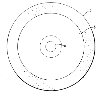
Figure l is a representation of the anatomic
relationship of the inlay lenses of the invention to
the cornea. A and B are, respectively, front and side
views of the inlaid lens and surrounding anatomical
features. Feature a is the sclera, b the cornea, c the
- location of the inlay of the invention, and d the point
of surgical incision.
DETAILED DESCRIPTION OF THE INVENTION
The invention comprises corneal implant corrective
lenses of novel dimension and configuration, adapted to
be surgically inserted into stromal pockets via a very
small incision in the corneas of patients suffering
W O 93/05731 2 1 1 9 1 2 1 P~r/U592/07762
from refractive error, the dimensions and configuration
of the lenses being such as not to impede the flow of
nutrients and gases through the layers of the cornea.
The surgical procedure involves making a stromal
cut parallel to the limbus of about 2mm in length to
approximately 75~ thickness, using a blunt spatula to
make a pocket in the stroma to the center of the
corneal optic zone (pupillary aperture), inserting the
corrective lens, then resealing the incision (Figure
10 1).
It is an important aspect of the invention that
the inlay lenses of the embodiments are either of a
diameter smaller than that of the optic zone of the
cornea or of a configuration so that the implanted
lenses, regardless of composition, water content or
index of refraction, are designed not to impede the
transport of fluids, nutrients and gases to all layers
of the cornea.
The invention relates to two embodiments. In one
embodiment, the inlay lenses are of a diameter
substantially smaller than that of the optic zone of a
normal cornea, e.g., about 1 mm to 2 mm. Such lenses
create regions of different refractive indices within
the optic zone, one created by the lens and the other
by the neighboring stroma tissue, thereby providing a
useful bifocal capability. The brain is capable of
sorting out the different signals and using the
information appropriately. This embodiment is not
limited to a single small diameter lens; a mosaic of
such lenses may be implanted in the same plane, thereby
providing for additional multifocality.
In another embodiment, which bears some
resemblance to the so-called "bull's eye' intraocular
lens, the diameter of the inlay lens may be that of the
WO93/05731 PCT/US92/07762
~,g~ 10
minimum pupillary aperture, i.e., about 3 mm to about 4
mm, or even larger, but the center of the lens
corresponding to the center of the optic zone is
drilled out so that the lens resembles an annulus or
washer in configuration. The hole in the center of the
lens ring permits unimpeded passage of nutrients and
gases through the corneal layers. Advantageously, the
lens and the adjacent stroma are of different
refractive index, thereby providing useful bifocal
capability. Thus, for a nearsighted individual, i.e.,
a myope, the hole in the center of the inlay permits
light to image on a portion of the optic zone of the
normal cornea, providing for near vision, while light
impinging on the peripherally located lens material is
refracted, thereby correcting for far vision.
Presbyopes will benefit from the multifocality of
the cornea which is generated by its central zone being
altered by the small lens of the first embodiment for
near vision, while the unaltered peripheral zone
remains responsible for distance vision. Myopic
patients can benefit in the reverse way by implanting a
negative lens in the center, rendering the small
central zone optically less powerful.
An enormous number of refractive corrections are
possible with the lenses of this invention. Positive
and negative lenses of all useful diopters may be
- employed. The lenses may be of a refractive index
greater or less than that of the neighboring corneal
tissue. Thus this invention can be applied to
presbyopes and myopes, possibly hyperopes and perhaps
other corrections as well.
As noted above, the lenses made in accordance with
this invention avoid the problems of nutrient and gas
passage attendant upon prior art corneal implant
11 2ll9l2l
lenses. Thus, the invention provides a great deal of
flexibility in the selection of lens composition,
refractive index and water content. For example, one
may use a hydrogel lens of low water content, a
diameter of about 2 mm, a center thickness of only
about 0.02 - 0.05 mm, an index of refraction (R.I.) of
1.42 to 1.43, and a power of +2.5 D in the stroma to
correct for presbyopia. High water content materials
of R.I. slightly greater than or less than the R.I. of
the stroma may also be used by an appropriate choice of
design. Also suitable are non-water containing
polymeric material such as the high R.I., relatively
rigid polysulfones (e.g., Udel~, Union Carbide Corp.,
R.I. typically 1.633) whose high R.I. allows
lS corrections of up to +10 D with a lens 0.04 mm thick,
and a correction of -10 D with a differently shaped
lens with a thickness of only 0.01 mm at its center,
polyethersulfones (Victrex~, ICI), polyarylsulfones,
Perspex CQ~ or Perspex CQUV (ICI) (R.I. 1.49),
polycar~onates, silicones, fluoropolymers, PMMA,
cellulose acetate butyrate, or other like materials.
The following examples are merely exemplary of the
invention and are in no way int~n~eA to limit the scope
of the invention which is defined by the specification
and the appended claims.
EXAMPLE 1
INSERTION OF A PMMA LENTICULE IN
-- THE CORNEA OF RABBITS' EYES
Physical Parameters:
Material: PMMA, Meniscus
Diameter: 2.0 mm; edge thickness: 0.02 mm; center
thic~ness: 0.022 mm; Base curve: 7.6 mm; power:
+2.SD.
WO93/05731 ~ l PCT/US92/07'62
1~-
terilization: gamma radiation 2.5 - 3 Mrad due to the
thinness of the lenticule, the slight
yellowing of the PMMA is negligible.
Implant Procedure:
1.1 Surgical Procedure
Perform a 2 mm incision approximately 75~ of
the stromal thickness about 1 mm central from
the limbus in clear cornea. Using a blunt
spatula, make a pocket to the center of the
cornea.
1.2 Intraoperative Drug Treatment
The resulting wound is then rinsed with
irrigating solution.
1.3 Lens Placement
Prior to placing the lens, several drops of
irrigating solution are placed on the eye.
The appropriate lens is poured into a wire
strainer and rinsed with sterile saline.
Several drops of irrigating solution are
placed on the lens. The lens is carefully
picked up with a non-toothed forceps and
inserted in the pocket. The lens is then
moved to the center of the cornea. Care must
be taken to ensure that the lens is well
centered.
1.4 Completion
Flush the eye well with irrigating solution.
Suture if necessary. Apply two (2) drops of
postoperative drug solution.
1.5 Postoperative Treatment
Give Maxidex 2X daily (weekend treatment is
once daily), and antibiotics as necessary.
EXAMPLE 2
INSERTION OF A HYDROGEL LENTICULE IN THE
CORNEA OF RABBITS' EYES
Physical Parameters:
Material: Hefilcon A; Meniscus
Water content: 45%; Refractive Index: 1.425
Diameter: 2.0 mm; edge thickness: 0.02 mm; center
thickness: 0.023 mm; Base curve: 7.6 mm; power:
+2.5D.
WO93/0~731
PCT/US92/07762
l321191~i
Sterilization method: Autoclaving
Implant Procedure:
2.1 Surgical Procedure
Perform a 2 mm incision approximately 75% of
the stromal thickness about 1 mm central from
the limbus in clear cornea. Using a blunt
- spatula, make a pocket to the center of the
cornea.
2.2 Intraoperative Drug Treatment
The resulting wound is then rinsed with
irrigating solution.
2.3 Lens Placement
Prior to placing the lens, several drops of
irrigating solution are placed on the eye.
The appropriate lens is poured into a wire
strainer and rinsed with sterile saline.
Several drops of irrigating solution are
placed on the lens. The lens is carefully
picked up with a non-toothed forceps and
- 20 inserted in the pocket. The lens is then
moved to the center of the cornea. Care must
be taken to ensure that the lens is well
centered.
2.4 Completion
Flush the eye well with irrigating solution.
Suture if necessary. Apply two (2) drops of
postoperative drug solution.
2.5 Postoperative Treatment
Give Maxidex 2X daily (weekend treatment is
once daily), and antibiotics as necessary.
EXAMPLE 3
INSERTION OF A HYDROGEL LENTICLE IN THE
CORNEA OF CATS' EYES
Physical Parameters:
Material: Hefilcon A; Biconvex
Water content: 45~; Refractive Index: 1.425
Diameter: 2.0 mm; edge thickness: 0.02 mm; center
thickness: 0.04 mm; Anterior radius: 7.0 mm;
Posterior radius: 9.8 mm; Power: +2.5D.
Sterilization method: Autoclaving
WO93/05731 PCT/US92/07762
~9~ 14
Implant Procedure:
3.l Surgical Procedure
Perform a 2 mm incision approximately 90% of
the stromal thickness about l mm central from
the limbus in clear cornea. Using a blunt
spatula, make a pocket to the center of the
cornea.
3.2 Intraoperative Drug Treatment
The resulting wound is then rinsed with
irrigating solution.
3.3 Lens Placement
Prior to placing the lens, several drops of
irrigating solution are placed on the eye.
The appropriate lens is poured into a wire
lS strainer and rinsed with sterile saline.
Several drops of irrigating solution are
placed on the lens. The lens is carefully
picked up with a non-toothed forceps and
inserted in the pocket. The lens is then
moved to the center of the cornea. Care must
be taken to ensure that the lens is well
centered.
3.4 Completion
Flush the eye well with irrigating solution.
Suture if necessary. Apply two (2) drops of
postoperative drug solution.
3.5 Postoperative Treatment
Give Maxidex 2X daily (weekend treatment is
once daily), and antibiotics as necessary.