Note: Descriptions are shown in the official language in which they were submitted.
WO93/21&~ PCT/US93/03849
~ q 3 ~
APPARATUS FOR SEALING
VASCULAR PUNCTURES
FIELD OF THE INVENTION
The present invention relates to an apparatus and
method for closing and sealing vascular punctures.
More particularly, the present invention relates to a
novel apparatus and method for sealing a vascular
puncture resulting from the use of a medical device,
catheter system or the like by using radio frequency or
other energy to effect closure and thermal fusing of a
puncture.
BACKGROUND OF THE INVENTION
Many medical procedures require access into the
vascular system of the patient. Although various means
may be used to obtain access into a vein or artery,
typically access is obtained by inserting a cannula or
introducer sheath through the skin and into the
selected blood vessel. A medical device or diagnostic
instrument, such as a guide wire, guiding catheter,
balloon angioplasty device, atherectomy device, or the
like is then inserted into the vascular system through
the cannula or introducer sheath.
In percutaneous transluminal coronary angioplasty,
for example, it is customary to introduce a catheter
into the femoral artery at an entry site in a patient's
leg and to advance the catheter through the artery to
the coronary region. The artery, which may be located
B
2134071
WO93/21&~ ~ PCT/US93/03~9
t
- 2 - _
one half inch or more beneath the skin, is punctured
with a needle or similar device. A guide wire is
inserted through the needle and the needle is removed.
An introducer sheath and dilator together are threaded
over the guide wire. The i--L~Gd~cer sheath is often
twisted and otherwise manipulated as it is inserted
into the vessel, thereby causing further enlargement of
the vascular puncture. The dilator is then removed and
the catheter is inserted.
To permit the insertion of a medical device or
instrument therethrough, the introAllc~r sheath must be
of a relatively large diameter. IllL-od~cer sheaths
typically have a diameter in the range between one
millimeter and six millimeters, thus creating a
significant puncture in the artery. After the
intravascular medical procedure is completed, this
puncture must be closed and bleeding from the blood
vessel stopped.
At present, such ble~ing is stopped by the
application of direct digital pressure over the
puncture site by a trained physician or other suitably
trained medical personnel. Such direct pressure must
be applied for a sufficiently long time for hemostasis
to occur so that the opening is effectively closed and
further bleeding is prevented. In the case of
punctures into the femoral artery, the pressure is
generally applied for twenty to thirty minutes, but it
may be n~cess~ry to apply pressure for as long as one
hour. Further, twelve pound sandbags may then be
placed on the puncture site for an additional two to
six hours.
The time required to stop bleeding using digital
pressure is not an efficient use of medical
professional services. Not only is this direct digital
pressure application procedure wasteful of time by
highly skilled medical professionals, the procedure
results in a substantial reduction, if not virtual
~093/21&~ 2 1 3 4 0 7 1 PCT/US93/03~9
~ - 3
- arrest, of blood flow through the vessel. Since
thrombosis is one of the major problems that can occur
in the immediate post-operative period, any reduction
in blood flow, caused by the application of digital
pressure, is undesirable. Furthermore, when digital
pressure is applied, an undesirable bruise or hematoma
can form at the entry site, since internal blPe~ing of
the punctured artery continues until clotting blocks
the puncture. There is also a significant chAnce that
upon movement by the patient, the puncture will reopen
and begin bleeding again, resulting in a hematoma or
other complications. In addition, when anticoagulants
used in the medical proceduLe are left active in the
body, the i"LLod~cer sheath is generally left inside
the patient for twelve to twenty four hours in order
for the anticoagulants to clear from the blood.
Because the patient may be required to remain immobile
and because of the risk of complications, patients are
usually required to remain overnight in the hospital
for observation, thus greatly increasing the cost of
the overall procedure.
One prior device for stopping bleeding from a
puncture in a blood vessel is a type of eYr~n~Ahle
plug. An example of such a device is shown in U.S.
Patent 4,890,612 (Kensey). The plug is pushed through
the puncture into the blood vessel and into the blood
stream. Once exposed to blood, it eYr~n~s. The
eYrAn~ed plug is then pulled back against the puncture
where, because of its ~Yr~n~ed size, it plugs the
orPning. A similar device is an PYr~n~Ahle closure,
such as that described in U.S. Patent 4,852,568
(Kensey). Such devices may work satisfactorily, but
require inserting and leaving a foreign object in the
vessel for a period of time. It is usually medically
preferable to avoid leaving objects in a vessel, even
if they eventually biodegrade.
2134071
W093/21&~ ! ~ PCT/US93/03~9
Another device for stopping bleeding from a
puncture is disclosed in U.S. Patent 4,929,246
(Sinofsky). This patent relates to a method for
closing an artery using laser energy while simultane-
ously applying pressure directly to the artery through
the use of a balloon placed on the exterior of the
artery over the puncture site.
SUMMARY OF THE lN Vh~ llON
An apparatus for closing and sealing a puncture at
a puncture site in a vessel located beneath the skin
using radio frequency or other energy to cauterize the
puncture has been developed. In one aspect, the
invention constitutes a probe sized to be
~e~ aneously inserted adjacent the vascular op~in~
and a conn~ctor for connecting the probe to an energy
supply source; the probe being configured to conduct
energy directly to tissue adjacent the probe to cause
heating of tissue s~oul.ding the vascular opening to
close the opening.
In another aspect, the apparatus comprises a
cautery device having a means for forcing together
biological tissue surro~ g a percutaneous vascular
puncture and at least one electrode connectable to a
radio frequency power source such that an electrical
~ el.L may flow through the tissue, thermally fusing
the tissue together.
In yet another aspect, the invention is an
apparatus for the percutaneous medical treatment of
biological tissue, comprising a plurality of electrodes
connectable to a radio frequency power source, the
electrodes adapted to engage biological tissue at
spaced points; and a lumen connected to the electrodes
for guiding the electrodes to the biological tissue at
said spaced points.
In one specifically disclosed emhoAiment, the
apparatus comprises a radio frequency cautery device
'~093/21&~ 2 1 3 ~ 0 7 1 PCT/US93/03~9
~._
- 5 -
having forceps adapted to grasp vascular tissue
~ul~o~nding the puncture site. The forceps, when
conn~cted to a radio fre~uency power source, also serve
as bipolar electrodes for fusing the tissue ~l.oul.ding
the puncture.
A backstop element, such as a balloon occluder
assembly or a T-shAr~~ occluder, may also be used in
conjunction with the cautery device. The balloon
occluder assembly essentially comprises a balloon at
the distal end of a balloon shaft and a means for
inflating the balloon. The balloon occluder assembly
temporarily occludes the puncture while providing a
backstop against which the forceps may grasp the
v~c~ r tissue. The balloon occluder assembly also
has utility separate from its use with the disclosed
cautery device, as ~;F~11C~0~ more fully hereafter.
In another aspect, the invention is a method of
~e~ g a vascular opening comprising the step of
delivering energy to the vA~clll~r wall, resulting in
local heating of bodily material external to the intima
layer of the vessel to achieve hemostasis without
substantially heating the intima layer of the vessel.
In yet another aspect, the method of the invention
comprises the steps of percutaneously inserting a probe
adjacent to the vascular opening; conducting energy
from the probe directly to tissue adjacent the probe in
an amount sufficient to cauterize the tissue to thereby
close the vascular or~n; ng; and removing the probe.
The invention in still another aspect is a method
of sealing a vascular puncture comprising the steps of
holding the vascular tissue ~ur~ounding the puncture
site in a contacting position and applying energy to
that tissue, the energy being sufficient to thermally
fuse the tissue together, thus sealing the puncture.
Preferably, this method of sealing a puncture includes
the steps of advancing a balloon into the lumen of a
vessel, inflating the balloon and withdrawing it to
21~071
WO93/21&~ PCT/US93/03~9
6 _
abut the puncture from within the vessel, inserting a
cautery device having forceps connected to a radio
frequency power source, grasping and bringing the
va~cular tissue into a contacting position, causing an
electrical current to flow from one forceps, through
the vascular tissue to the other forceps, thus
effecting a closure by thermally fusing the vA~c~lAr
tissue together.
In another aspect of the invention, a balloon
occluder need not be used. Instead, pressure is
applied to the vessel to restrict blood flow there-
through, an electrode is percutaneously inserted to a
position proximate the puncture site, and radio
frequency energy is used to cause thrombosis of the
blood to seal the puncture site.
The present invention thus provides an apparatus
which is simple to use and which overcomes the
disadvantages of the prior art, including the need for
the application of digital pressure for long periods of
time and the possibility of a substantial reduction of
blood flow through the vessel. The present invention
also provides methods that are effective for closing
off a puncture or other opening in a blood vessel by
using radio frequency or other energy to thermally fuse
the vascular tissue or form a seal by causing
thrombosis of the blood. The puncture is
hemostatically sealed almost immediately after the
medical procedure is performed, thus avoiding any
potential complications associated with re-opening of
the puncture or long hospital stays while antico-
agulants remain active in the body.
The forgoing has outlined rather broadly the
advantages of the present invention. Additional
benefits of the invention will be described herein-
after. These advantages, as well as the invention
itself, are more easily understood in view of the
'V093/21&~ 2 1 ~ ~ 0 7 1 PCT/US93/03~9
~, _
- 7 -
attached drawings, a brief description of which
follows.
B~TEF DESCRIPTION OF THE DR'~NGS
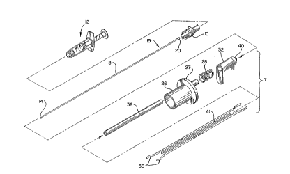
FIG. 1 is an exploded ~ ~ of the first preferred
apparatus emho~iment of the present invention.
FIG. 2 is an enlarged cross-sectional view of the
distal portion of the device of the first preferred
emho~l i ment .
FIG. 3 is an enlarged perspective view of the
distal end of a fO~c~a of the first preferred
embodiment.
FIG. 4 is an enlarged cross-sectional view of the
distal end of a fo ~e~a of the first preferred
embodiment.
FIG. 5 is an enlarged cross-sectional view of a
check valve assembly and hub used in conjunction with
the inflation means of the first preferred embodiment.
FIG. 6 through FIG. 8 illustrate alternate
emho~imentS of the actuating mec~Anism.
FIG. 9 through FIG. 18 are partial cross-sectional
views illustrating the method of using the first
preferred embodiment of the present invention.
FIG. 9A is a partial cross-sectional view taken
along line 9A-9A of FIG. 9 showing the relationship of
the arterial sheath to the femoral artery and
associated anatomy.
FIG. 15A is an enlarged cross-sectional view of
the region of FIG. 15 showing the various layers of the
vascular tissue being contacted by the electrodes.
FIG. 17A is an enlarged cross-sectional view of
the region of FIG. 17 where the seal is made.
FIG. l9A and l9B illustrate an alternate embodi-
ment of the backstop element of the present invention.
~ 1 ~ 4 ~ ~ 1
WO93/21&~ ;~ PCT/US93/03~9
- 8 -
DET~TT~n DESCRIPTION OF THE DRAWINGS AND PREFERRED
EMBODIMENTS OF THE lN V ~:N~l lON
Before describing the apparatus of the present
invention, a brief description of a typical intra-
vascular surgical procedure, e.g., catheter instru-
mentation of an artery using a percutaneous incision or
puncture, will be given to best appreciate the features
of the cautery apparatus of the present invention. In
such a pro el~e a c~nnlll A of an instrument, such as an
angiographic needle, is inserted percutaneously through
the skin and arterial sheath and into the artery. The
needle cannula of an instrument is held in place and
the flexible end of a guide wire is then passed through
the cannula into the artery to the desired depth (i.e.,
longitl1~inAl position therealong). Once the guidewire
is in place the needle cannula is removed leaving the
guidewire in place. A conventional i~ Gducer sheath
combined with an arterial dilator are then pA~ over
the guidewire through the puncture and into the artery.
The guidewire and the dilator are then removed leaving
the sheath in place. The catheter is then inserted
through the introducer sheath and threaded down the
artery to the desired intravascular location, e.g., the
situs of the atherosclerotic occlusion, usually the
coronary region. Once the intravascular procedure has
been completed, the catheter is removed. Thereafter,
once anticoagulants have been inactivated or cleared
from the body, the usual procedure has been to remove
the sheath and to have a surgeon or other trained
person apply digital pressure to the percutaneous
puncture until hemostasis has occurred. As noted
above, the stopping of bleeding from a puncture was
previously a difficult and time consuming task.
The apparatus of the present invention effects the
hemostatic closure of a percutaneous or other type of
puncture, incision or opening in a body vessel without
necessitating the application of digital pressure
'VO93/21&~ - 2 1 3 ~ 0 7 1 PCT/V593/03~9
_ g _
thereto. In accordance with the preferred embodiment
of the present invention, the introducer sheath is left
in place after the catheter is removed and a balloon
occluder is adv~n~ through the i~.LLGd~cer sheath into
the vessel lumen. In additional preferred embodiments,
any backstop element, such as a T-~h~r~ occluder, can
be used to ~POL~ the tis~~le ~urlvul,~ing the puncture.
A cautery device having forceps which are connected to
a radio frequency power source are then inserted into
the skin to the puncture site, where the forceps grasp
the vAsc~ r tissue ~ o~nding the puncture. The
balloon or T-shaped occluder is withdrawn and the
device is then energized, causing a cauterizing
discharge to pass from the device to the vasc~ r
tissue surrol~A i ng the puncture, thereby thermally
fusing the puncture.
Referring now in greater detail to the various
figures of the drawing wherein like reference
characters refer to like parts, FIG. 1 generally
illustrates a cautery apparatus of the first preferred
emho~iment. This apparatus consists essentially of
three components: a cautery device 7, a balloon
occluder assembly 15 and a radio frequency power source
(not shown). The apparatus functions to close and seal
a puncture or other opening in a blood vessel, duct or
lumen in a living being. The apparatus thus has
particular utility when used in connection with
intravascular prore~res such as angioplasty and other
types of recanalization of atherosclerotic arteries,
etc. However, it should be appreciated that the
apparatus can be used to hemostatically close a
puncture or other opening within a body. Thus, it is
to be understood that while the description of the
invention contained herein is directed to closing and
sealing percutaneous punctures in vessels, the
apparatus has other applications.
21~4071
WO93/21~ PCT/US93/03~9
; I b .
~ ,.
-- 10 -- '_
The cautery device or probe 7 of the first
preferred embodiment comprises a gripping handle 26, a
tllh~lAr retaining housing 38, a spring 28, a thumb rest
32, forceps 50, a cap 40, an inner tubular housing 41
and detachable electrical leads 42. The gripping
handle 26 is preferably cylindrical, but may be of any
shape or size which allows it to be conveniently
grasped with one hand. The gripping handle 26, for
example, may in~o~o.ate an outwardly projecting
annular ledge 27 or any other additional element which
allows it to be easily grasped and held. The gripping
handle 26, as well as the cap 40 and the thumb rest 32,
can be constructed from any suitable material,
preferably a lightweight plastic, such as polycarbonate
or acrylonitrile-butadiene-styrene copolymer (ABS).
The cap 40 is located at the proximal end of the thumb
rest 32 and provides outlets for the balloon shaft 8
and the detachable electrical leads 42.
In the first preferred emho~iment~ the thumb rest
32, the spring 28 and the gripping handle 26 comprise
the actuator element. While holding the gripping
handle 26, the thumb rest 32 is used to oppose the
spring force of the spring 28, actuating the forceps
50. Actuating the forceps 50 causes the forceps to
move from a first stored position to a second open
position, as ~iscll-cced more fully hereafter.
The t~h~lAr ret~ining housing 38, the distal end
of which is also referred to as an elongated cautery
probe or a cautery probe tip, is preferably an
elongated, thin-walled tube or lumen made of any common
plastic, including but not limited to PTFE, poly-
ethylene, polyurethane, polycarbonate, polyester, nylon
or ABS. The wall of the housing 38 is preferably
0.010" thick, but may be between 0.005" and 0.030".
The inner diameter of the housing 38 is preferably
about 0.158" and may vary from approximately 0.010" to
0.250". The tubular retaining housing 38 has an inner
'~093/21&~ 2 1 ~ 4 0 7 1 PCT/US93/03~9
tubular housing 41 inside, which provides a guide
lumen. The inner tubular housing 41, along with the
tllhlil Ar retaining housing 38, are used to guide the
forceps 50 to the puncture site.
Detachable electrical leads 42 connect the
proximal end of the forceps 50 to the power source,
allowing the forceps 50 to act as electrodes. Any
connector element, however, that co~nects the folce~s
to the power supply is contemplated by the present
invention. Further, the connector element may also
include an activating switch element, such as a thumb
switch, which allows the electrical current to flow
only when said switch element is activated. Alter-
natively, a foot switch associated with the power
source may be used. The activating switch element may
also include a timing feature which allows the
physician to energize the device for a predetermined
amount of time, regardless of how long the switch
element is engaged.
In their first position, the fo~ce~ 50 reside
substantially inside the t~-h~llAr retaining housing 38
(FIG. 2). The forceps 50 are insulated, preferably
with plastic insulation 51, except for the distal end
where the gripping of tissue occurs (FIG. 4). Any
suitable insulating material may also be used. The
distal end of the forceps 50 of the first preferred
embodiment form an arc of approximately 160~ and have a
serrated gripping portion 52 (FIG.3). The forceps are
preferably up to 2 mm wide at their gripping
portion 52. The gripping portion 52 of the forceps 50
will preferably almost touch when just out~ide the
distal end of the tubular ret~; ni ng housing 38. When
in use, the vascular tissue is disposed in this gap.
The forceps 50 are preferably uneven in length to
accommodate the angle of entry of the cautery device 7
into the skin (as shown in FIG. 14), the angle ideally
being 45~ to the surface of the vessel. For additional
2134071
WO93/21&~ PCT/US93/03~9
preferred emboA;~ents, the forceps are preformed into
any shape that is advantageous for gripping tissue and
may be of even or uneven length. The forceps 50 are
preferably made of a metal alloy s~ch as Elgiloy~,
manufactured by Elgiloy Partners~ip, Ltd., MP-35N~ or
hardened stainless steel, but may be made of any
material suitable for the purpose of gripping
biological tissue.
Preferably, the forceps comprise bipolar
electrodes. Thus, at any one time, one forceps will
function as the anode and the other as the cathode.
Although the first preferred embodiment contemplates
the use of only two forceps, emho~iments including a
plurality of forceps are also contemplated. In these
embodiments, the firing or activating of the c~-e11t
can be controlled electronically to occur in sequence.
As best shown in FIGS. l and 2, the inner tubular
housing 41, also referred to as a guide lumen, is a
thin tube preferably made of any common plastic,
including but not limited to PTFE, polyethylene,
polyurethane, polycarbonate, polyester, nylon or ABS.
It is located between the substantially parallel arm
portions of the insulated forceps 50 and extends
through the gripping handle 26 and the tubular
ret~ining housing 38. The inner tubular housing 41
allows the balloon shaft 8 of the balloon occluder
assembly 15 to pass through the forceps 50 and out
through the proximal end of the cautery device 7. In
additional preferred emho~iments~ conventional triple
lumen tubing comprising an inner hollow tube connected
to the inside of an outer hollow tube by two longi-
tl1~in~lly exten~ing flat sections can be used in place
of the combination of the tubular retaining housing 38
and the inner tubular housing 4l. The triple lumen
tubing is advantageous in that it isolates the forceps
from each other and from the balloon shaft and avoids
the need for constructing the tubular retaining housing
vog3/21&~ 2 1 3 ~ 0 7 1 PCT/US93/03~9
,._
- 13 -
38 and the inner tubular housing 41 from separate
elements.
The balloon occluder assembly 15 of the first
preferred embodiment consists of a elongated balloon
shaft 8 having spaced markings 24 on the distal portion
thereof, a balloon 14 at the distal end of shaft 8, a
check valve assembly 20 on the proximal portion of the
shaft 8, a removable hub 10 and a syringe 12.
The balloon shaft 8 is essentially a thin tube or
lumen made of plastic or metal. The balloon shaft has
an outer diameter of a~oximately 0.050" and an inner
diameter of approximately 0.040". The balloon 14,
disposed at the distal end of the balloon shaft 8, may
be made with any suitable material including, but not
limited to, latex, polyurethane, silicone, polyethylene
terephthalate (PETP) and polyethylene copolymer, and
may be compliant or non-compliant. Preferably, the
balloon is made from a natural rubber latex material
and is shaped in the form of a flat disk, though
spherical and cylindrical forms are also acceptable.
The balloon may be of any shape and size suitable to
occlude the puncture being sealed. The balloon 14 may
also be fitted with a balloon protector (not shown).
The protector is a lumen or tube, made of plastic,
PTFE, PETP or any other suitable material, which fits
around the balloon 14 to protect the balloon from being
torn or ripped and also, if neC~sc~ry~ to alter the
shape of the inflated balloon by radially compressing
certain areas of the balloon.
The check valve assembly 20 at the proximal end of
balloon shaft 8 provides a means for inflating and
keeping the balloon 14 inflated for the desired period
of time. The diameter of both the balloon shaft 8 and
the check valve assembly 20 is preferably smaller than
approximately 0.12" (9 French), although both can be of
any size which allows the cautery device to be easily
inserted over them. As best seen in F~G. 5, the
213~071
WO93~21&~ ! PCT/US93/03~9
_
- 14 -
preferred embodiment of the check valve assembly 20
consists essentially of housing 60 into which the
proximal end of the balloon shaft 8 enters, an air
paCc~e 62 connecting the balloon shaft 8 to a chamber
64. The chamber 64 has a conical portion at the
proximal end and a shelf 68 at the distal end thereof.
The chamber also contains a spherical member 70, which
is movable between a first and cecon~ position within
the chamber 64. When in a first position (as shown in
FIG. 5), the spherical member 70 is in a contacting
position with the shelf 68, which prevents the
spherical member 70 from blocking the air passage 62.
The spherical member 70 is held in this position by the
pin element 72, discussed below. Thus air is allowed
to pass through the assembly to inflate or deflate the
balloon 14. At a second position, the spherical member
70 lodges against the conical portion of the chamber
64, completely preventing any air from passing through
the assembly. Also contemplated by this invention are
other conventional check valve assemblies.
A removable hub 10 with a stAn~rd female luer
fitting is adapted to attach to the check valve
assembly 20. The hub 10 generally provides a means for
deflating the balloon 14, and, in conjunction with a
syringe 12, for inflating the balloon. In the first
preferred embodiment, a pin element 72 in the hub 10
provides a means for moving the spherical member 70 of
the check valve assembly 20 from a position where it
blocks the flow of air through the assembly to a
position where the flow of air is unimpe~e~. The hub
10 may be made from any suitable material, such as
polycarbonate or high-density ABS, and may be of any
shape and size suitable for accomplishing the desired
task.
A syringe 12 attaches to the removable hub 10 via
a st~n~rd female luer fitting on the proximal end of
the hub 10 and provides a means for inflating the
~093/21&~ 2 1 3 4 0 7 1 PCT/US93/03~9
- 15 -
balloon 14. Preferably, a 1 ml syringe is used. A
liquid or a gas may be used to inflate the balloon 14,
though a solution of saline is preferable.
A suitable radio frequency power source (not
shown) is the Wet Field II made by Mentor O&O, Inc.
The power source may be either alternating current (AC)
or direct current (DC).
The cautery apparatus of the first preferred
embodiment also includes other secondary components,
such as a conventional i"L~ cer sheath 2, a
dilator 34, a cautery sheath 30 and an introducer (not
shown). The i~ Gd~cer sheath 2 comprises a hollow
tube which extends into the vessel lumen 6 (FIG. 9).
It is left in the artery after the catheterization or
other percutaneous intravascular pro~eduLe and is
stAn~l~rd and well known in the art. It is generally
made from a suitable, flexible material, such as
polyurethane, PTFE or polyethylene. Typical introducer
sheaths range in diameter from 5 to 20 French and
contain a ~ hragm at the pr~ximal end thereof to
prevent the fluid in the lumen of the vessel from
escaping through the sheath 2 once it is inserted into
the vessel. Any suitably sized and constructed
i~,LLoducer sheath may be used.
The ill~Gd~cer (not shown), which is also
conventional, is a small hollow tube having a tapered
distal end. The illL~oducer is adapted to be inserted
into the proximal end of the introducer sheath 2. The
introducer spreads apart the walls of the diaphragm in
the illL~od~cer sheath 2 to allow a portion of an
instrument, such as a guide wire, to be inserted into
the introducer sheath without damaging the instrument.
When used in practicing the method of the present
invention, the introducer is used to allow insertion of
the distal end of the balloon occluder assembly, which
contains a relatively fragile balloon 1~, into the
~ ~d~cer sheath and hence into the vessel lumen 6.
~1~ 4~ (1
WO93/21&~ ~ PCT/US93/03~9
The cautery sheath 30 is similar to the introducer
sheath 2, except that it is larger in diameter and not
designed to extend into tl.~ vessel lumen 6 (FIG. 12).
The cautery sheath 30 is a hollow tube which is adapted
to be inserted into the skin after the introducer
sheath 2 has been removed and around the balloon shaft
8 already in place. The cautery sheath 30 spreads and
holds the skin and subcutaneous tissue above the
vascular puncture away from the balloon shaft 8 and
allows the tubular retaining housing 38 containing the
forceps 50 to be inserted into the body without
contacting the surface of the skin or any subdermal
tissue. It may be made of any suitable material
including polyethylene, polyurethane and PTFE and may
have an inner diameter of approximately O.lO" to
0.250", but in any case, must be larger in diameter
than the tllh~ r ret~i n; ng housing. The cautery sheath
30 of the first preferred embodiment is capable of
spreading the tissue to an opening dimension that is
both larger than the opening in the vessel wall and
larger than the dimension of the portion of the energy
delivery probe used to contact the tissue 5~L ~ ounding
the opening. The cautery sheath 30 is also generally
about 3"-4" in length. The distal end of the cautery
sheath 30 is preferably cut at a 45~ angle, but any
suitable angle is also acceptable. The cautery sheath
30 has markings 36, which correspond to the markings 24
on the balloon shaft 8. These markings could be
numbers or a sequence of color bands. Also
contemplated are other marking systems where the
physician is able to identify and locate the exact
depth of the puncture.
The dilator 34 is a hollow tube portion having a
blunted tapered distal end portion (FIG. 12). The
tapered distal end is adapted to be inserted into the
skin above the puncture site and over the balloon shaft
8 to gradually spread the skin apart. The tapered
V093/21~ 2 1 3 4 0 7 1 PCT/US93/03~9
- 17 -
distal end is blunted, however, so that it abuts the
exterior surface of the vessel surrounding the
puncture. The dilator 34 is generally longer than the
cautery sheath 30 so that it may be conveniently
removed from the cautery sheath. Prior to insertion
into the skin, the dilator 34 is fitted inside the
cautery sheath 30, with the blunted tapered distal end
of the dilator extending beyond the di~tal end of the
cautery sheath. In use, the distal end of the dilator
34 is inserted first, followed by the distal end of the
cautery sheath 30. Once the cautery sheath 30 is in
place, i.e., its distal end contacting the exterior
surface of the vessel wall, the dilator 34 is removed
(FIG. 12).
The cautery device 7, the balloon occluder
assembly 15 and all the secondary components mentioned
above may be disposed of after one use. The ~OWeL
supply, however, may be reused.
Generally, the present invention contemplates
various methods of using radio frequency and other
energy to seal a ~e.~u~aneous vascular puncture.
Operation of the first preferred embodiment of the
cautery apparatus may be explained with reference to
FIGS. 9 - 18.
FIG. 9A show the location of the vascular
sheath 21 with respect to the vessel wall 5, in this
case the femoral artery. The v~ ~c~ r sheath 21 is
actually made of an outer layer 22 that comprises
collagen, a fatty layer 23 and a thin connective tissue
25 in contact with the artery wall 5. At the point in
the body where punctures are made for ~eL~aneous
transluminal coronary angioplasty procedures, the outer
layer 22 of the arterial sheath 21 is actually a
continuation of the iliac facia combined with the facia
transversalis, which come together at the femoral
triangle to form the sheath. The fatty layer 23 is a
funnel shaped areolar tissue which encapsulates the
wo~ 7 ~ PCT/US93/0~49
- 18 - -
vascular bundle (the femoral artery 5, the femoral
vein 9 and lymph canal 13). The fatty areolar tissue
is made of clusters of fat cells linked together by
collagenous connective fibers. As used herein and in
the claims, the term vascular tissue includes the
vessel wall and any associated vascular sheath. It has
been found that the vascular sheath 21, as explained
more fully below, plays a role in properly closing the
puncture site in the vessel wall 5.
In use, a catheter i~l~L od~cer sheath 2, if not
already in place from a prior medical p~ G~edU1 e, is
inserted into a vessel, such that it extends from the
interior of the vessel lumen 6, through the vessel wall
5 and out through the vascular sheath 21, subcutaneous
tissue and skin surface 4 of the patient (FIG. 9). The
distal portion of the balloon occluder assembly 15 is
inserted into the introducer sheath 2 through the
diaphragm using the i~ od~cer (not shown), and pllc
until the distal end of the balloon shaft 8 extends
beyond the distal end of the introducer sheath 2
(FIG. 10).
The syringe 12 and the removable hub 10 are
attached to the check valve assembly 20, and the
balloon 14 is inflated with a predetermined volume of
fluid, preferably saline. The balloon 14 is inflated
to a size sufficient to occlude the puncture and
preferably in the form of a sphere as shown, or more
preferably in the form of a flat disk. Preferably, the
syringe 12 is sized such that full displacement of its
piston will provide the exact amount of fluid to
properly inflate the balloon 14. The removable hub 10,
together with the syringe 12, are then removed from the
balloon occluder assembly 15. The check valve assembly
20 prevents deflation of the balloon.
The balloon 14 is withdrawn (i.e., pulled out of
the body) until the inflated balloon abuts the distal
end of the introducer sheath 2, and then both are
' i
-V093/21&~ 2 1 3 4 ~ 7 1 PCT/US93/0~9
,i.,
-- 19 --
withdrawn until the balloon abuts the puncture. At
this point, the introducer sheath 2 is totally removed
from the body, exposing the color bands or marking 24
on the balloon shaft 8 (FIG. 11). The balloon 14
temporarily occludes the puncture site to prevent
ble~A ~ ng . Digital pressure is thus not required.
The physician notes the markings 24 on the shaft 8
at the point where the shaft meets the surface of the
skin (FIG. 11). The balloon occluder assembly 15, in
addition to temporarily occluding the puncture, also
functions to (a) identify for the physician the exact
depth of the puncture, (b) provide positioning ~u~oLL
for the area ~uLlo~..Aing the puncture so that the
forceps 50 may more easily grasp the vascular tissue
(i.e., a backstop element), (c) act as a guide for a
hemostatic device, including, but not limited to the
cautery device 7 of the present invention and (d) to
keep the vascular tissue through which the puncture has
been made separated from the tissue of the opposite
vessel wall. The importance of the various functions
of the balloon occluder assembly 15 will become more
evident as the subsequent steps in the preferred method
are expl A in~. It will be understood that backstop
elements of additional preferred embodiments will also
perform some or all of these functions.
The cautery sheath 30 and dilator 34 are inserted
over the shaft 8 of the balloon occluder assembly 15
and into the skin. Based on the depth markings, the
tapered distal end of the dilator 34 and cautery
sheath 30 are inserted so that they do not penetrate
the vessel, but merely abut it (FIG. 12). Once the
cautery sheath 30 is in place, the dilator 34 is
removed.
Referring to FIG. 13, the cautery device 7 is
inserted over the shaft 8 of the balloon occluder
assembly 15 and into the cautery sheath 30. As can be
seen in FIG. 13, the check valve assembly 20, located
, 2l3~u7l
W093/21&~ ;i PCT/US93/03~9
~ .
-
- 20 -
at the proximal end of the shaft 8, is small enough in
diameter to thread the cautery device 7 over it. The
markings on the balloon shaft 8 and the cautery
sheath 30 provide a means for placing the cautery
device 7 at a predetermined distance from the puncture
site.
The thumb rest 32 on the cautery device 7 is then
depressed, causing the spring 28 to actuate the
fo~ 50 (FIG. 14). Upon actuation, the forceps 50
extend beyond the tubular retAin;~g housing 38 and
eYrA~ slightly due to the lack of radial compression
provided by the retaining housing 38. The balloon
occluder assembly is withdrawn slightly so as to bring
the vascular tissue into proper position. The serrated
gripping portion 52 of the fo~ 50 grasps the
vascular tissue ~LLo~ g the puncture at spaced
points (FIG. 14). The balloon 14 provides, among other
things, a backstop against which the vascular tissue is
grasped. Referring to FIG. 15, the thumb rèst 32 is
released, causing the forceps 50 to retract or withdraw
into the retA;n;ng housing 38, thus pulling the grasped
tissue together until stopped by the balloon occluder
assembly 15.
As shown in detail in FIG. 15A, the vessel wall 5
is made of three layers. The innermost layer is the
intima 16, which is the most delicate and important
layer for vessel health and healing. It is preferred
that any heat conducted to or generated in the vessel
wall be limited to the other layers so that the intima
layer is not substantially heated so as to preserve the
cells in the intima layer. The second layer is the
media 17. The media is dense and will resist being
pulled by the forceps 50. The outer layer is the
adventitia 18. The adventitia is fibrous and somewhat
loose. It is easier to grasp and is more flexible and
elastic than the other layers. If the forceps 50
anchor in the adventitia layer 18, the adventitia can
~093/21&~ 2 1 3 4 0 7 1 PCT/US93/03~9
_
- 21 -
be pulled closed without drawing the media layer 17
together.
Preferably the fo~e~ 50 penetrate through the
vascular sheath 21 and anchor in the adventitia
layer 18 as shown in FIG. 15A. The balloon 14 is then
deflated by putting the hub 10 back onto the end of the
check valve assembly 20 (FIG. 16). The deflated
balloon pAcc~c through the grasped ti~ e. The entire
balloon occluder assembly 15 i6 fully withdrawn from
the cautery device 7. The fo~e~ 50 continue to grasp
the tissue, pulling the vascular sheath 21 and
adventitia layer 18 surrounding the puncture together
(FIG. 16).
The radio frequency power supply (not shown) is
then activated and the ele~-ludes are energized. In
the first preferred method, a thumb or foot switch is
used to activate the power. The tissue in between the
fG-~e~ 50, which serve as electrodes, acts as a high
resistance conductor. It will be understood that the
parameters of the electrical energy applied to the
V~C111Ar tissue S~~LO1~ ng the puncture site must be
selected to thermally fuse the puncture without causing
widespread damage to the tissue or coagulating blood in
the vessel. The frequency of the alternating
electrical energy can be anywhere in the radio
frequency range (10 kHz to 300 GHz). For medical
reasons, the frequency should be above 25 kHz. For
most applications, a high frequency energy range,
generally 300 kHz to 1,000 kHz, may be used, with the
frequency preferably being in the range of 300 kHz to
600 kHz, more preferably between 450 kHz and 550 kHz,
and most preferably 500 kHz. In other applications,
frequencies in the short wave range (10 MHz to 100
MHz), or in the microwave range (1 GHz to 300 GHz),
will be more useful. A duration of application of the
energy will generally be between about one and ten
seconds.
2134071
WO93/21&~ PCT/US93/03~9
- 22 -
- It has been found preferable to start the
cauterization procedu-e before the fo~e~s 50 get too
close to one another to prevent shorting out between
them. In fact, it may be preferable to energize the
electrodes while the balloon occluder assembly 15 is
still between the forceps 50. The vascular tissue is
instantaneously heated as the ~LL-l~L rACce~ from one
electrode to the other. It is believed that the
generated heat denatures or melts the collagen in the
tissue, causing the tissue to fuse together and close
the puncture. In addition, the heat generated may
cause thrombosis or coagulation of blood which seals
the puncture. After the vascular tissue has been
thermally fused, the electrodes are deenergized.
FIG. 17A shows in detail how a puncture may be
sealed if the forceps 50 are anchored as shown in FIG.
15A. The tissue from the femoral sheath 21 and
adventitia 18 is drawn together and fused. The fused
tissue forms a cap or plug over the puncture. The plug
may include a weld 19 of the sheath 21 as well as a
weld 29 of the adventitia layer 18, or the cap may be a
homogenous mass of fused collagen. The gap between the
media layers 17 is quickly closed with an arterial
clot, and the intima layer 16 starts to grow closed a
short time later.
If the forceps 50 only grasp the arterial sheath
21, it is possible that a cap or weld 19 of the sheath
will only occur in the sheath, but that a plug will
form below the sheath 21 and above the or~n; ng in the
vessel wall to seal the puncture. Also, even though
current may flow only between grasped portions of
sheath 21, heat generated thereby may be conducted to
the vessel wall 5 to also heat and fuse the adventitia
layer 18.
After the seal has been formed, the thumb rest 32
is depressed once again, causing the forceps 50 to
expand slightly, thus releasing the vascular tissue
-~'093/21&~ 2 1 3 4 0 7 1 PCT/US93/03~9
- 23 -
(FIG. 17). The cautery device 7, followed by the
cautery sheath 30, are removed from the body, leaving
the vascular puncture hemostatically sealed (FIG. 18).
Additional preferred embodiments of the actuator
element of the cautery device 7 are shown in FIGS. 6 -
8. FIG. 6 illustrates a cautery device 107 comprising
a gripping handle 126, which pivots about a screw,
causing a portion of the gripping handle to compress a
spring and actuate the forceps 50. Similarly, FIG. 7
illustrates an additional preferred embodiment of the
cautery device 207 comprising a rack and pinion
mech~nism 226 for actuating or moving the forceps 50
from a first position to a second position. FIG. 8
shows another preferred embodiment of the cautery
device 307 wherein the gripping handle comprises a
wedge which acts against an inclined plane 326 and
compresses a spring when squeezed, actuating the
forceps 50. Also contemplated by this invention are
cautery devices comprising additional suitable
mech~nicms for actuating the fol~e~ 50.
In addition to the balloon occluder assembly of
the first preferred embodiment, the present invention
contemplates the use of any other device, assembly or
mechAn;sm which will provide a backstop for the tissue
~u~oul.ding the vascular puncture. The backstop
element, the distal portion of which is located inside
the puncture, essentially functions as an anchor or a
positioning mechAni~m to provide positioning su~oLL
and to help guide a hemostatic device to the ~ul.~L~e
site.
In an additional preferred embodiment, the
backstop element is a T-shaped occluder 114 adapted to
be inserted into the vessel lumen 6 to provide
positioning support for the tissue surrounding the
vascular puncture and to temporarily occlude the
puncture (FIGS. l9A & l9B). The ~u~yose of providing
positioning ~u~po~ to the tissue surrounding the
213~071
.. .
WO93/21&~ ~ ' PCT/US93/03~9
vascular puncture is to allow the forceps to more
easily grasp the vascular tissue and to grasp only the
proper tissue, i.e., to prevent the cautery forceps
from grasping and sealing the entire vessel. The
purpose of temporarily occluding the puncture is
obviously to prevent blood or fluid loss.
The backstop element may be connected to a guiding
shaft, such as the guiding shaft 108 as shown in FIGS.
l9A & l9B. The guiding shaft 108, similar to the
balloon shaft 8, allows the backstop element to be
manipulated and controlled from outside the body and
also provides a means for determining the depth of the
puncture.
The T-chApe~ occluder 114 is made of a flexible,
springy material. It may be either plastic pre-bent
into a T shape or a coiled wire similar to that of
conventional guide wires. The T-shaped occluder may
have more horizontally ex~n~ing legs than just the two
shown. Prior to insertion (FIG. l9A), the T-shaped
occluder is disposed in the guiding shaft 108 similar
to the balloon shaft 8 of the first preferred embodi-
ment. The radial compression of the guiding shaft 108
causes the horizontal portion of the T-shaped occluder
to fold up. The folded-up horizontal portion forms the
distal end of the T-ch~p~ occluder. In use, the
distal end of the occluder is pushed out of the guiding
shaft 108, causing the folded-up portion to unfold and
contact the interior surface of the vessel wall
immediately proximate the puncture (FIG. l9B). The
perpendicular vertical portion of the occluder extends
out from the vessel lumen 6 through the puncture, into
the guiding shaft 108 and to the skin surface. A
spring 112 is used to move the T-shaped occluder from a
first position to a second position. A locking
mechAnism 120 particularly a locking pin 122, is used
to keep the T-shaped occluder in its first or second
position.
213~071
'093/21&~ PCT/US93/03~9
..~,..~
- 25 -
Although it is preferable to use a backstop
element which functions to provide positioning -~ulL
and to temporarily occlude the puncture, it is not
nece~C~ry. That is, another aspect of the present
invention provides a method of sealing a vascular
puncture wherein the il-L~ucer sheath is withdrawn
from the vascular puncture, a cautery sheath is
inserted and the distal end of the cautery device is
then inserted into the cautery sheath and activated as
previously described. If no backstop element is used,
however, digital pressure may be required to
temporarily stop the bleeding from the puncture.
In additional preferred emhoA~ents, the means for
forcing together biological tissue may include any
conventional system or me~h~nism suitable for pulling,
p-lCh i ng or cau~ing tissue to come together. In
addition to forceps, one such means may be a vacuum
system. In a vacuum system, the force of the suction
causes the vascular tissue to be pulled into a
contacting position. Other mec~nical systems which
push the tissue together may also be used.
In some methods of the invention, the tissue may
not need to be grasped, or at least not pulled all the
way together. It has been found that as heat is
generated in, or thermally conducted to, the tissue
~u~-o~ ing the puncture, the tissue undergoes a
sphinctering effect, closing upon itself to seal the
artery. De~ending on the size of the puncture, a radio
frequency cautery device could be percutaneously
inserted such that its electrode or electrodes were
proximate the puncture site and then the radio
frequency energy would cause this sphinctering effect
and thrombosis of the blood to seal the opening. In
this method, pressure would be applied to the vessel to
restrict blood flow therethrough while the
cauterization was performed.
213~071
WO93/21&~ PCT/US93/03~9
- 26 -
Bipolar electrodes are preferred, although
monopolar electrodes are also contemplated by the
present invention. One of the ~O~ of the fo~ep 50
may thus comprise a monopolar electrode, or a separate
monopolar electrode may be located proximate to the
forceps, such that radio frequency energy can be
applied to the biological tissue which is held in a
contacting position by the forceps. When a monopolar
electrode is used, the patient is ~L oul.ded using a
grounding pad. Alternately, a monopolar electrode may
be placed in the center of the forceps 50, or used
without the forceps 50 where the tissue can be treated
without being grasped. When a monopolar electrode is
used, most of the energy is concentrated, and most of
the heat generation occurs, in the tissue contacting
the electrode. However, energy is transmitted to
deeper layers (such as through the arterial sheath 21
and into the vessel wall 5) as the current dissipates
and moves toward the grounding pad, and this ~Le.lL
then produces heating at the sites near the electrode
where the current density is still sufficiently high.
Since the use of heat is the operative element in
the process, the invention also contemplates delivering
heat to the tissue by thermal conduction from a heated
probe. Thus the energy that is directly conducted to
the tissue may be electrical energy (either alternating
current or direct current, including pulsed direct
current) or thermal energy. Microwave energy may also
be used to generate heat in the tissue, particularly if
a probe is constructed with a microwave source or
receptor at its operative tip.
Depending on how the heat is conducted to or
generated in the tissue, and whether the tissue is
grasped together, the heat will fuse the tissue in a
variety of mec~n;sms, including fusing and cross-
linking of collagen, coagulation of blood, and
combinations thereof.
~093/21&~ 2 1 3 ~ 0 7 i PCT/US93/0~9
"~,~
- 27 -
An additional preferred embodiment of the present
invention contemplates the use of an internal plunger
mec~A~ism as a means for inflating the balloon 14. The
internal plunger mec~A~i~m would fit within the shaft 8
and would use the air already present in the shaft to
inflate the balloon. The me~hAn;sm would incoL~o~ate a
check valve to keep the balloon inflated and would thus
alleviate the need for the removable hub 10, syringe 12
and check valve assembly 20 which comprise the
inflation means of the first preferred embodiment.
The present invention incorporates an assembly for
temporarily occluding a vascular puncture, as discllsse~
above, which, when used with a hemostatic device or
composition, effectively and efficiently seals a
vascular or other percutaneous puncture. Additional
aspects of the present invention include the use of any
suitable hemostatic device or composition known in the
art in conjunction with the occluding assembly
mentioned above. Although the preferred hemostatic
means of the present invention is the cautery device 7,
additional devices or compositions which are capable of
hemostatically sealing a vascular puncture, such as a
tissue adhesive, a thrombolic agent, a vA~cl~lAr clip,
sutures or a suturing device, are contemplated for use
with the occluder assembly.
Another aspect of the ~L e_ent invention is to
provide an assembly adapted to guide a hemostatic means
to a puncture site. The first preferred embodiment
disclosed the use of a balloon occluder assembly. Any
assembly, however, comprising an elongated shaft having
a positioning mechAnism at the distal end thereof and a
means for ~oll~Lolling or manipulating the positioning
mechAnism at the proximal end thereof, wherein the
distal end of the elongated shaft is insertable into
the lumen of a vessel and the positioning mechA~ism is
configured to anchor the distal end of the assembly
inside the vessel is contemplated. Any such assembly
213gO71
WO93/21&~ PCT/US93/03~9
- 28 -
should further prevent entry of the hemostatic means
into the vessel through the puncture site. Preferred
embodiments of such an assembly include the balloon
occluder assembly and the T-chAreA occluder device.
Another aspect of the present invention is to
provide an assembly adapted to determine the depth of a
~e~ aneous vascular puncture comprising an elongated
shaft having markings thereon and a positioning
mechAnism at the distal end thereof. Any such assembly
adapted to measure the depth of a percutaneous vascular
puncture from the level of the skin when the distal end
of the elongated shaft is inserted into the lumen of
the vessel and the positioning mechanism is anchored in
the vessel is acceptable.
An additional aspect of the present invention is
to provide a method of sealing a vascular puncture
which does not require the use of a cautery sheath or
dilator. Instead, the original introducer sheath may
be used in place of the cautery sheath if it is
withdrawn slightly from the puncture site so that it is
not in the vessel lumen 6.
It should be appreciated that the apparatus and
methods of the present invention are capable of being
incorporated in the form of a variety of embodiments,
only a few of which have been illustrated and described
above. The invention may be embodied in other forms
without departing from its spirit or essential
characteristics. In some aspects of the invention,
other energy sources could be used to generate heat in
the tissue or cause thrombosis of the blood to seal the
puncture. The described embodiments are to be
considered in all respects only as illustrative and not
restrictive and the scope of the invention is,
therefore, indicated by all the appended claims rather
than by the foregoing description. All changes which
come within the meaning and range of equivalency of the
claims are to be embraced within their scope.