Note: Descriptions are shown in the official language in which they were submitted.
2 ~ ~
This invention relates to magnetic resonance
procedures performed on a body wherein an antenna is detecting
magnetic resonance response signals, the antenna being intended
to be inserted into the body for interacting with a magnetic
resonance procedure for calculating the position of the antenna
in the body.
Tracking of catheters and other devices positioned
within a body may be achieved by means of a magnetic resonance
imaging system in order to avoid using X-rays and the risk of
accumulated X-ray dose to the patient and long term exposure to
the attending medical staff.
Typically, such a magnetic resonance imaging system
may be comprised of magnet means, pulsed magnetic field
gradient generating means, a transmitter for electromagnetic
waves in radio-frequency, a radio-frequency receiver, a
processor, and a controller. The device to be tracked has
attached to its end a small coil of electrically conductive
wire. The patient is placed into the magnet means and the
device is inserted into the patient. The magnetic resonance
imaging system generates electromagnetic waves in radio-
frequency and magnetic field gradient pulses that are trans-
mitted into the patient and that induce a resonant response
signal from selected nuclear spins within the patient. This
response signal induces current in the coil of electrically
conductive wire attached to the device. The coil thus detects
the nuclear spins in the vicinity of the coil. The radio-
frequency receiver receives this detected response signal and
7S490-7
~,~
processes it and then stores it ~ith the controller. This is
repeated in three orthogonal directions. The gradients cause
the frequency of the detected signal to be directly proportional
to the position of the radio-frequency coil along each applied
gradient. The position of the radio-frequency coil inside the
patient
- la -
A 75490-7
2141271
-
-- 2
may therefore be calculated by processing the data using
Fourier transformations so that a positional picture of
the coil is achieved. Since however the coil only reacts,
literally not a positional picture of the coil but in
fact a positional picture of the position of the response
signals inside the patient is achieved. Since this
positional picture contains no information yet on the
region surrounding the immediate vicinity of the coil,
this positional picture can be superposed with a magnetic
resonance image of the region of interest. In this case
the picture of the region may have been taken and stored
at the same occasion as the positional picture or at any
earlier occasion.
Radio-frequency antennas in the form of a coil couple
inductively to the electromagnetic field and they allow
obtaining a substantially spatially uniform magnetic field
which results in a relatively uniform image intensity
over a wide region. The problem is however that coil
configurations are bulky (the received signal is determined
by the loop diameter) and cannot be implemented for use
in narrow vessels, whereby their use for the placement
of medical appliances such as catheters may be critical.
Furthermore, the spot image which is provided for by the
coil antenna does not allow knowing or even evaluating
the orientation of the device; as a result, the magnetic
resonance imaging system cannot be used for steering the
device into tortuous areas such as blood vessels.
European Patent N~ 0165742 describes a catheter for use
with magnetic resonance imaging systems. This catheter
comprises a sheath which has embedded within the wall
thereof a pair of conductors preferably formed of a foil
composite obtained by plating of conductive materials
of selected magnetic susceptibility to yield a composite
of desired susceptibility substantially matching that
of the sheath. In this way, the magnetic invisibility
2141271
- 3 -
of the catheter is maintained. The tip of the catheter
contains a loop connecting the conductors, the plane of
such a loop being preferably transverse to the catheter
symmetry axis. As explained in the document, when excited
by a weak pulse source, the loop supports a dipole magnetic
field which locally distorts the magnetic resonance image
providing an image cursor on the magnetic resonance imaging
display, and a low magnetic susceptibility functional
element such as a light pipe threaded into the catheter
0 sheath allows direction of the catheter through selected
blood vessels. The essence of this structure is thus the
accurate location and monitoring of the catheter tip.
However, this is achieved within the environment of a
bulky configuration which cannot be advanced through narrow
vessels and which cannot be steered by reference to the
magnetic resonance imaging system.
The document WO 87/04080 shows surgical catheters composed
of alternating annular segments of non-magnetic materials
which are highly opaque to nuclear magnetic resonance
examination and less opaque, respectively. These catheters
have thin coatings of silicone rubber on their external
surface as well as on the internal surface of their main
central lumen. A plurality of further lumens are
distributed circumferentially within the catheter wall
and guidance wires are housed in said lumens, secured
at the distal end of the catheter wall and coupled to
a joystick at the prcximal end of the catheter for
individual tightening and relaxing to permit radial
guidance of the distal end of the catheter. The central
lumen of the catheter and still further secondary lumens
arranged in the catheter wall are for the distribution
of various drugs or for surgical tools such as optic fiber
for laser surgery or suturing devices or still stitching
grippers. By these arrangements, location of the catheters
is apparent under nuclear magnetic resonance examination,
visually at the distal end. These structures are however
2191271
-- 4 --
bulky and they have the same drawbacks as outlined
hereinbefore.
European Patent Application published under N~ 0385367
shows an insertable prostate pick-up probe devised for
being a nuclear magnetic resonance receiving device capable
of imaging spectra from the human prostate and surrounding
tissue; this probe may also be used as the transmit coil
for radio-frequency excitation. This probe is intended
0 to be used with an interface network providing the tuning,
impedance matching, and decoupling functions, and including
a connection to a magnetic resonance imaging scanner.
The probe includes a shaft supporting a patient interface
balloon at its distal end, comprising an inner balloon
and an outer balloon, the inner balloon being capable
of being inflated with air supplied through a lumen within
the shaft. A non-stretchable plane formed of an adhesive
backed cloth material partly covers the inner balloon
and serves as a guide for a flexible receiving coil
arranged between the inner balloon and the outer balloon,
this coil being electrically connected to the interface
via an insulated cable extending through the shaft. Upon
inflation, the non-stretchable plane rises and forces
the receiving coil and outer balloon against the region
of interest so that the receiving coil is in position
to receive the best possible radio-frequency signal from
the region of interest. Special indentations forming a
shell are provided on the outer balloon to act as coil
positioners when the balloon is in its uninflated state
so that the coil may be repeatedly positioned relative
to the shell inside the outer balloon for numerous clinical
inflation and deflation cycles. A colored stripe is marked
on the shaft, possibly including a scale, for indicating
the distance which the shaft has been inserted into the
patient and also the radial orientation of the balloon
for proper alignment with the region of interest. In
operation, the probe is inserted while the patient
7~ 7 ~
interface balloan is in the uninflated state; the alignment
stripe marked on the shaft is used to radially and
longitudinally position the probe within the region of interest.
Once the probe is correctly placed, the patient interface
balloon is inflated and the receiving coil is forced against the
region of interest. The probe is then connected to the inter-
face network via the insulated cable. This particular arrange-
ment of the radio-frequency coil does not reduce the bulk of the
system which cannot be used for narrow or tortuous vessels.
Furthermore, the system does not provide for any information as
to orientation of the probe for steering purposes.
The document DE-3937052 Al shows a biopsy tube for use
in a magnetic resonance imaging procedure, comprising
longitudinally extending coaxial conductor tubes separated by
insulator tubes and extending the length of the biopsy tube.
In a further embodiment, the conductor tubes are replaced by
gutter like portions of coaxial conductor tubes which are
separated by an insulator filling. Here again, the result is a
bulky configuration which cannot be advanced to narrow vessels.
In addition, that kind of assembly is substantially stiff,
thereby further preventing the applicability of the instrument
in tortuous vessels.
The object of this invention is to improve the
possibilities of using magnetic resonance imaging procedures by
means of a magnetic resonance imaging system for tracking a
medical appliance which is simple and efficient, which may
continuously provide a full information as to its position and
75490-7
.
orientation., which occupies a minimal space and which has a
great flexibility so as to be capable of reaching narrow and
tortuous vascuIar configurations, which may be actually steered
under magnetic resonance imaging, which may be used as an
interventional means, and which may also prove efficient in the
determination of the vascular configurations.
To this effect, the invention provides in a magnetic
resonance imaging system for tracking a medical appliance, means
for performing magnetic resonance imaging procedures on a body,
whereby electromagnetic waves in radio-frequency and magnetic
field gradient pulses are generated and transmitted into the
body to induce a resonant response signal from selected nuclear
spins within the body, a wire antenna detecting magnetic
resonance response signals, said antenna being included in said
medical appliance and intended to be inserted into the body for
obtaining a positional picture of said antenna which can be
superposed with a magnetic resonance image of the body for
calculating the position of the antenna in the body, said
antenna being formed of a distally open length of wire which
couples capacitively with the electromagnetic field, and said
antenna forming at least part of a guidewire for vascular
procedures.
As opposed to the coil configuration, the open wire
length antenna couples capacitively to the electromagnetic
field and as the received signal originates from the immediate
neighbourhood of the open wire length, it becomes possible to
obtain an image of the antenna, of its position, as well as of
75490-7
i ". .
its orientation. Steering of tke appliance is thus actually
possible. The open wire length antenna may be extremely thin
and it may also kave a high flexibility, allowing safe driving
and passage through vascular configurations, even in tortuous
and restricted areas thereof. This opens way to using magnetic
resonance imaging procedures in interventional conditions where
time and precision are of the essence. By repeatedly measuring,
reconstructing, and displaying the image with a very short image
repetition time, a magnetic resonance imaging fluoroscopy
system can be created. And one could also use the open wire
length antenna to make a high resolution image of a vessel wall.
According to a simple inexpensive embodiment, the
open wire length antenna may be formed by a coaxial cable.
According to an embodiment aiming very thin configurations, the
open wire length antenna may be made of a coaxial cable in
which the shield and insulators are respectively made of a
conductor coating and insulating coatings. In both these cases,
the first and second conducting elements of the coaxial
configuration may have the same length or unlike lengths.
According to a further embodiment, also aiming very
thin configurations, the open wire length antenna may be made
of two conducting strands insulated from one another,
- 6a -
75490-7
,_, 2141271
twisted or parallel to one another. And these strands
may have the same length or unlike lengths.
The open wire length antenna may be included in a catheter
5 and the like. As opposed to coil antennas for which the
received signal depends on the loop diameter, the diameter
of the open wire length antenna is of secondary relevance
and, therefore, the open wire length antenna may be devised
to form the whole or part of a guidewire as used in
10 vascular procedures for the positioning of catheters and
the like.
These and other objects will become readily apparent from
the following detailed description with reference to the
15 accompanying drawings which show, diagrammatically and
by way of example only, four embodiments of the invention.
Figure 1 is a block diagram of a system environmental
to the present invention.
Figure 2 is a longitudinal part section of a first
embodiment of the appliance according to the invention.
Figure 3 is a longitudinal part section of a second
25 embodiment of the appliance according to the invention.
Figures 4 and 5 are longitudinal views of two further
embodiments of the appliance according to the invention.
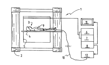
30 The system shown in Figure 1 is a magnetic resonance
imaging apparatus 1 comprising a magnet system 2 for
generating a homogeneous magnetic field on a subject 3
placed on a support table 4. Inside the magnet system
2 is a coil structure 5 to produce around the subject
35 a magnetic field obtained from radio-frequency energy
source 6. Receiver 7 responds to the resonance signal
and processor 8 reconstitutes the integers of the
_ 2141271
projection which will be shown on display 11. The medical
appliance 9, inserted into subject 3, is connected via
conductor 10 to control station 12. Such a general
configuration is familiar to those skilled in the art
5 and it will not be described in further detail.
The appliance 9, as exemplified in Figure 2, is a guidewire
including an open wire length antenna formed by a coaxial
cable comprising a central conductor 13 enclosed in an
10 insulator 14 surrounded by a shield 15 encased in an
insulator 16. The shield 15 or outer conductor and the
outer insulator 16 of the coaxial cable has been removed
some length from the tip or distal end 17. The proximal
end (not shown) of the coaxial cable is for connection
15 to the standard antenna input of control station 12 as
generally shown in Figure 1.
The appliance 9 of Figure 2 is also a guidewire including
an open wire length antenna formed by a coaxial cable.
20 However, the insulator 14 surrounding the central conductor
130 is replaced by an insulating coating 140, while the
shield 15 is replaced by a conductor coating 150 and the
insulator 16 by an insulator coating 160. As for the
embodiment of Figure 1, the conductor coating 150 and
25 insulator coating 160 have been removed some length from
the distal end of tip 170. Also, the proximal end (not
shown) of this coaxial cable is adapted to connection
to the standard antenna input of control station 12
(Figure 1).
Variants may be envisaged.
For instance, the outer conductor and insulator, 15-16
resp. 150-160, need not being removed some length from
35 the distal end 17 resp. 170. Similarly, the outer conductor
and insulator may be removed a far greater length from
the distal end 17 resp. 170, being also possible to have
21~1271
g
them removed up to proximal end of the guidewire, outside
of the patient.
Subject to the precautions or requirements inherent to
5 patient protection, it would be also possible to have
the guidewire comprised of a naked conductor 13 or 130,
while the insulator 14 or 140 and outer conductor 15,
150 and insulator 16, 160 would be installed towards the
proximal end of the guidewire, outside of the patient.
1 0
Similarly, the coaxial configuration shown is not
compulsory, being possible to have the open wire length
antenna as a naked or insulated wire with appropriate
polarities arranged for connection thereof to the antenna
5 input of the control station.
Figure 4 shows one such possibilities, in which the open
wire length antenna is made of two twisted conducting
strands 18 and 19 insulated from one another by appropriate
20 coatings 20 and 21.
Figure 5 also shows one such possibilities, in which the
open wire length antenna is made of two conducting strands
22 and 23 parallel to one another and separated by
25 insulator coatings 24 and 25.
As for the previous embodiments, the strands 18 and 19,
respectively 22 and 23, may have the same length or unlike
lengths.
In both the embodiments of Figure 4 and Figure 5, the
channels 30 which are left open along the insulated strands
may be used for further investigation purposes when the
open wire length antenna is placed in the lumen of a
35 catheter, for example for pressure readings.