Note: Descriptions are shown in the official language in which they were submitted.
CA 02162586 2004-07-08
WO 94126303 PCTIUS94105265
PREVENTION AND TREATMENT OF
PATHOLOGIES ASSOCIATED WITH ABNORMALLY PROLIFERATIVE
SMOOTH MUSCLE CELLS
Field of the Invention
This invention relates generally to the prevention and
treatment of conditions characterized by abnormal smooth
muscle cell proliferation. More specifically, mechanisms for
in vivo vascular smooth muscle cell proliferation modulation
and agents that impact those mechanisms are discussed.
Background of the Invention
Many pathological conditions have been found to be
associated with smooth muscle cell proliferation. Such
conditions include restenosis, atherosclerosis, coronary heart
disease, thrombosis, myocardial infarction, stroke, smooth
muscle neoplasms such as leiomyoma and leiomyosarcoma of the
bowel and uterus, uterine fibroid or fibroma, and obliterative
disease of vascular grafts and transplanted organs. The
mechanism of abnormal smooth muscle cell proliferation is not
yet well understood.
For example, percutaneous transluminal coronary
angioplasty (PTCA) is widely used as the primary treatment
modality in many patients with coronary artery disease. PTCA
can relieve myocardial ischemia in patients with coronary
artery disease by reducing lumen obstruction and improving
coronary flow. The use of this surgical procedure has grown
rapidly, with 39,000 procedures performed in 1983, nearly
150,000 in 1987, 200,000 in 1988, 250,000 in 1989, and
,over 500,000 PTCAs per year are estimated by 1994. Stenosis
following PTCA remains a significant problem, with from 25~
WO 94/26303 PCTlUS94/05265
2
to 35~ of the patients developing restenosis within
1 to 3 months. Restenosis results in significant morbidity
and mortality and frequently necessitates further
interventions such as repeat angioplasty or coronary bypass
surgery. No surgical intervention or post-surgical treatment
(to date) has proven effective in preventing restenosis.
The processes responsible for stenosis after PTCA are not
completely understood but may result from a complex interplay
among several different biologic agents and pathways. Viewed
in histological sections, restenotic lesions may have an
overgrowth of smooth muscle cells in the intimal layers of the
vessel. Several possible mechanisms for smooth muscle cell
proliferation after PTCA have been suggested.
Compounds that reportedly suppress smooth muscle
proliferation in vitro may have undesirable pharmacological
side effects when used in vivo. Heparin is an example of one
such compound, which reportedly inhibits smooth muscle cell
proliferation in vitro but when used in vivo has the potential
adverse side effect of inhibiting coagulation. Heparin
peptides, while having reduced anti-coagulant activity, have
the undesirable pharmacological property of a short
pharmacological half-life. Attempts have been made to solve
such problems by using a double balloon catheter, i.e., for
regional delivery of the therapeutic agent at the angioplasty
site (ela., U.S. Pat. No. 4,824,436), and by using
biodegradable materials impregnated with a drug, i.e., to
compensate for problems of short half-life (ea., U.S. Pat.
No. 4,929,602).
At least five considerations would, at first blush,
appear to preclude use of inhibitory drugs to prevent stenosis
resulting from overgrowth of smooth muscle cells. First,
inhibitory agents may have systemic toxicity that could create
an unacceptable level of risk for patients with cardiovascular
disease. Second, inhibitory agents might interfere with
vascular wound healing following surgery and that could either
delay healing or weaken the structure or elasticity of the
PCT/US94/05265
WO 94/26303
3
newly healed vessel wall. Third, inhibitory agents that kill
smooth muscle cells could damage surrounding endothelium
and/or other medial smooth muscle cells. Dead and dying cells
also release mitogenic agents that might stimulate additional
smooth muscle cell proliferation and exacerbate stenosis.
Fourth, delivery of therapeutically effective levels of an
inhibitory agent may be problematic from several standpoints,
such as the following: a) delivery of a large number of
molecules into the intercellular spaces between smooth muscle
cells may be necessary to establish favorable conditions for
allowing a therapeutically effective dose of molecules to
cross the cell membrane; b) delivery of an inhibitory drug
into the intracellular compartment where its action is exerted
may be difficult to control; and c) optimizing the association
of the inhibitory drug with its intracellular target (e~cx. ,
a ribosome) while minimizing intercellular redistribution of
the drug (ea., to neighboring cells) may be difficult.
Fifth, because smooth muscle cell proliferation takes place
over several weeks it would appear a priori that the
inhibitory drugs should also be administered over several
weeks, perhaps continuously, to produce a beneficial effect.
As is apparent from the foregoing, many problems remain
to be solved in the use of inhibitory drugs to effectively
treat smooth muscle cell proliferation. It would be highly
advantageous to develop new compositions or methods for
inhibiting stenosis due to proliferation of vascular smooth
muscle cells following, for example, traumatic injury to
vessels rendered during vascular surgery.
Summary of the Invention
TGF-beta activators and TGF-beta production stimulators
may be employed in the practice of the present invention to
prevent or treat conditions characterized by inappropriate
proliferation of smooth muscle cells, such as the prevention
or reduction of restenosis following angioplasty or other
vascular trauma. Such TGF-beta activators and production
WO 94/26303 PCTIUS94/05265
a
~~ ~ ~ t~,~.v
4
stimulators inhibit abnormal proliferation of smooth muscle
cells. A preferred TGF-beta activator/production stimulator
is trans-2-[4-(1,2-diphenyl-1-butenyl)phenoxy]-N,N-dimethyl
ethylamine as well as functional equivalents, analogs or
derivatives thereof.
The amount of TGF-beta activator or production stimulator
administered is selected to treat vascular trauma of differing
severity, with smaller doses being sufficient to treat lesser
vascular trauma such as in the prevention of vascular
rejection following graft or transplant. TGF-beta activators
or production stimulators that are not characterized by an
undesirable systemic toxicity profile at a prophylactic dose
are also amenable to chronic use for prophylactic purposes
with respect to disease states involving proliferation of
vascular smooth muscle cells over time (e. a., atherosclerosis,
coronary heart disease, thrombosis, myocardial infarction,
stroke, smooth muscle neoplasms such as leiomyoma and
leiomyosarcoma of the bowel and uterus, uterine fibroid or
fibroma and the like). For prevention of restenosis, a large
dose is preferably administered before or during the traumatic
procedure (e~ct. , angioplasty) . After the traumatic procedure
is conducted, a series of smaller doses is administered over
time to maintain an anti-proliferative effect for a time
sufficient to substantially reduce the risk of or to prevent
restenosis. A preferred therapeutic protocol duration for
this purpose is from about 3 to about 26 weeks.
Further provided is a method for upregulating cellular
mRNA coding for TGF-beta. Cells (e-Q., smooth muscle cells)
amenable to such metabolic manipulation are identified in the
manner described herein and are exposed to an effective amount
of a TGF-beta mRNA regulator (i.e., a subset of TGF-beta
production stimulators). In this manner, TGF-beta production
is stimulated, thereby inhibiting the abnormal proliferation
of smooth muscle cells.
In addition, methods for using TGF-beta to maintain and
increase vessel lumen diameter in a diseased or injured
WO 94126303 ~ ~ ~ ~ PCT/US94/05265
mammalian vessel are described. Further, methods for
preventing or reducing atherosclerosis in a mammal are
provided. Methods for determining TGF-beta in vitro are also
presented.
5
Description of the Drawings
Figures 1 and 2 depict pathways for the modulation of
vascular smooth muscle cell proliferation in vivo.
Detailed Description of the Invention
As used herein the following terms have the meanings as
set forth below:
"Proliferation," means an increase in cell number, i.e.,
by mitosis of the cells.
"Abnormal or Pathological or Inappropriate Proliferation"
means division, growth or migration of cells occurring more
rapidly or to a significantly greater extent than typically
occurs in a normally functioning cell of the same type.
"Expressed" means mRNA transcription and translation with
resultant synthesis, glycosylation, and/or secretion of a
polypeptide by a cell, e.g., chondroitin sulfate proteoglycan
(CSPG) synthesized by a vascular smooth muscle cell or
pericyte.
"Tamoxifen" includes traps-2-[4-(1,2-diphenyl-1
butenyl)phenoxy]-N,N-dimethylethylamine which is capable of
enhancing the production or activation of TGF-beta. The
activated form of TGF-beta, in turn, inhibits vascular smooth
muscle cell proliferation. Functional equivalents and
derivatives of the aforementioned chemical compound are also
included within the scope of the term "tamoxifen" for the
purposes of this disclosure. Exemplary tamoxifen functional
equivalents are plasmin, heparin, angiotensin II,
hexamethylene bisacetamide (HMBA), compounds capable of
reducing the level or inactivating the lipoprotein Lp(a) or
the glycoprotein apolipoprotein(a) and derivatives or analogs
WO 94/26303 PCTlUS94105265
~~,'b a~~~
thereof. Tamoxifen is used herein as a prototypical TGF-beta
activator/production stimulator.
"TGF-beta" includes transforming growth factor-beta as
well as functional equivalents, derivatives and analogs
thereof. The TGF-beta isoforms are a family of
multifunctional, disulfide-linked dimeric polypeptides that
affect proliferation and differentiation of various cells
types. TGF-beta is a polypeptide produced in a latent
propeptide form having, at this time, no identified biological
activity. To be rendered active and, therefore, capable of
inhibiting vascular smooth muscle cell proliferation, the
propeptide form of TGF-beta must be cleaved to yield active
TGF-beta.
"TGF-beta activator" includes moieties capable of
directly or indirectly activating the latent form of TGF-beta
to the active form thereof. Plasmin, plasmin activators,
tamoxifen as well as analogs, derivatives or functional
equivalents thereof are exemplary TGF-beta activators useful
in the practice of the present invention.
."TGF-beta, production stimulator" includes moieties
capable of directly or indirectly stimulating the production
of TGF-beta (generally the latent form thereof). Such TGF-
beta production stimulators may be TGF-beta mRNA regulators
(i.e., moieties that increase the production of TGF-beta
mRNA), enhancers of TGF-beta mRNA expression or the like.
"Direct" action implies that the TGF-beta activator acts
on the latent form of TGF-beta. Such direct action, when
applied to TGF-beta production stimulators, indicates that
cells upon which the production stimulator acts increase TGF-
beta mRNA production or expression of TGF-beta.
"Indirect" action implies that the TGF-beta activator
acts on a moiety that itself or through one or more other
moieties acts on latent TGF-beta. Such indirect action, when
applied to TGF-beta production stimulators, indicates that the
stimulators act on a moiety that itself or through one or more
~~~2~:~~
WO 94/26303 PCTlUS94/05265
7
other moieties acts on a population of cells to stimulate the
production of TGF-beta mRNA or the expression of TGF-beta.
For the purposes of this description, the prototypical
cells, upon which the effects of TGF-beta activators or
production stimulators are felt, are smooth muscle cells and
pericytes derived from the medial layers of vessels and
adventitia vessels which proliferate in intimal hyperplastic
vascular sites following injury, such as that caused during
PTCA. TGF-beta activators and production stimulators are not
restricted in use for therapy following angioplasty; rather,
the usefulness thereof will be proscribed by their ability to
inhibit abnormal cellular proliferation, for example, of
smooth muscle cells and pericytes in the vascular wall. Thus,
other aspects of the invention include TGF-beta activators or
production stimulators used in early therapeutic intervention
for reducing, delaying, or eliminating (and even reversing)
atherosclerotic plaques and areas of vascular wall hypertrophy
and/or hyperplasia. TGF-beta activators and production
stimulators also find utility for early intervention in
pre-atherosclerotic conditions, e.g., they are useful in
patients at a high risk of developing atherosclerosis or with
signs of hypertension resulting from atherosclerotic changes
in vessels or vessel stenosis due to hypertrophy of the vessel
wall.
TGF-beta activators or production stimulators of the
invention are useful for inhibiting the pathological
proliferation of vascular smooth muscle cells, e.g., for
reducing, delaying, or eliminating stenosis following
angioplasty. As used herein the term "reducing" means
decreasing the intimal thickening that results from
stimulation of smooth muscle cell proliferation following
angioplasty, either in an animal model or in man. "Delaying"
means delaying the time until onset of visible intimal
hyperplasia (e. a., observed histologically or by angiographic
examination) following angioplasty and may also be accompanied
by "reduced" restenosis. "Eliminating" restenosis following
WO 94/26303 PCT/US94/05265
8
angioplasty means completely "reducing" intimal thickening
and/or completely "delaying" intimal hyperplasia in a patient
to an extent which makes it no longer necessary to surgically
intervene, i.e., to re-establish a suitable blood flow through
the vessel by repeat angioplasty, atheroectomy, or coronary
artery bypass surgery. The effects of reducing, delaying, or
eliminating stenosis may be determined by methods routine to
those skilled in the art including, but not limited to,
angiography, ultrasonic evaluation, fluoroscopic imaging,
fiber optic endoscopic examination or biopsy and histology.
High levels of lipoprotein Lp(a) are known to constitute
a major risk factor for atherosclerosis, coronary heart
disease and stroke. One symptom associated with such
conditions and other problems, such as restenosis following
balloon angioplasty and other pathogenic conditions, is the
proliferation or the migration of smooth muscle cells. No
direct link between Lp (a) and proliferation of vascular smooth
muscle cells had been established in the prior art.
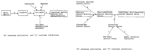
An in vivo pathway for the modulation of vascular smooth
muscle cell proliferation is shown in Fig. 1. This mechanism
is believed to constitute a portion of the mechanism that
maintains vascular smooth muscle cells in a non-proliferative
state in healthy vessels.
Vascular smooth muscle cell proliferation is inhibited
by an active form of TGF-beta. Tamoxifen has been shown by
the experimentation detailed in Example 1 hereof to stimulate
both the production and the activation of TGF-beta. Heparin
stimulates the activation of TGF-beta by affecting the release
of the active form of TGF-beta from inactive complexes present
in serum. TGF-beta neutralizing antibodies inhibit the
activity of TGF-beta, thereby facilitating the proliferation
of vascular smooth muscle cells. The apparent in vivo
physiological regulator of the activation of TGF-beta is
plasmin. Plasmin is derived from plasminogen through
activation by, for example, tPA (tissue plasminogen
activator). Plasminogen and, therefore, plasmin activity is
WO 94/26303 ~ ~ ~ PCT/US94/05265
9
inhibited by the lipoprotein Lp(a) or apolipoprotein(a)
(apo(a) ) , thereby decreasing the activation of the latent form
of TGF-beta and facilitating proliferation of vascular smooth
muscle cells.
An additional pathway for the modulation of vascular
smooth muscle cell proliferation is shown in Fig. 2. Resting
smooth muscle cells constitute cells in their normal,
quiescent non-proliferative state. Such resting smooth muscle
cells may be converted to proliferating smooth muscle cells
through activation by platelet derived growth factor (PDGF),
fibroblast growth factor (FGF) or other stimulatory moieties.
The proliferating smooth muscle cells may be converted to
continual proliferating smooth muscle cells (i.e., smooth
muscle cells capable of generating a pathological state
resulting from over-proliferation thereof) by an autocrine
growth factor. This growth factor is believed to be produced
by proliferating smooth muscle cells. An increased level of
autocrine growth factor, which can be inhibited by the active
form of TGF-beta or an appropriately structured (i.e.,
designed) small molecule inhibitor, is believed to mediate the
production of continual proliferating smooth muscle cells.
Lp(a) consists of low density lipoprotein (LDL) and
apo(a). Apo(a) shares approximately 80% amino acid identity
with plasminogen (see MacLean et al. , Nature, 330: 132, 1987) .
Lp(a) has been found to inhibit cell-associated plasminogen
activity (see, for example, Harpel et al., Proc. Natl. Acad.
Sci. USA, 86: 3847, 1989). Experiments conducted on human
aortic vascular smooth muscle cells derived from healthy
transplant donor tissue, cultured in Dulbecco's modified
Eagles medium (DMEM) + 10% fetal calf serum (FCS) as described
in Grainger et al. , Biochem. J. , 283: 403, 1992, indicated the
following:
1) Addition of Lp(a) to sub-confluent human vascular
smooth muscle cells stimulated their proliferation in a dose
dependent manner (addition of 500 nM Lp(a) to human vascular
WO 94/26303 PCTIUS94/05265
to
smooth muscle cells caused a reduction in doubling time from
82 +/- 4 hours to 47 +/- 4 hours);
2) Addition of apo(a) had a similar effect, although a
higher concentration of apo(a) appeared to be required
therefor;
3) Addition of LDL at varying concentrations up to 1
micromolar had no effect on proliferation.
One possible mode of action for Lp(a) and apo(a) is
competitive inhibition of surface-associated plasminogen
activation, which in turn inhibits the subsequent activation
of TGF-beta by plasmin. TGF-beta is a potent growth inhibitor
of a number of anchorage-dependent cells, including smooth
muscle cells. TGF-beta is produced as a latent propeptide
having a covalently linked homodimer structure in which the
active moiety is non-covalently linked to the amino-terminal
portion of the propeptide. Latent TGF-beta must be cleaved
(e~ct: , in vitro by acid treatment or in vivo by the serine
protease plasmin) in order to become capable of inhibiting the
proliferation of vascular smooth muscle cells. Plasmin is
therefore a leading candidate to be a physiological regulator
of TGF-beta.
The hypothesis that Lp(a) and apo(a) were acting on
cultured human vascular smooth muscle cells by interfering
with activation of latent TGF-beta was tested. In support of
this hypothesis, an observation was made that plasmin activity
associated with vascular smooth muscle cells was reduced 7-
fold by Lp(a) and 5-fold by apo(a). The plasmin activity in
the conditioned medium was also reduced by Lp(a) and apo(a)
by about 2-fold, but was much lower than cell-associated
plasmin activity in vascular smooth muscle cell cultures.
These observations are consistent with previous findings that
Lp (a) is a more potent inhibitor of surface-associated, rather
than fluid phase, plasminogen activation.
To exclude the possibility that Lp(a) was affecting the
synthesis of plasminogen activators rather than plasminogen
activation, plasminogen activator levels in human vascular
PCT/US94I05265
W0 94126303
11
smooth muscle cell cultures were measured in the presence and
absence of the lipoproteins and in the presence of a large
excess of plasminogen, so that the lipoproteins present would
not significantly act as competitive inhibitors. Total
plasminogen activator activity was not affected by the
presence of any of the lipoproteins in the vascular smooth
muscle cell cultures. For example, plasminogen activator
activity in the conditioned medium remained at 0.7 +/- 0.6
mU/ml with Lp(a) additions up to 500 nM.
Lp(a) and apo(a) both reduced the level of active TGF-
beta by more than 100-fold compared to control or LDL-treated
cultures. The level of total latent plus active TGF-beta
measured by ELISA as described in Example 1 was unaffected by
the presence of Lp(a) or apo(a), however. These facts lead
to the conclusion that Lp(a) stimulates proliferation of human
vascular smooth muscle cells by inhibiting plasmin activation
of latent TGF-beta to active TGF-beta.
To further test this conclusion and exclude the
possibility that Lp(a) was acting by binding active TGF-beta
as well as reducing plasmin activity, human vascular smooth
muscle cells were cultured in the presence of Lp(a). These
cells had a population doubling time of 47 +/- 3 hours.
Addition of plasmin was able to overcome the population
doubling time reducing effect of Lp(a) and reduce the cell
number to control levels, with the population doubling time
increased to 97 +/- 4 hours.
The role of plasmin in the pathway was confirmed by
studies in which inhibitors of plasmin activity were added to
human vascular smooth muscle cells. Like Lp(a), these
protease inhibitors increased cell number. Aprotinin, for
example, decreased the population doubling time from 82 +/-
4 hours in control cultures to 48 +/- 5 hours, and alpha2-
antiplasmin decreased the population doubling time to 45 +/-
2 hours. 500 nM Lp(a) and aprotinin addition resulted in only
a slight additional stimulation of proliferation, with the
population doubling time for cultures of this experiment being
WO 94/26303 , PCTIITS94/05265
~~~i"~
12
45 +/- 6 hours. Neutralizing antibodies to TGF-beta similarly
decreased population doubling time in vascular smooth muscle
cells (see, for example, Example 1). In summary, Lp(a),
plasmin inhibitors and neutralizing antibody to TGF-beta
stimulate proliferation of vascular smooth muscle cells, while
plasmin nullifies the growth stimulation of Lp(a). These
results support the theory that the mode of action of Lp(a)
and apo(a) is the competitive inhibition of plasminogen
activation.
Experimentation conducted to ascertain the impact of
tamoxifen on TGF-beta and vascular smooth muscle cell
proliferation is set forth in detail in Example 1. The
results of those experiments are summarized below.
1) Addition of tamoxifen decreased the rate of
proliferation, with maximal inhibition observed at
concentrations above 33 micromolar. 50 micromolar tamoxifen
concentrations produced a cell number 96 hours following the
addition of serum that was reduced by 66% +/- 5.2% (n=3) as
compared to cells similarly treated in the absence of
tamoxifen.
2) Tamoxifen did not significantly reduce the proportion
of cells completing the cell cycle and dividing. Inhibition
of vascular smooth muscle cells caused by tamoxifen therefore
appears to be the result of an increase in the cell cycle time
of nearly all (>90%) of the proliferating cells.
3) Tamoxifen decreases the rate of proliferation of
serum-stimulated vascular smooth muscle cells by increasing
the time taken to traverse the Gz to M phase of the cell
cycle.
4) Tamoxifen decreased the rate of proliferation of
vascular smooth muscle cells by inducing TGF-beta activity.
5) Vascular smooth muscle cells produced TGF-beta in
response to tamoxifen. Tamoxifen appears to increase TGF-beta
activity in cultures of rat vascular smooth muscle cells by
stimulating the production of latent TGF-beta and increasing
the proportion of the total TGF-beta which has been activated.
WO 94/26303 ~ ~ PCT/US94/05265
13
6) Tamoxifen, unlike heparin, does not act by releasing
TGF-beta from inactive complexes present in serum.
7) TGF-betal mRNA was increased by approximately 10-fold
by 24 hours after addition of tamoxifen (10 micromolar) . This
result suggests that the expression of TGF-beta mRNA by the
smooth muscle cells will be increased, thereby facilitating
decreased proliferation thereof by activated TGF-beta. This
mechanism can be exploited using cells incorporating nucleic
acids encoding TGF-beta mRNA, which cells are identifiable by
persons skilled in the art employing known techniques.
8 ) Tamoxifen is a selective inhibitor of vascular smooth
muscle proliferation with an EDso (a concentration resulting
in 50% inhibition) at least 10-fold lower for vascular smooth
muscle cells than for adventitial fibroblasts.
Additional experimentation has shown that the addition
of Lp(a) or apo(a) substantially reduced the rat vascular
smooth muscle cell proliferation inhibitory activity of
tamoxifen, with the population doubling time in the presence
of tamoxifen and Lp(a) being 42 +/- 2 hours (as compared to
a population doubling time of 55 +/- 2 hours for tamoxifen
alone, and a time of 35 +/- 2 hours for the control). Also,
the presence of Lp(a) reduced the levels of active TGF-beta
produced in response to the addition of tamoxifen by about 50-
fold. Addition of plasmin to rat vascular smooth muscle cells
treated with tamoxifen and Lp(a) resulted in most of the TGF-
beta being activated, and proliferation was again slowed (with
the population doubling time being 57 +/- 3 hours) . These
observations are consistent with the theory that Lp(a) acts
by inhibiting TGF-beta activation.
Identification of therapeutic agents (direct or indirect
TGF-beta activators or production stimulators) that act to
inhibit vascular smooth muscle cell proliferation by the
pathway shown in Fig. 1 can be identified by a practitioner
in the art by conducting experiments of the type described
above and in Example 1. Such experimental protocols
facilitate the identification of therapeutic agents useful in
WO 94/26303 PCT/iJS94/05265
~a 14
the practice of the present invention and capable of one of
the following activities:
1) production or activation of TGF-beta;
2) having TGF-beta activity;
3) activation of plasmin;
4) activation of plasminogen; and
5) reduction of Lp(a) or apo(a) level.
Identification of therapeutic agents (direct or indirect
TGF-beta activators or production stimulators) that act to
inhibit vascular smooth muscle cell proliferation by the
pathway shown in Fig. 2 can be identified by a practitioner
in the art by conducting experimentation using known
techniques that are designed to identify growth factors made
by proliferating smooth muscle cells, which growth factors
also act on those cells (i.e., autocrine growth factors).
Known techniques for rational drug design are then used to
screen small molecules for the ability to inhibit the
production or activity of such autocrine growth factors. Such
experimental protocols facilitate the identification of
therapeutic agents useful in the practice of the present
invention and capable of one of the following activities:
1) production or activation of TGF-beta;
2) having TGF-beta activity; and
3) inhibit the activity or production of an autocrine
growth factor produced by proliferating smooth muscle cells.
Smooth muscle cell proliferation is a pathological factor
in myocardial infarctions, atherosclerosis, thrombosis,
restenosis and the like. Therapeutic/prophylactic agents of
the present invention, including tamoxifen and the like,
having at least one of the activities recited above and
therefore being capable of inhibiting proliferation of
vascular smooth muscle cells, are useful in the prevention or
treatment of these conditions. Manipulation of the
proliferation modulation pathway for vascular smooth muscle
cells to prevent or reduce such proliferation removes or
reduces a major component of the arterial lesions of
WO 94/26303 ~ g A~ PCT/US94/05265
atherosclerosis and the restenosed arteries following
angioplasty, for example.
More specifically, chronically maintaining an elevated
level of activated TGF-beta reduces the probability of
5 atherosclerotic lesions forming as a result of vascular smooth
muscle cell proliferation. Consequently, administration of
TGF-beta activators or TGF-beta production stimulators
protects against atherosclerosis and subsequent myocardial
infarctions that are consequent to coronary artery blockage.
10 Also, substantially increasing the activated TGF-beta level
for a short time period allows a recipient to at least
partially offset the strong stimulus for vascular smooth
muscle cell proliferation caused by highly traumatic injuries
or procedures such as angioplasty. Continued lower dose
15 delivery to the traumatized site further protects against
restenosis resulting from vascular smooth muscle cell
proliferation in the traumatized area.
Tamoxifen, for example, is commercially available from
ICI Pharmaceuticals (Macclesfield, England). The prevalent
commercially available form is a 10 mg tablet. Such tablets
or portions thereof can be employed in the prophylactic and
treatment protocols described herein.
Prevention or treatment relating to a traumatized or
diseased vascular site, for example, the TGF-beta activators
or production stimulators may also be administered in
accordance with the present invention using an infusion
catheter, such as produced by C.R. Bard Inc., Billerica, MA,
or that disclosed by Wolinsky (7; U.S. Patent No. 4,824,436)
or Spears (U.S. Patent No. 4,512,762). In this case, a
therapeutically/prophylactically effective dosage of the TGF-
beta activator or production stimulator will be typically
reached when the concentration thereof in the fluid space
between the balloons of the catheter is in the range of about
10'3 to 10'lzM. It is recognized by the present inventors that
TGF-beta activators or stimulators may only need to be
delivered in an anti-proliferative therapeutic/prophylactic
WO 94/26303 PCTIUS94/05265
16
dosage sufficient to expose the proximal (6 to 9) cell layers
of the intimal or tunics media cells lining the lumen thereto.
Also, such a dosage can be determined empirically, e.g., by
a) infusing vessels from suitable animal model systems and
using immunohistochemical methods to detect the TGF-beta
activator or production stimulator and its effects; and
b) conducting suitable in vitro studies.
It will be recognized by those skilled in the art that
desired therapeutically/prophylactically effective dosages of
a TGF-beta activator or production stimulator administered by
a catheter in accordance with the invention will be dependent
on several factors, including, e.g.. a) the atmospheric
pressure applied during infusion; b) the time over which the
TGF-beta activator or production stimulator administered
resides at the vascular site; c) the nature of the therapeutic
or prophylactic agent employed; and/or d) the nature of the
vascular trauma and therapy desired. Those skilled
practitioners trained to deliver drugs at therapeutically or
prophylactically effective dosages (e. g., by monitoring drug
levels and observing clinical effects in patients) will
determine the optimal dosage for an individual patient based
on experience and professional judgment. In a preferred
embodiment, about 0.3 atm (i.e., 300 mm of Hg) to about 5 atm
of pressure applied for 15 seconds to 3 minutes directly to
the vascular wall is adequate to achieve infiltration of a
TGF-beta activator or production stimulator into the smooth
muscle layers of a mammalian artery wall. Those skilled in
the art will recognize that infiltration of the TGF-beta
activator or production stimulator into intimal layers of a
diseased human vessel wall will probably be variable and will
need to be determined on an individual basis.
While two representative embodiments of the invention
relate to prophylactic or therapeutic methods employing an
oral dosage for or infusion catheter administration, it will
be recognized that other methods for drug delivery or routes
of administration may also be useful, e.g., injection by the
PCTIUS94105265
W0 94/26303
17
intravenous, intralymphatic,intrathecal, intraarterial, local
delivery by implanted osmotic pumps or other intracavity
routes. Administration of TGF-beta activators or production
stimulators in accordance with the present invention may be
continuous or intermittent, depending, for example, upon the
recipient's physiological condition, whether the purpose of
the administration is therapeutic or prophylactic and other
factors known to skilled practitioners.
In the practice of certain embodiments of the present
invention, catheter and other administration routes are
preferably conducted using a TGF-beta activator or production
stimulator dispersed in a pharmaceutically acceptable carrier
that is in liquid phase. Useful pharmaceutically acceptable
carriers include generally employed carriers, such as
phosphate buffered saline solution, water, emulsions (e~cr. ,
oil/water and water/oil emulsions) and wetting agents of
various types.
For TGF-beta activators or production stimulators, such
as tamoxifen, several exemplary dosing regimens are
contemplated, depending upon the condition being treated and
the stage to which the condition has progressed. For
prophylactic purposes with respect to atherosclerosis, for
example, a low chronic dose sufficient to elevate in vivo TGF-
beta production is contemplated. An exemplary dose of this
type is about 0.1 mg/kg/day (ranging between about 0.1 and
about 10 mg/kg/day). Another exemplary dose range is from
about 0.01 to about 1000 micrograms/ml. Such low doses are
also contemplated for use with respect to ameliorating
stenosis following relatively low trauma injury or
intervention, such as vein grafts or transplants or organ
allografts, for example. No adverse side effects (e. a.,
nausea as experienced by recipients of higher dose
administrations when tamoxifen has been employed in the
treatment of breast cancer) are anticipated with respect to
these chronic or low dosing regimens.
WO 94/26303 PCTILTS94105265
18
For prevention of restenosis following angioplasty, an
example of a higher trauma injury or intervention resulting
in a stronger acute proliferative stimulus to smooth muscle
cells, a higher dose would be required. For example, a dosing
regimen is contemplated which involves a single "pre-loading"
dose (or multiple, smaller pre-loading doses) given before or
at the time of the intervention, with a chronic smaller
(follow up) dose delivered daily for two to three weeks or
longer following intervention. For example, a single pre-
loading dose may be administered about 24 hours prior to
intervention, while multiple preloading doses may be
administered daily for several days prior to intervention.
Alternatively, one or more pre-loading doses may be
administered about 1-4 weeks prior to intervention. These
doses will be selected so as to maximize TGF-beta activator
or production stimulator activity, while minimizing induction
of synthesis and secretion of extracellular matrix proteins.
An exemplary single pre-loading dose is about 50 mg/kg
(ranging between about 10 and about 1000 mg/kg), while an
exemplary multiple pre-loading individual dose is about 10
mg/kg/day (ranging between about 0.01 and 10 mg/kg/day).
It will be recognized that where the TGF-beta activator
or production stimulator is to be delivered with an infusion
catheter, the therapeutic dosage required to achieve the
desired inhibitory activity can be anticipated through the use
of in vitro studies. In a preferred aspect, the infusion
catheter may be conveniently a double balloon or quadruple
balloon catheter with a permeable membrane. In one
representative embodiment, a therapeutically effective dosage
of a TGF-beta activator or production stimulator is useful in
treating vascular trauma resulting from disease (e. g.,
atherosclerosis, aneurysm, or the like) or vascular surgical
procedures such as angioplasty, atheroectomy, placement of a
stent (e. g., in a vessel), thrombectomy, and grafting.
Atheroectomy may be performed, for example, by surgical
excision, ultrasound or laser treatment, or by high pressure
WO 94/26303 ~ PCT/US94/05265
19
fluid flow. Grafting may be, for example, vascular grafting
using natural or synthetic materials or surgical anastomosis
of vessels such as, e.g., during organ grafting. Those
skilled in the art will recognize that the appropriate
therapeutic dosage for a given vascular surgical procedure
(above) is determined in in vitro and in vivo animal model
studies, and in human preclinical trials.
While two representative embodiments of the invention
relate to therapeutic methods employing an oral dosage for or
infusion catheter administration, it will be recognized that
other methods for drug delivery or routes of administration
may also be useful, e.g., injection by the intravenous,
intralymphatic, intrathecal, intraarterial, local delivery by
implanted osmotic pumps or other intracavity routes.
Administration of TGF-beta activators or production
stimulators in accordance with the present invention may be
continuous or intermittent, depending, for example, upon the
recipient's physiological condition, whether the purpose of
the administration is therapeutic or prophylactic and other
factors known to skilled practitioners.
In the practice of certain embodiments of the present
invention, catheter and other administration routes are
preferably conducted using a TGF-beta activator or production
stimulator dispersed in a pharmaceutically acceptable carrier
that is in liquid phase. Useful pharmaceutically acceptable
carriers include the generally employed carriers, such as
phosphate buffered saline solution, water, emulsions (e. a.,
oil/water and water/oil emulsions) and wetting agents of
various types.
Human vascular smooth muscle cells (VSMC) are more
difficult to grow in culture than VSMC derived from other
species, such as rat (doubling time for adult human VSMC =
70-85 h; for adult rat VSMC = 35 h). Medium conditioned on
human VSMC decreased the proliferation of rat VSMC in vitro.
Entry of rat VSMC into S phase of the cell cycle was not
affected. However, the duration of GZ and/or M phase was
WO 94/26303 PCT/US94/05265
y.~3~~~~~ 20
extended. Anti-TGF-beta antibody reversed the delayed entry
into M phase caused by exposure to human VSMC conditioned
medium (HCM). An examination of the HCM showed that 64~12%
of the TGF-beta present in the medium was already activated.
In contrast, rat VSMC conditioned medium displayed very low
levels of latent TGF-beta and no detectable TGF-beta activity.
Human VSMC were f ound to produce t-PA activity in culture .
The t-PA leads to an increase in plasmin activity, which in
turn activates TGF-beta. This was confirmed by culturing
human VSMC in the presence of aprotinin, a plasmin inhibitor.
Aprotinin increased the rate of proliferation of human VSMC
to almost the same extent as neutralizing anti-TGF-beta
antibodies and a2-antiplasmin. Thus, growth of human VSMC in
culture is determined by the production of TGF-beta activated
by plasmin, which feeds back in an autocrine loop to increase
the duration of the cell cycle.
Subcultured human aortic VSMC remain more differentiated
in culture than rat aorta VSMC (i.e., they contain higher
levels of the smooth muscle-specific isoforms of myosin heavy
chain (SM-MHC) and a-actin). TGF-beta likely plays a role in
maintaining SM-MHC and a-actin content, and thus may be
responsible for maintaining cells in a more differentiated
phenotype. In view of these data, heparin, which is believed
to release TGF-beta from inactive complexes in the serum,
would be predicted to have little effect on the rate of
proliferation of human VSMC, which is already inhibited by
endogenous active TGF-beta production. Such observations may
explain why human clinical trials of heparin administered
after PTCA have failed to demonstrate any beneficial effect.
Freshly dispersed rat aortic VSMC lose SM-MHC and a-SM
actin as they start to proliferate. After 7 days in culture
when the cells reach confluence. When serum is removed,
approximately 40% of the VSMC reexpress SM-MHC and a-SM actin
at levels comparable to those present in freshly dispersed
cells. If the cells were subcultured for more than five
passages and allowed to reach confluence, less than 1%
WO 94126303 ~ ~ ~ c~ ~ ~ ~ PCTlUS94/05265
21
reexpress SM-MHC even after prolonged serum withdrawal. These
cells represent proliferating de-differentiated VSMC.
When primary cultures of rat aortic VSMC are exposed to
TGF-beta, the loss of the 204 kD (SM-1) and 200 kD (SM-2) SM
MHC isoforms is substantially inhibited. However, TGF-beta
did not induce re-expression of SM-MHC in subcultured cells
that have very low levels of this protein. Therefore, TGF-
beta can maintain a cell's differentiated state (as defined
by SM-MHC content), but cannot induce re-differentiation in
a de-differentiated proliferating cell. Since TGF-beta
extends the GZ phase of the cell cycle in both primary and
passaged VSMC cultures, the data suggest that the pathways
that mediate proliferation and differentiation are regulated
independently.
Specific markers of both differentiated and proliferating
VSMCs have been isolated. Four cell populations were probed
using generated cDNAs: (a) freshly dispersed rat aortic cells;
(b) freshly dispersed rat aortic VSMC after 7 days in culture
(D7 cells); (c) freshly dispersed rat aortic VSMC after
subculturing 12 times (S12 cells); and (d) rat fibroblasts.
Five classes of gene markers were defined. Class 1 cDNAs were
expressed to a similar level in all of the RNAs. Class 2
cDNAs were highly expressed in RNA from freshly dispersed
aortic cells, but were barely detectable in D7 or S12 cells
and were not detectable in rat fibroblasts. Class 3 cDNAs
were expressed at similar levels in freshly dispersed aortic,
D7 and S12 cells. Class 4 cDNAs showed higher expression in
freshly dispersed aortic and D7 cells than in S12 cells and
fibroblasts. Class 5 cDNAs were expressed more strongly in
S12 cells than in freshly dispersed aortic cells, D7 cells and
fibroblasts. Class 4 genes included a-SM actin, y-SM actin,
SM22a, calponin, tropoelastin, phospholamban and CHIP28. In
addition, previously defined markers of the differentiated
phenotype include SM-MHC, integrin and vinculin. Class 5
genes included matrix Gla (MGP) and osteopontin. When
passaged cells were made quiescent by removal of serum, the
WO 94126303 ~; PCT/LTS94/05265
22
levels of MGP and osteopontin did not change significantly,
indicating that high expression of these two genes occurs in
VSMC that have undergone proliferation, but does not depend
on the cells being in the cell cycle.
Such studies of gene expression provide insight into the
processes of de-differentiation that occur during
proliferation of VSMC. In situ hybridization analysis of
balloon-injured rat carotid arteries suggests that dividing
intimal cells present 7 days after injury express high levels
of both osteopontin and MGP RNA. In contrast, osteopontin is
only weakly expressed in the media of intact rat aorta and
carotid arteries. Osteopontin and MGP may play a role in
regulating calcification, which can occur rapidly in vascular
lesions.
In the course of investigating potential heterogeneity
of cells from rat aortas, three groups of VSMC clones have
been identified. One group consists of small cells that have
an epithelioid or cobblestone morphology and proliferate
without the need for added growth factors, suggesting
production of an autocrine growth factor(s). The second group
consists of intermediate size, spindle shaped cells that grow
in a characteristic "hills and valleys" pattern and are
dependent on exogenous growth factors. These cells resemble
the predominant cell morphology in standard cultures of adult
aortic VSMC. The third group consists of large, often
multinucleate, cells with limited proliferative capacity.
These large cells express high quantities of smooth muscle
specific proteins.
All three types of cells could be isolated from neonatal
and adult rat aortae. However, aortas from young rats yielded
high proportions of the small cell clones, while those from
adult rats yielded high proportions of intermediate and large
cell clones. Clones of small VSMC can be induced to convert
to intermediate sized cells by treatment with TGF-beta. A
proportion of these cells, in turn, converts to large cells
if plated at low density. The small cells may represent a
PCT/US94/05265
WO 94/26303
23
progenitor cell and the large, non-proliferating cells may
represent mature VSMC.
VSMC derived from neonatal rat aortas differ from normal
adult VSMC in several ways : ( a ) they do not require exogenous
growth factors for sustained growth; (b) they secrete PDGF
like growth factors; (c) they grow with a characteristic
epithelioid morphology; and (d) they express high levels of
cytochrome P450IA1, elastin and osteopontin (J. Biol. Chem.
266:3981-86, 1991; Biochem. Biophys. Res. Comm. 177:867-73,
1991; Nature 311:669-71, 1984). After intimal damage,
neointimal lesions grow with an epithelioid morphology,
secrete a PDGF-like protein and display increased expression
of osteopontin in the vascular wall (Proc. Natl. Acad. Sci.
USA 83:7311-15, 1986). These data are consistent with the
presence in vivo of a subpopulation of VSMC that comprises a
diminishing proportion of the total cell population with age
and which proliferates preferentially.
TGF-beta is released by platelets, macrophages and VSMC
at sites of vascular injury. Since VSMC and endothelial cells
at the site of vascular injury can synthesize and release t
PA, a local mechanism for activating secreted TGF-beta exists.
The level of t-PA activity depends on expression of
plasminogen activator inhibitor-1 (PAI-1) which is also
synthesized in the vessel wall, and may be up-regulated by
TGF-beta. In addition, TGF-beta binds with high affinity to
a2-macroglobulin. Such binding renders TGF-beta unable to
bind to cell surface receptors for TGF-beta. Polyanionic
glycosaminoglycans, such as heparin, are also normally present
in the vessel wall, and these moieties can reverse the
association of TGF-beta with a2-macroglobulin. The phenotypic
state of the VSMC may affect the VSMC response to activated
TGF-beta. The phenotypic state of the VSMC may be influenced
by their extracellular environment. Accordingly, the
biological effects of TGF-beta are subject to a variety of
regulatory mechanisms.
WO 94/26303 PCT/US94I05265
. 1 ~~n~~
~, 1
24
TGF-beta inhibits DNA synthesis in rat aortic VSMC
stimulated with either PDGF or EGF. In serum stimulated
cells, however, TGF-beta has little effect on DNA synthesis.
Instead, TGF-beta exerts its anti-proliferative effect by
prolonging the GZ phase of the cell cycle. Likewise, heparin
inhibits proliferation of serum-stimulated rat VSMC by
extending the Gz phase of the cell cycle. This effect of
heparin can be eliminated by anti-TGF-beta antibody. These
observations suggest that the anti-proliferative effect of
heparin on VSMC in vitro and possibly in vivo may be exerted
through the release of TGF-beta.
When VSMC are dispersed in cell culture, they lose
contractile proteins and modulate to a "synthetic" phenotype
as they proliferate. The majority of VSMC in atheromatous
plaques appear to have this synthetic phenotype also. Since
loss of smooth muscle-specific proteins occurs spontaneously
in cell culture in the absence of mitogens where no
proliferation occurs, this phenotypic change is not
attributable to mitogenic stimulation, but rather to removal
of the cells from their extracellular matrix. The matrix
contains large quantities of collagen and glycosaminoglycans
that may maintain VSMC in a contractile state. TGF-beta does
not exert its anti-proliferative effect through inhibition of
phenotypic modulation, however, since it is effective at
slowing proliferation of passaged cells that can no longer
express contractile proteins. Thus, TGF-beta displays the
independent properties of (1) maintaining differentiated adult
VSMC in the contractile phenotype; (2) causing maturation of
small VSMC to intermediate size, spindle-shaped VSMC; and (3)
inhibiting VSMC proliferation regardless of phenotype. Change
from a contractile to synthetic phenotype is not obligatory
for proliferation.
Cultured VSMC synthesize and secrete large quantities of
extracellular matrix proteins. TGF-beta enhances production
of extracellular matrix proteins, which favors maintenance of
the synthetic phenotype in cells that have been allowed to
WO 94/26303 ~ ~ ~ PCTlUS94/05265
modulate. In addition, TGF-beta increases expression of
numerous protease inhibitors, which also increase accumulation
of extracellular matrix proteins.
In hypertension, there is increased thickness of the
5 vessel media, with a consequent decrease in maximum lumen
diameter, leading to increased vascular resistance. The
increased thickness of the vessel media is due to growth of
VSMC within the media. In large conductance vessels, such as
the aorta, the VSMC growth is believed to be attributable
10 primarily to VSMC hypertrophy (i.e., enlargement of the cell
without proliferation). In hypertensive animals, these
vessels display an increased incidence of polyploid cells
within the aortic media. In resistance vessels, such as the
mesenteric arteries, however, VSMC proliferation may
15 contribute to the increased thickness of the vessel media.
Previously, VSMC growth in hypertension was believed to result
from elevated blood pressure. Current data suggest that
increased vascular tone and VSMC hypertrophy and/or
hyperplasia may be caused independently by a common stimulus.
20 For instance, under certain circumstances, the vasoconstrictor
peptide All may be mitogenic for VSMC. Further, VSMC
stimulated with All also synthesize TGF-beta. Thus, any
mitogenic effect of All might be inhibited by TGF-beta, with
the net effect of All stimulation being arrest in G1 and
25 hypertrophy without proliferation. All may induce activation
of TGF-beta by stimulating expression of t-PA by VSMC.
The VSMC involved in hypertension remain within the media
of the vessel and are surrounded by a heparin-containing
extracellular matrix. Therefore, any TGF-beta produced is
freely available and will maintain VSMC in a contractile
state.
In obliterative vascular disease, such as
atherosclerosis, VSMC migrate from the media and proliferate
in the intima. There they secrete extracellular matrix
proteins and form a lipid-rich plaque that encroaches on the
vascular lumen. This process is similar to, but slower than,
WO 94126303 ~ PCT/L1S94/05265
26
the process that occurs following PTCA, leading to restenosis.
Such inappropriate intimal VSMC proliferation also occurs in
vascular bypass grafts and the arteries of transplanted
organs, leading to graft occlusion and organ failure,
respectively. In atherosclerosis, the VSMC involved in the
lesion are generally of the synthetic phenotype and localized
in the intima, in contrast to the VSMC involved in
hypertension.
For medial VSMC involved in atherosclerosis, VSMC
migration is accompanied by an increase in synthesis and
secretion of matrix proteins and by proliferation. TGF-beta
may reduce or prevent the VSMC proliferative response to
mitogens and/or may induce synthesis and secretion of
extracellular matrix proteins. The effect of TGF-beta in this
case would be reduction of cellularity and increase of the
matrix component of an atherosclerotic plaque.
Alternatively, VSMC in the intima may arise from a
population of neonatal-like VSMC that are capable of migration
and preferential proliferation following vascular injury.
This intimal phenotype may be either induced or selected in
response to vessel injury. When these cells are exposed to
TGF-beta, the neonatal-like, small cell phenotype should
convert into intermediate sized, spindle-shaped cells that no
longer produce an autocrine growth factor. Thus, cells of the
intermediate size should have a decreased tendency to
proliferate. Over time, a portion of this intermediate sized
population of cells would convert to the large, non-
proliferative VSMC phenotype.
If VSMC are producing autocrine TGF-beta, tamoxifen has
minimal or no further inhibitory effect on VSMC proliferation.
Moreover, these TGF-beta-producing VSMC exhibit responses to
mitogenic stimuli that may differ from those of VSMC that are
not producing TGF-beta. Such data provides further evidence
of a complex interaction between the elements that are likely
involved in atherosclerosis and vascular injury or trauma.
WO 94/26303 ~ ~ ~ ~ ~ PCT/US94105265
w
27
Transgenic mice that express the human apo(a) gene are
useful tools for studying TGF-beta activation, VSMC
proliferation and vascular lesions that mimic early human
atherosclerotic lesions. In these mice, the apo(a)
accumulates in focal regions in the luminal surface of vessel
walls. These foci of apo(a) inhibit plasminogen activation,
which leads to a decrease in production of plasmin. A low
local concentration of plasmin results in reduced activation
of TGF-beta. This inhibition of TGF-beta activation is
greatest at sites of highest apo(a) accumulation. Further,
these effects are observed whether the transgenic mice are fed
a normal diet or a lipid-rich diet. Serum levels of activated
TGF-beta correlate with the immunofluorescence determinations
performed on tissue sections. Osteopontin, a marker of
activated VSMC, co-localized with focal apo(a) accumulation
and regions of very low TGF-beta activation.
In general, atherosclerosis is a cardiovascular disease
in which the vessel wall is remodeled, compromising the lumen
of the vessel. The atherosclerotic remodeling process
involves accumulation of cells, both smooth muscle cells and
monocyte/macrophage inflammatory cells, in the intima of the
vessel wall. These cells take up lipid, likely from the
circulation, to form a mature atherosclerotic lesion.
Although the formation of these lesions is a chronic process,
occuring over decades of an adult human life, the majority of
the morbidity associated with atherosclerosis occurs when a
lesion ruptures, releasing thrombogenic debris that rapidly
occludes the artery. When such an acute event occurs in the
coronary artery, myocardial infarction can ensue, and in the
worst case, can result in death.
The formation of the atherosclerotic lesion can be
considered to occur in five overlapping stages. Each of these
processes can be shown to occur in man and in animal models
of atherosclerosis, but the relative contribution of each to
the pathology and clinical significance of the lesion is
unclear.
WO 94/26303 PCTlUS94/05265
" ~ 28
1. MIGRATION. In a healthy vessel, most or all of the
smooth muscle cells (SMC) are contained in the vessel media.
The appearance of SMC in the enlarged intima during lesion
formation must therefore require migration of the SMC from the
media to the intima of the vessel. Inhibition of this SMC
migration would significantly alter the nature of the lesion,
and may ameliorate the pathology associated with lesion
formation.
2. LIPID ACCUMULATION. Medial SMC in healthy vessel
walls do not significantly accumulate lipid. However, intimal
SMC have an increased capacity for lipid uptake and storage.
When exposed to elevated levels of circulating lipid
(particularly low density lipoprotein; LDL), SMC may become
saturated with fatty lipid and die. The accumulation of lipid
is necessary for the progression of the lesion to clinical
significance, since it forms the thrombogenic necrotic core
of the lesion. Inhibition of lipid accumulation in the SMC
should significantly reduce or prevent lesion formation and/or
progression, thus reducing or preventing atherosclerosis and
resultant myocardial infarction.
3. RECRUITMENT OF INFLAMMATORY CELLS. Human lesions
contain many macrophage-derived cells. The process of
recruitment, the function of these cells, and their
contribution to pathology are unclear. An oversimplified
mechanism suggests that macrophages are attracted to the lipid
accumulating in the lesion, in order to remove the lipid from
the vessel wall. While inhibition of recruitment of
macrophage-derived cells might reduce lesion pathology, it may
also speed progression to the lipid-filled, rupture-prone
state.
4. PROLIFERATION. Intimal SMC accumulation is
accompanied by medial thinning in many cases. Therefore,
total SMC number may not increase significantly at the lesion
site. Furthermore, the chronic nature of atherosclerosis
makes it difficult to detect stimulation of proliferation in
these lesions. Data obtained from transgenic apo(a) mice
WO 94/26303 PCT/US94/05265
29
suggest that apo(a) may stimulate SMC proliferation. However,
evidence that SMC hyperplasia is a major contributor to
atherosclerosis is lacking. Thus, the ultimate effect that
inhibition of apo(a) has on atherosclerosis is dependent on
the contribution of SMC proliferation to initiation or
progression of an atherosclerotic plaque.
5. EXTRACELLULAR MATRIX DEPOSITION. Atherosclerotic
lesions are also rich in extracellular matrix (ECM), and in
particular, collagen fibers. Increased ECM synthesis may
increase plaque stability. Early plaque rupture, leading to
myocardial infarction, may be associated with low ECM
deposition and resultant weakening of the fibrous cap that
overlays the necrotic, lipid-rich core of the lesion.
Accordingly, atherosclerosis involves the complex
interplay of various processes, some of which may be yet
unidentified. Targeting a single process in an effort to
reduce or prevent atherosclerosis depends on knowledge of the
relative contribution of each process to the manifested
pathology. For these reasons, a coordinated, therapeutic
strategy is preferred. An exemplary strategy involves
inhibition of SMC migration, lipid accumulation and
proliferation, with possible beneficial effects of increasing
ECM deposition.
A diagnostic assay for identifying patients at risk for
atherosclerosis, and therefore for identifying suitable
candidates for therapy, finds use within this invention. In
addition, this diagnostic assay provides a means to monitor
patients that are being treated for atherosclerosis. In one
format, a sandwich ELISA for determining total TGF-beta, ELISA
plates are coated with a rat antibody that binds both latent
and active TGF-beta. Patient sera are incubated with these
ELISA plates, then the plates are washed to remove unbound
components of the patients' sera. Rabbit anti-TGF-beta
antibody, capable of binding both latent and active TGF-beta,
is then added to the plates and incubated. The plates are
then washed to remove unbound antibody, and peroxidase-labeled
WO 94126303 PCT/US94/05265
anti-rabbit IgG is added. After incubation and washing, the
plates are exposed to the chromogenic substrate,
orthophenylenediamine. The presence of total TGF-beta in
patients' sera is then determined colorimetrically at A4~ by
5 comparison to a standard curve. In patients treated with an
agent that modifies TGF-beta, a pretreatment determination of
TGF-beta can be compared with post-treatment timepoints to
monitor treatment results and effectiveness.
In an alternate format, TGF-beta type II receptor
10 extracellular domain, which recognizes the active form of TGF
beta, is coated onto ELISA plates. Patient sera are added to
the plates, and processed as above. This assay measures
active TGF-beta present in sera.
In another alternate format, fluorescent-labeled anti
15 TGF-beta antibody or TGF-beta type II receptor extracellular
domain is used in place of peroxidase labeled second antibody
to detect the presence of TGF-beta in patients' sera. In yet
another alternate format, anti-TGF-beta antibody or TGF-beta
type II receptor extracellular domain is labeled with a
20 radioactive moiety capable of detection by standard means.
These latter two assays may be performed in an ELISA format,
with or without using the additional anti-TGF-beta antibody
described above. In addition, these latter two assays are
amenable to other automated or non-automated assay and
25 detection methods.
The invention will be better understood by making
reference to the following specific examples.
30 EXAMPLE 1
Impact of Tamoxifen on Vascular Smooth Muscle Cells
and the Relationship thereof to TGF-Beta Production
and Activation
Cell culture DNA synthesis assay and cell countinct. Rat
vascular smooth muscle cells were cultured after enzymatic
WO 94126303 ~ ~ ~ PCTIUS94/05265
31
dispersion of the aortic media from 12-17 week old Wistar rats
as described in Grainger et al., Biochem. J., 277: 145-151,
1991. When the cells reached confluence (after about 6 days)
the cells were released with trypsin/EDTA (available from
Gibco) and diluted 1:2 in Dulbecco's modification of Eagle's
medium (DMEM; available from ICN/Flow) supplemented with 100
U/ml penicillin and 10% fetal calf serum (FCS). The cells
were then replated on tissue culture plastic (available from
ICN/Flow) at approximately 1 x 10⁴ cells/cm2. The cells were
subcultured repeatedly in this way when confluence was
attained (about every 4 days) , and the celis were used between
passages 6 and 12.
Rat adventitial fibroblasts were cultured as described
in Grainger et al., Biochem. J., 283: 403-408, 1992.
Briefly, the aortae were treated with collagenase (3 mg/ml)
for 30 minutes at 37'C. The tunica adventitia was stripped
away from the media. The adventitia was dispersed for 2 hours
in elastase (1 mg/ml) and collagenase (3 mg/ml) dissolved in
medium M199 (available from ICN/Flow). The cells were then
spun out (900 x g, 3 minutes), resuspended in DMEM + 10% FCS
and plated out at 8 x 10⁴ cells/cmz on tissue culture plastic.
When the cells reached confluence (after about 10 days), they
were subcultured as described for vascular smooth muscle
cells. Adventitial fibroblasts were subcultured every 3 days
at 1:3 dilution and used between passages 3 and 9.
DNA synthesis was assayed by [3H] -thymidine incorporation
as described in Grainger et al., Biochem. J., 277:145-151,
1991. Vascular smooth muscle cells were subcultured, grown
in DMEM + 10% FCS for 24 hours, made quiescent in serum-free
DMEM for 48 hours and restimulated with 10% FCS at "0" hours.
[3H]-thymidine (5 microcuries/ml; available from Amersham
International) was added 12 hours after restimulation and the
cells were harvested after 24 hours. DNA synthesis by
adventitial fibroblasts was determined similarly, except that
the cells were made quiescent in serum-free DMEM for 24 hours.
WO 94/26303 PCT/US94/05265
t.~~
32
Cells were prepared for counting by hemocytometer from
triplicate culture dishes as described in Grainger et al.,
Biochem. J., 277:145-151, 1991. Cells were also counted by
direct microscopic observation of gridded culture dishes. The
grids were scored into the plastic on the inner surface, so
that the cells could not migrate into or out of the area being
counted during the experiment. Cells in each of four squares
in two separate wells were counted at each time point. All
cell counting experiments were repeated on at least three
separate cultures.
A stock solution of tamoxifen (5 mM; available from ICI
Pharmaceuticals) was made up in 10% ethanol (EtOH) and diluted
in DMEM and 10% FCS to give the final concentration. The
effects of each tamoxifen concentration were compared with the
effects observed in control wells containing the same final
concentration of the ethanol vehicle. Recombinant TGF-beta
(available from Amersham International) was dissolved in 25
mM Tris/C1 to give a 5 microgram/ml stock solution and sterile
filtered through a Spinnex Tube (such as a Centrex Disposable
Microfilter Unit available from Rainin Instrument Company,
Inc., Woburn, MA). Neutralizing antiserum to TGF-beta (BDA19;
available from R & D Systems) was reconstituted in sterile
MilliQ water (available from Millipore Corporation, Bedford,
MA). At 10 micrograms/ml, this antibody completely abolished
the activity of 10 ng/ml recombinant TGF-beta on subcultured
(8th passage) vascular smooth muscle cells.
Assays for TGF-Beta. The TGF-beta activity present in
medium conditioned on various cells was determined by DNA
synthesis assay on mink lung endothelial (MvLu) cells; a
modification of the assay described in Danielpour et al., J.
Cell. Physiol., 138: 79-83, 1989. MvLu cells were subcultured
at 1:5 dilution in DMEM + 10% FCS. After 24 hours, the medium
was replaced with the conditioned medium to be tested in the
absence or presence of the neutralizing antiserum to TGF-beta
at 10 micrograms/ml. DNA synthesis during a 1 hour pulse of
[3H)-thymidine (5 microcuries/ml) was determined 23 hours
WO 94/26303 ~ ~~ PCT/US94/05265
33
after addition of the test medium. TGF-beta activity was
calculated as the proportion of the inhibition of DNA
synthesis which was reversed in the presence of neutralizing
antibody, using a standard curve to convert the inhibition
values into quantities of TGF-beta. The TGF-betal standards
and conditioned media both contained 10% FCS in DMEM.
The total latent and active TGF-beta present was
determined by a sandwich ELISA. Maxisorb 96-well ELISA plates
(available from Gibco) were coated with neutralizing antiserum
against TGF-beta (BDA19; available from R & D Systems) at 2
micrograms/cm2 in phosphate buffered saline (PBS) overnight at
room temperature. The plates were washed between each step
with tris-buffered saline containing 0.1% Triton X-100
(available from Sigma Chemical Company). The plates were
incubated with samples for 2 hours, with a second antibody to
TGF-beta (BDA5; available from R & D Systems) at 0.1
micrograms/ml for 2 hours, with anti-rabbit IgG peroxidase-
conjugated antibody (available from Sigma Chemical Co.) for
1 hour, and with the chromogenic substrate o-phenylenediamine
(Sigma) , made up according to manufacturer's instructions, for
15 minutes. Absorbances at 492 nm were converted into
quantities of TGF-beta protein using a standard curve. Both
conditioned media and standards were assayed in the presence
of 10% FCS in DMEM. This assay was linear for TGF-beta
concentrations in the range from 0.1 ng/ml to 20 ng/ml in the
presence of 10% FCS in DMEM.
RNA Preparation and Northern Analysis. Total cytoplasmic
RNA was isolated from cultured vascular smooth muscle cells
as described in Kemp et al., Biochem. J., 277: 285-288, 1991.
Northern analysis was performed by electrophoresis of total
cytoplasmic RNA in 1.5% agarose gels in a buffer containing
2.2 M formaldehyde, 20 mM 3-(N-morpholino)propanesulfonic
acid, 1 mM EDTA, 5 mM sodium acetate and 0.5 micrograms/ml
ethidium bromide. The integrity of the RNA was checked by
visualizing the gel under W illumination prior to transfer
onto Hybond N (available from Pharmacia LKB) as specified by
WO 94126303 ~ ~, ~~~ PCTlUS94105265
'~,1~ N~
34
the manufacturer. Filters were hybridized as described in
Kemp et al., Biochem. J., 277: 285-288, 1991, using a [32P]-
oligolabeled mouse TGF-betal probe corresponding to amino
acids 68-228 in the precursor region of the TGF-betal
polypeptide as set forth in Millan et al., Development, 111:
131-144.
Results . Vascular smooth muscle cells from the aorta of
adult rats proliferate with a cell cycle time of approximately
35 hours in DMEM + 10% FCS (see, for example, Grainger et al. ,
Biochem. J., 277: 145-151, 1991). Addition of tamoxifen
decreased the rate of proliferation with maximal inhibition
at concentrations above 33 micromolar. 50 micromolar
tamoxifen concentrations produced an increase in cell number
(96 hours following the addition of serum) that was reduced
by 66% +/- 5.2% (n=3). The slower rate of proliferation was
hypothesized to stem from a complete blockage of proliferation
for a proportion of the vascular smooth muscle cells or from
an increase in the cell cycle time of all of the cells. To
distinguish between these possibilities, the proportion of the
cells passing through M phase and the time course of entry
into cell division were determined.
Quiescent vascular smooth muscle cells were stimulated
with DMEM + 10% FCS in the absence or presence of 33
micromolar tamoxifen, with the cell number being determined
at 8 hour intervals by time lapse photomicroscopy. In the
presence of ethanol vehicle alone, more than 95% of the
vascular smooth muscle cells had divided by 40 hours, whereas
there was no significant increase in cell number in the
presence of tamoxifen until after 48 hours. By 64 hours,
however, more than 90% of the cells had divided in the
presence of tamoxifen. The time taken for 50% of the cells
to divide after stimulation by serum was increased from 35 +/-
3 hours (n=7) to 54 +/- 2 hours (n=3) by 33 micromolar
tamoxifen. Since tamoxifen did not significantly reduce the
proportion of cells completing the cell cycle and dividing,
inhibition of vascular smooth muscle cells caused by tamoxifen
WO 94/26303 ~ ~l PCT/US94/05265
appears to be the result of an increase in the cell cycle time
of nearly all (>90%) of the proliferating cells.
To determine whether tamoxifen increased the duration of
the cell cycle of vascular smooth muscle cells by increasing
5 the duration of the Go to S phase, the effect of tamoxifen on
entry into DNA synthesis was analyzed. Tamoxifen at
concentrations up to 50 micromolar did not significantly
affect the time course or the proportion of cells entering DNA
synthesis following serum stimulation of quiescent vascular
10 smooth muscle cells (DNA synthesis between 12 hours and 24
hours after stimulation was measured by [3H]-thymidine
incorporation: control at 17614 +/- 1714 cpm; 10 micromolar
tamoxifen at 16898 +/- 3417 cpm; and 50 micromolar tamoxifen
at 18002 +/- 4167 cpm). Since the duration of S phase is
15 approximately 12 hours (unpublished data), tamoxifen does not
appear to have significantly impacted the time course of entry
into DNA synthesis. These results therefore imply that
tamoxifen decreases the rate of proliferation of serum-
stimulated vascular smooth muscle cells by increasing the time
20 taken to traverse the GZ to M phase of the cell cycle.
Based upon these results, it appeared that tamoxifen
exhibited effects similar to those previously described for
TGF-beta (see, for example, Assoian et al., J. Cell. Biol.,
109: 441-448, 1986) with respect to proliferation of
25 subcultured vascular smooth muscle cells in the presence of
serum. Tamoxifen is known to induce TGF-beta activity in
cultures of breast carcinoma cell lines as described, for
example, in Knabbe, et al., Cell, 48: 417-425, 1987.
Consequently, experimentation was conducted to determine
30 whether tamoxifen decreased the rate of proliferation of
vascular smooth muscle cells by inducing TGF-beta activity.
When quiescent vascular smooth muscle cells were stimulated
with 10% FCS in the presence of 50 micromolar tamoxifen and
10 micrograms/ml neutralizing antiserum against TGF-beta, the
35 cells proliferated at the same rate as control cells in the
presence of ethanol vehicle alone.
WO 94/26303 PCT/US94105265
36
To confirm that the vascular smooth muscle cells produced
TGF-beta in response to tamoxifen, such cells were treated
with tamoxifen for 96 hours in the presence of 10% FCS. The
conditioned medium was then collected and TGF-beta activity
was determined by the modified mink lung epithelial (MvLu)
cell assay described above. Tamoxifen increased the TGF-beta
activity in the medium by > 50-fold. Addition of tamoxifen
(50 micromolar) in fresh DMEM + 10% FCS to the MvLu cells had
no effect on DNA synthesis, demonstrating that tamoxifen did
not induce production of active TGF-beta by the MvLu cells.
TGF-beta is produced as a latent propeptide which can be
activated outside the cell by proteases such as plasmin. To
determine whether tamoxifen increased TGF-beta activity by
promoting the activation of latent TGF-beta or by stimulating
the production of the latent propeptide which was subsequently
activated, the total latent plus active TGF-beta present in
the conditioned medium was determined by sandwich ELISA as
described above. After 96 hours in the presence of tamoxifen
(50 micromolar), the total TGF-beta protein present was
increased by approximately 4-fold. Furthermore, the
proportion of the TGF-beta present in active form was
increased from < 5% in the medium conditioned on vascular
smooth muscle cells in the presence of ethanol vehicle alone
to approximately 35% in the medium conditioned on cells
treated with tamoxifen. Thus, tamoxifen appears to increase
TGF-beta activity in cultures of rat vascular smooth muscle
cells by stimulating the production of latent TGF-beta and
increasing the proportion of the total TGF-beta which has been
activated.
Heparin increases TGF-beta activity in medium conditioned
on vascular smooth muscle cells (unpublished data). The
mechanism of action of heparin in this regard appears to
involve the release of TGF-beta from inactive complexes
present in serum, because pretreatment of serum with heparin
immobilized on agarose beads is as effective as direct
addition of free heparin to the cells. To determine whether
PCT/US94105265
WO 94/26303
37
tamoxifen acts to release TGF-beta from sequestered complexes
in serum which are not immunoreactive in the ELISA assay, 10%
FCS + DMEM was treated with 50 micromolar tamoxifen for 96
hours at 37 ° C in the absence of cells. Medium treated in this
way contained similar levels of TGF-beta protein and activity
to untreated medium. It appears, therefore, that tamoxifen,
unlike heparin, does not act by releasing TGF-beta from
inactive complexes present in serum.
The content of TGF-betal mRNA was also analyzed by
Northern analysis at various time points after addition of
tamoxifen. Subcultured rat vascular smooth muscle cells (6th
passage in exponential growth) in the absence or presence of
ethanol vehicle alone contain very little mRNA for TGF-betas.
By 24 hours after addition of tamoxifen (10 micromolar), TGF
betal mRNA was increased approximately 10-fold.
Although TGF-beta decreases the rate of proliferation of
vascular smooth muscle cells, it does not affect the rate of
proliferation of fibroblasts. Tamoxifen at concentrations of
up to 50 micromolar did not reduce the rate of proliferation
of subcultured adventitial fibroblasts. Tamoxifen is
therefore a selective inhibitor of vascular smooth muscle
proliferation with an EDT at least 10-fold lower for vascular
smooth muscle cells than for adventitial fibroblasts.
EXAMPLE 2
Heparin Effect on VSMC Proliferation and Differentiation
Heparins. An unfractionated, high molecular weight,
anticoagulant pig mucosal heparin, fragments of heparin devoid
of anticoagulant activity, and fragments of heparin with
anticoagulant activity were tested. In addition, heparin
coupled to agarose beads (Sigma Chemical Co., St. Louis, MO)
was examined (see also Grainger et al., Cardiovascular Res.
27:2238-47, 1993).
Effect on proliferation. Freshly dispersed rat VSMC,
prepared as in Example 1, were cultured in medium containing
WO 9412 3~~ ~ ~ PCTIUS94/05265
38
serum (as in Example 1) in the presence or absence of heparin.
The cells were counted at intervals. Depending on the heparin
used, the increase in cell number at 144 hours (when control
cells enter stationary phase) was reduced by between 27~4.2%
and 76~3.2% (p<0.0005 compared with cell number in control
wells for all heparins tested). Although the effects of the
heparins at 100 ~g/ml were similar, there was a trend to
greater effectiveness with increasing molecular size. The
four heparins of 20 kD or above inhibited proliferation by 60-
76%, and the four heparins of 12.6-3 kD inhibited
proliferation by 27-45%.
Entry into cell cycle phases. Heparin had no effect on
the entry of cells into S phase, as determined by growing the
cells in the presence of 10 ~,M bromodeoxyuridine from 0-72 h.
Similar results were obtained when the cells were pulse-
labeled with [3H]-thymidine.
The proportion of cells completing mitosis in the
presence or absence of heparin was determined. Defined fields
of cells were photographed at eight hour intervals by time
lapse microscopy of gridded culture dishes. The grids were
scored into the plastic on the inner surface so that the cells
could not migrate into or out of the area being counted. In
the absence of heparin, 92~1% of primary cells divided by 60
h, but there was no detectable cell division in the presence
of heparin until 72 h. By 88 h, however, 96~2% of the cells
had divided in the presence of heparin. In the presence or
absence of heparin, the time to complete mitosis was less than
3 h. The total cell cycle times in the presence and absence
of heparin were determined. The data showed that the major
effect of heparin was to extend selectively the duration of
Gz to M phase of the cell cycle.
The concentration of heparin required to inhibit S phase
entry decreased as the serum concentration was reduced. This
observation is consistent with the removal by heparin of
components of serum required for progression to S phase.
WO 94/26303 PCT/US94/05265
39
Heparin and TGF-beta. To determine whether TGF-beta
mediated the effects of heparin, anti-TGF-beta antibody (10
~,g/ml; R&D Systems) was added. Anti-TGF-beta antibody alone
had no effect on VSMC proliferation stimulated by 10% FCS.
This antibody completed reversed the inhibition of VSMC
proliferation observed when cells were incubated in the
presence of heparin. Heparin coupled to agarose beads at an
extracellular concentration of 100 ~g/ml was as effective as
free heparin (100 ~Cg/ml) at inhibiting VSMC proliferation.
Agarose beads alone at the same concentration had no effect.
These results are consistent with extracellular action of
heparin on VSMC to inhibit proliferation. Further cell cycle
studies indicated that heparin must be present within the
first 12 hours of G~ to inhibit VSMC proliferation.
Heparin and smooth muscle-specific myosin heaw chain
expression. Previous studies demonstrated that primary VSMC
in culture lose both the 204 kD (SM-1) and the 200 kD (SM-2)
isoforms of SM-MHC, whether the VSMC are cultured in serum or
in serum-free medium onto fibronectin. In primary cultures
stimulated by serum, 100 ~,g/ml heparin substantially inhibited
the loss of both SM-1 and SM-2 proteins in all cells, as
assayed by direct immunoperoxidase staining or Western
blotting (Cell Tissues Res. 257:1137-39, 1989; Biochem. J.
277:145-51, 1991). If the cells were plated in serum-free
medium onto fibronectin, the normal loss of SM-1 and MS-2
proteins was unaffected by the presence of heparin. The
effect of heparin in preventing the de-differentiation of
primary VSMC in serum was completely reversed by the addition
of anti-TGF-beta antibody (10 ~g/ml), indicating that this
heparin effect was also mediated by TGF-beta-like activity.
Although heparin prevented the loss of smooth muscle-specific
myosin heavy chain from primary VSMC in the presence of serum,
it did not promote its reexpression. Moreover, heparin did
not promote reexpression of SM-MHC in subcultured cells that
exhibit very low levels of this protein. Thus, the effects
WO 94/26303 PCTIUS94/05265
of heparin and TGF-beta on the expression of SM-MHC in primary
VSMC are similar.
EXAMPLE 3
5 Comparison of Enzyme-Dispersed and
Explant-Derived Human VSMC
Materials. Collagenase (C-0130), elastase (E-0258),
anti-rabbit IgG peroxidase-conjugated antibody, the
10 chromogenic substrate orthophenylenediamine, and streptomycin
sulfate were obtained from Sigma. Tamoxifen (free base) was
purchased from Aldrich. Dulbecco's modified Eagle's Medium
(D-MEM) and medium M199 were purchased from Flow Laboratories.
6-[3H]-thymidine and the cell proliferation kit were obtained
15 from Amersham International. Anti-TGF-beta antibodies (BDA19
and BDA47) were purchased from R&D Systems. EGF, PDGF-AA and
PDGF-BB were obtained from Bachem, and were dissolved in
filter-sterilized 25 mM Tris-HC1, pH 7.5, containing 1% fatty
acid-free bovine serum albumin (BSA). Basic fibroblast growth
20 factor and insulin-like growth facter 1 (N-mer) were obtained
from Bachem and dissolved in sterile MilliQ water.
Antiotensin II and endothelin 1 were obtained from Sigma and
dissolved in sterile MilliQ water. TGF-beta (0.5 ~cg,
lyophilized solid) was purchased from Peninsula, dissolved in
25 5 mM HC1 to yield a 5 ~g/ml stock, and diluted with PBS + 0.2%
BSA.
Human aortic VSMC cultures. Adult human VSMC were
obtained from 6 transplant donors (either sex, age range from
3 to 54 years) using the enzyme dispersal or explant
30 technique. In one case, the same donor (a 24 year old male)
was used to establish both an enzyme-dispersed (ED) and
explant-derived (EX) cell culture. Prior to enzyme-dispersion
or explanting treatment, human aortas were obtained within 18
h of death. The endothelium layer was removed with a scalpel
35 blade and strips of smooth muscle cells (tunics media) were
removed with forceps and chopped into small pieces (1 mm3).
PCT/L1S94105265
WO 94126303
41
ED Cultures. The aortic pieces were washed once with
serum-free Hanks Balanced Salt Solution, then enzyme-dispersed
with collagenase and elastase, as described in Example 1. The
cells were plated at an initial density of 1.5 x 10⁵ cells/cmz
and incubated in a humidified atmosphere at 37°C in 5% COZ in
air. The cells were subcultured every 6-7 days (at stationary
phase) by releasing them with trypsin/EDTA and diluting them
1:1.5 in D-MEM + 10% FCS. Subcultured ED cells were cultured
with D-MEM + 20% FCS 24 h after plating, and thereafter at 48
h intervals.
EX Cultures. The aortic pieces were washed once with D-
MEM + 10% FCS, resuspended in a small volume of fresh D-MEM
+ 10% FCS, and transferred to culture flasks or Petri dishes.
The pieces were allowed to sediment onto the plastic and were
evenly distributed (~ 4 pieces/cmz). Cells started to grow
out from the explants after 3-7 d in culture. The aortic
pieces were removed during the third week in culture, and the
cells adhering to the plastic were allowed to grow to
confluence for a further week. The cells were then
subcultured every 4-5 days by releasing them with trypsin/EDTA
and diluting them 1:2 in D-MEM + 10% FCS. Subcultured cells
were incubated with fresh D-MEM + 20% FCS as described for ED
cultures.
ED and EX subcultures were used between passage 5-20.
Cell counting, DNA synthesis assays and assays for total
and active TGF-beta were performed as described in Example 1.
Results.
ED and EX cultures prepared from the aorta of a single
individual displayed distinct morphologies and growth
characteristics. The EX culture proliferated much more
rapidly than the ED culture. After 6 weeks of subculturing
the ED and EX culture whenever confluence was attained, the
total yield of cells was 4 fold higher per g wet weight of
aorta in the EX culture than the ED culture. The ED culture
had a longer population doubling time in D-MEM + 20% FCS (71~5
h) than the EX culture (35~2 h).
WO 94/26303 PCT/US94/05265
42
The VSMC in the EX culture were spindle-shaped and grew
to confluence with a characteristic "hills and valleys"
pattern at confluence. The EX culture VSMC reached stationary
phase at a high saturation density (2.0 - 4.0 x 10⁴ cells/cm2) .
In contrast, the VSMC in the ED culture had a stellate
morphology with numerous long cytoplasmic projections. They
reached stationary phase at a low saturation density (0.7 -
2.0 x 10⁴ cells/cmz) without reaching monolayer coverage of the
substrate. The VSMC in the ED culture contained high levels
of both SM-MHC and a-actin, while the VSMC in the EX culture
contained much lower levels of both of these protein markers.
The longer population doubling time of human ED cultures
compared to ED cultures from the rat aorta is due to autocrine
production of active TGF-beta. These human ED cultures
produced 15.2~1.6 ng/ml total TGF-beta protein, of which
64~12% was in the active form. In contrast, the human EX
cultures did not produce detectable amounts of TGF-beta.
Medium conditioned for 48 h on EX cultures during exponential
growth contained <1 ng/ml total TGF-beta. When TGF-beta
production was compared using ED and EX cultures obtained from
the same donor, the ED culture produced 8.5 ng/ml total TGF-
beta, of which 57% was in the active form. The corresponding
EX culture produced <1 ng/ml total TGF-beta protein.
Exogenous TGF-beta (10 ng/ml) was added to EX cultures
24 h after subculturing and cell number was determined at 24
h intervals. After 96 h in the presence of exogenous TGF
beta, the increase in cell number was inhibited by 34~2%. The
population doubling time of the EX cultures increased from
32~1 h to 42~3 h in the presence of exogenous TGF-beta.
Because the addition of exogenous TGF-beta extended the
population doubling time of EX cultures by less than 12 h,
TGF-beta activity alone cannot account for the difference in
population doubling time between the ED and EX cultures.
Therefore, the fraction of cells that entered DNA synthesis
in a 6 day period was compared using bromodeoxyuridine
incorporation with a cell proliferation kit. The proportion
WO 94/26303 PCT/US94/05265
43
of EX culture nuclei demonstrating bromodeoxyuridine
incorporation after a 6 day pulse was 86~4%, but for ED
culture cells was 48~4%. Therefore, the population doubling
time of ED cultures was further increased over that of EX
cultures, because less of the ED cells than the EX cells were
cycling in the presence of D-MEM + 20% FCS.
Tamoxifen (TMX) inhibits proliferation of rat ED VSMC by
inducing TGF-beta production with a half-maximal inhibition
of proliferation at 2-5 ~M TMX. Because human ED cultures
already produce autocrine TGF-beta, the addition of TMX would
not be expected to reduce the rate of VSMC proliferation
further. To confirm this prediction, various concentrations
of TMX (1 nM to 100 ~tM) or ethanol vehicle only (20 ppm to
0 . 2 % ) were added to the human VSMC f or 9 6 h , and the ce 11
number was determined by cell counting. Concentrations of TMX
>33 ~M caused cell death, but concentrations below 10 ~cM did
not affect the rate of proliferation.
EX cultures of human VSMC did not produce autocrine TGF
beta, so TMX would be predicted to inhibit VSMC proliferation.
Concentrations of >33~CM TMX caused cell death in human EX
cultures, as observed with human ED cultures. The half-
maximal inhibitory dose for EX cultures was 30-100 nM TMX.
At 5 ACM TMX, the increase in cell number in human EX cultures
was inhibited 33~8%.
To confirm these observations, quiescent EX cultures were
restimulated and cultured for 96 h in D-MEM + 20% FCS
containing TMX (0.5 ~M) in the presence or absence of anti-
TGF-beta antibody (25 ~tg/ml). The increase in cell number in
the presence of TMX alone was inhibited by 27~2%, as compared
to control cells incubated with ethanol vehicle alone. The
presence of anti-TGF-beta antibody completely reversed the
inhibition of proliferation due to TMX. ELISA assays for TGF-
beta confirmed that medium conditioned on human EX cultures
in the presence of 5 ~,M TMX contained 6.0~2.0 ng/ml total TGF-
beta protein, of which 55~5% was activated.
WO 94/26303 4-. PCT/US94/05265
s~ ~.c~ ~c3~'~-~
44
The effect of heparin on proliferation of human ED and
EX cultures was examined. Heparin IC86-1771, known to inhibit
proliferation of rat ED VSMC by releasing a TGF-beta-like
activity from serum, partially inhibited the proliferation of
human EX cultures, but not ED cultures. At 100 ~Cg/ml and at
48 h after addition, heparin inhibited the increase in cell
number in EX cultures by 51~10%; at 96 h after addition, by
71~15%. In ED cultures at 96 h after addition of 100 ~g/ml
heparin, the increase in cell number was inhibited by 8~5%.
Anti-TGF-beta antibody did not abolish the ability of heparin
to inhibit the proliferation of human EX cultured VSMC.
Therefore, human EX VSMC may release more TGF-beta from 20%
FCS than could be neutralized by added antibody, or heparin
affected TGF-beta DNA synthesis as well as TGF-beta activation
at the heparin concentrations tested.
The effect of mitogens on the entry of ED and EX cells
into DNA synthesis was examined. Quiescent ED and EX VSMC
were restimulated with either 20% FCS or 100 ng/ml PDGF-BB in
D-MEM, and entry into DNA synthesis was monitored during
successive 8 h pulses using [3H]thymidine. EX cells entered
DNA synthesis in response to both mitogenic stimuli more
rapidly than ED cells. The EX cells reached peak rate of DNA
synthesis in response to FCS 16-24 h after stimulation. The
ED cells reached peak rate of DNA synthesis 24-32 h after
mitogenic stimulation.
Quiescent EX cells were then exposed to various mitogens,
and stimulation of DNA synthesis was determined by
incorporation of [3H]thymidine 16-32 h after stimulation. DNA
synthesis was stimulated by 20% FCS by 8.0~1.5 fold, compared
to control cells that remained in serum-free D-MEM throughout.
PDGF-BB and PDGF-AA caused a N 3.0 fold stimulation of DNA
synthesis. Insulin-like growth factor (IGF-1; 25 ng/ml)
provided a 1.2 fold stimulation. However, epidermal growth
factor (EGF; 100 ng/ml) , basic fibroblast growth factor (bFGF;
100 ng/ml), TGF-beta (10 ng/ml), angiotensin II (AII; 100 nM)
WO 94/26303 ~ '~ ~~ PCT/US94/05265
and endothelia-1 (ET-1; 100 nM) did not significantly
stimulate DNA synthesis.
Quiescent ED cells were exposed to various mitogens, and
stimulation of DNA synthesis was determined by [3H]thymidine
5 incorporation 16-40 h after stimulation. DNA synthesis was
stimulated by 20% FCS by 25~6 fold, compared to control cells
that remained in serum-free D-MEM throughout. PDGF-BB
stimulated ~3.0 fold, but PDGF-AA stimulated only 2.0 fold.
The latter response was also variable ( 1 of 3 cultures did not
10 respond to PDGF-AA), in contrast to the stimulation of EX
VSMC. IGF-1 and EGF stimulated DNA synthesis 1.3 fold, and
bFGF, TGF-beta, All and ET-1 did not stimulate DNA synthesis.
EXAMPLE 4
15 TGF-beta and Transgenic apo(a) Mice
Apo(a~ mice. Human apo(a) has been expressed in
transgenic mice (Nature 360:670-72,1992), a species that
normally lacks apo(a). These mice were used to study whether
20 inhibition of TGF-beta activation, resulting in enhanced VSMC
proliferation, represents a key step in atherogenesis.
Apo(a) transgenic mice, when fed a lipid-rich diet,
develop vascular lesions similar to the fatty streak lesions
in early human atherosclerosis. Immunoperoxidase labeling
25 showed that apo(a) accumulated in the vessel wall at strongly
staining focal regions in the luminal surface of the vessel.
This phenomenon was studied using the more sensitive technique
of immunofluorescence labeling.
Briefly, transgenic apo(a) mice, confirmed for the
30 presence of the apo(a) gene by Southern blotting, and normal
litter mates were obtained by continued crossing of transgenic
mice with C57/B16 x SJL hybrids. The heart and attached aorta
were dissected out, immediately frozen in liquid nitrogen,
embedded, and 6 ~m frozen sections were prepared. The
35 sections were fixed in ice-cold acetone for 90 seconds and
stored at -20°C until used. All fluorescent labeling
WO 94/26303 PCTIUS94/05265
46
procedures were performed at 4°C. For apo(a) immunolabeling,
sections were incubated with 3% BSA in Tris-buffered saline
(TBS) for 30 min, then with sheep anti-human Lp(a) antibody
that had been adsorbed against human plasminogen diluted
1:1000 in TBS containing 3% BSA. The anti-human Lp(a)
antibody had no detectable cross-reactivity with mouse
plasminogen. The bound primary antibody was detected using
fluorescein-conjugated rabbit anti-sheep IgG diluted 1:80 in
TBS containing 3% BSA, and visualized by fluorescence
microscopy at 400x magnification (~exc=440nm; hem=510nm);
photomicrographs were taken with 5 second exposures (ASA
1600). The tissue sections were indistinguishable whether the
mice were fed a normal diet (Techlad, Madison, Wisconsin; 4%
mouse/rat chow) or a lipid-rich diet containing 1.25%
cholesterol, 7.5% saturated fat as cocoa butter, 7.5% casein
and 0.5% soldium cholate.
Immunofluorescence labeling for apo(a) showed strongly
labeled foci of apo(a) in the luminal surface of the aortic
wall, but apo(a) was also labeled at a substantially lower
intensity throughout the media of the vessel. No apo(a)
labeling was detected in the aortic sections from the normal
litter mate mice. The serum concentration of apo(a) in the
transgenic mice was 3.8~1.2 mg/dl. Analysis of human arteries
and of mice injected with radiolabeled apo(a) showed that
plasma-derived apo(a) penetrates the vessel wall. In situ
hybridization suggested that little, if any, apo(a) in the
vessel wall of the apo(a) mice was derived from local
synthesis.
Total and activated plasminogen. Activation of
plasminogen in the aortic wall was assayed using the specific
inhibitor, a2-antiplasmin (a2-AP), which forms a stable
covalent conjugate with active plasmin, but does not bind
covalently to plasminogen, apo(a) or other proteins in the
vessel wall. Briefly, a2-AP (Sigma) was labeled with either
fluorescein isothiocyanate (Sigma) or trimethylrhodamine
WO 94/26303 PCT/US94105265
47
isothiocyanate (Experimentia 16:430, 1960), and separated from
unincorporated label by two gel filtrations on Sephadex G25.
For determination of activated plasminogen, sections were
incubated for 16 h with a2-AP-FITC (1 ~Cg/ml) and washed. For
determination of total plasminogen, the sections were
incubated with a2-AP-FITC, as above, washed thoroughly in TBS
containing 0.2% Nonidet-P40 (NP-40) and 300 mM NaCl (wash
buffer), and then incubated with 1 mg/ml recombinant human
tissue plasminogen activator (rt-PA) in TBS for 3 h to
activate the plasminogen. The sections were washed, incubated
for 16 h with a2-AP-TRITC (1 ~g/ml), then washed throughly in
wash buffer, followed by TBS. Bound labeled a2-AP was
visualized by fluorescence microscopy at 400x magnification
(~exc=440nm; hem=510nm for FITC label; ~exc=490nm; hem=580nm
for TRITC label). The low level of background
autofluorescence from the acetone-fixed sections was
subtracted for each section from the fluorscence of the label.
There were no significant differences in the autofluorescence
intensity either between sections from the same mouse aorta,
or between normal litter mate aortic sections and those from
transgenic apo(a) mice. Photomicrographs of bound a2-AP-FITC
to detect active plasmin were exposed for 10 sec, and of bound
a2-AP-TRITC to detect plasminogen were exposed for 1 sec ( 1600
ASA).
Quantitation of fluorescence. A Magiscan image analysis
system (Joyce-Loebl) with extended linear range TV camera
(Photonic Science) attached to a Nikon Diaphor inverted
fluorescence microscope was used to quantitate the
fluorescence. The gain control on the photomultiplier was set
so that the average pixel value over the area of the vessel
wall was between 2-5% of full scale. For each section, four
fields of aortic wall were selected randomly under phase
contrast (400x magnification), and separate fluorescence
images were captured using filters for fluorescein and
trimethylrhodamine. For TGF-beta and plasminogen/plasmin, the
average pixel value for the fluorescence intensity over the
WO 94/26303 PCTIIJS94/05265
48
whole area of the vessel media was calculated, and the mean
for the four sections from each mouse (i.e., 16 fields of
view) was computed. For osteopontin, the vessel media was
only partly labeled, and only pixels with intensity values >5~
of full scale were included in the calculation of average
pixel value. The number of pixels (x 10'2) above the threshold
is shown as the area labeled for osteopontin.
The a2-AP-FITC was detected in aortic sections of both
the normal and apo(a) mice, predominantly associated with the
elastic laminae of the vessels. Quantitation of the
fluorescent label showed approximately 3 fold less active
plasmin in the vessel wall of the apo(a) mice than in the
normal mice, regardless of whether the mice had been fed a
lipid-rich or normal diet, as shown in Table 1.
WO 94/26303 ~ ~ ~ ~ ~ ~ ~, PCT/US94/05265
49
TABLE 1
Quantitative
fluorescent
data
Normal mice Transgenic
apo(a) mice
Normal Lipid-rich Normal diet Lipid-rich
diet .
TGF-(3
Total 11217 95112 11511 10916
% active 90 6 90 5 36 3 * 46 8
Pl~~noen
Total 702 f 47 748 95 789 i- I21 688 133
% activc 6.3 I 6.1 0.6 L7 0.7 *~ 1.9 1.2
3 *
Osteopontin
Total 1.4 0.8 0.4 0.1 32.3 4.4 12.6 2.1
* * +
Arca 0.710.9 1.21 1.6 80.3 -~- 0.0 103 3
* L7 * +
* p <0.05 for apo(a) mice compared with normal litter
mate mice
+ p <0.05 for apo(a) mice on a lipid-rich diet
compared with apo(a) mice on a normal diet
(Student's unpaired t-test)
WO 94!26303 PCTILTS94/05265
~"_3
~~'~~~~'~ 50
Control experiments demonstrated that the a2-AP-FITC bound
only to active plasmin in the sections. No fluorescence was
detected in aortic sctions that were incubated with a2-AP-FITC
in the presence of a large excess (1 mU) of exogenous active
plasmin. Aortic sections were also incubated with a2-AP-FITC
after treatment with the plasmin inhibitor, aprotinin (100
~cg/ml), and no fluorescence was detected, demonstrating that
there was no interaction of the label with the sections in the
absence of active plasmin.
To assay for plasminogen, active plasmin was first
labeled with a2-AP-FITC, as described above, then the same
sections were treated with rt-PA to activate the plasminogen.
The sections were relabeled for active plasminogen using a2-
AP-TRITC. When the rt-PA was omitted, no further staining for
active plasmin with a2-AP-TRITC was observed. Quantitation
of the two fluorescent labels of active plasmin before and
after activation of the plasminogen provides a measure of the
total amount of plasminogen and of the proportion of
plasminogen that was already activated in the sections (see
Table 1). There was no significant difference in the total
amounts of plasminogen in the sections from the apo(a) mice
and normal mice. In the normal mice, ~6% of the plasminogen
was activated to plasmin, compared with only 2% in the apo(a)
transgenic mice. Thus, apo(a) inhibits plasminogen
activation.
TGF-beta. To determine whether the low plasmin
concentration in the aortic wall of the apo(a) mice resulted
in reduced activation of TGF-beta, immunofluorescent labels
were used to quantitate active TGF-beta and total TGF-beta
(active + latent). Briefly, sections prepared as described
above were labeled for total TGF-beta for 2 h with 25 ~,g/ml
of BDA47 (R&D Systems), a rabbit polyclonal antiserum to TGF-
beta that detects isoforms 1 and 3 with equal sensitivity, but
does not distinguish between latent and active TGF-beta. The
sections were washed 3 times in TBS, and incubated with goat
anti-rabbit IgG (Sigma; 1:50 dilutaion) conjugated with TRITC.
WO 94/26303 ;? ~ f° ~ a PCTlUS94/05265
51
Both antibodies were diluted in TBS containing 3% BSA. The
same section was then washed 3 times in TBS and labeled for
active TGF-beta with R2X (TGF-beta type II receptor
extracellular domain, which recognizes the active form of
isoforms 1 and 3 only) that was conjugated with FITC, as
described above. Sections were incubated for 16 h, then
washed 3 times in PBS. Bound label was visualized by
fluorescence microscopy, as described above. Photomicrograph
exposures were 5 sec (1600 ASA). To calibrate the
fluorescence intensities of the two labels, a solution
containing various proportions of active TGF-beta (6 ng/ml of
total TGF-beta) was spotted on gelatin-polylysine-coated
slides and allowed to dry at room temperature. The protein
spots were labeled for total and active TGF-beta, as described
for the aortic sections, and the fluorescence intensity ratios
(TRITC/FITC) were determined. False color images of the
proportion of TGF-beta in the active form were computed from
the fluorescence ratios of the aortic sections using the
calibration.
TGF-beta was present throughout the aortic media,
predominantly associated with the elastic laminae in both the
normal and apo(a) mice. No fluorescent label was bound to the
sections when the primary anti-TGF-beta antibody was omitted.
Quantitation of the fluorescent label showed no significant
difference in the total amount of TGF-beta present in the
aortic wall of normal and apo(a) mice (see Table 1).
Active TGF-beta was assayed using a truncated
extracellular domain of the type II TGF-beta receptor fused
to glutathione-S-transferase (R2X) that had been FITC labeled.
This label was detected in sections from both normal and
apo(a) mice in association with the elastic laminae. In the
presence of 100 mg/ml recombinant active TGF-beta-1, the
binding of R2X-FITC to the sections was completely blocked.
In addition, glutathione-S-transferase labeled with FITC did
not detectably bind to aortic sections from either normal or
apo(a) mice.
WO 94/26303 PCTIUS94105265
52
~_~~~~a,~'j
The TGF-beta present in the aortic wall from apo(a) mice
was significantly less active than the TGF-beta in the aortic
wall from normal mice, irrespective of whether the mice had
been fed a lipid-rich diet or normal diet (see Table 1).
Thus, TGF-beta activation in the aortic wall is significantly
inhibited by the presence of apo(a). Moreover, activation of
TGF-beta is most strongly inhibited at the sites of highest
apo(a) accumulation. Therefore, changes in the vessel wall
that are a consequence of reduced TGF-beta activity will occur
preferentially at the sites of focal apo(a) accumulation, but
will not be dependent on the accumulation of lipid.
The mouse serum was also assayed for inhibition of ZGF-
beta activation by apo(a), using ELISAs for total and active
TGF-beta (see Example 1). The total TGF-beta in the serum of
apo(a) mice was 14.4~4.7 ng/ml; in normal mice it was 14.2~3.5
ng/ml. However, the proportion of total TGF-beta that was
active in the serum of apo(a) mice was 34~19%, compared with
92~12% active TGF-beta in the serum of normal mice.
Osteopontin. Aortic sections were assayed for
osteopontin, a marker of activated smooth muscle cells.
Osteopontin was detected by incubating sections with
monoclonal antibody MPIIIB101 (National Institute of Health
Developmental Studies Hybridoma Bank) at 10 ~g/ml in TBS
containing 3% BSA for 16 h. The sections were washed 3 times
in TBS, and bound antibody was detected using goat anti-mouse
IgG conjugated to fluorescein (Sigma F-2012; 1:50 dilution;
2 h). Photomicrographs were obtained with 2.5 sec exposure
time (ASA 1600).
Fluorescent labeling of osteopontin was detected in the
aortic sections from apo(a) mice on either a lipid-rich or
normal diet. Although a small increase in labeling for
osteopontin was detected throughout the media of the aortae
from transgenic apo(a) mice, very high levels of osteopontin
labeling were co-localized with regions of focal apo(a)
accumulation and very low TGF-beta activation. Treatment of
apo(a) mice with bromodeoxyuridine for 24 h before sacrifice
WO 94126303 PCTIUS94/05265
53
showed no significant mitotic activity in the aortic media.
Thus, in the absence of physical injury, replication rates in
atheromatous plaques are low, reflecting the slow growth of
the lesions. Areas of aortic sections from normal mice that
showed high proportions of active TGF-beta did not show
detectable labeling for osteopontin. The total intensity and
area of osteopontin labeling in the normal mouse sections were
also very low compared with the apo(a) mouse sections.
Therefore, the presence of apo(a) induces osteopontin
expression in VSMC in the aortic wall, similar to the changes
that occur during the development of vascular lesions,
regardless of whether the mice are fed a lipid-rich or normal
diet. Accumulation of lipid into the vessel wall under
conditions where circulating lipid is elevated may be a
consequence, rather than a cause, of the changes in VSMC
activation marked by the expression of osteopontin. Previous
studies have shown that activated VSMC in culture accumulate
about 20 fold more lipid than contractile VSMC.
The results of these experiments link apo(a) to the
inhibition of plasminogen and latent TGF-beta activation. The
inhibition of TGF-beta activation likely contributes to the
subsequent development of fatty lesions when apo(a) containing
subjects (mice or human) are subject to a lipid-rich diet.
EXAMPLE 5
Tamoxifen Inhibits Migration and
Lipid Uptake in Atherosclerosis
Cell culture. Rat aortic SMCs from 12-20 week old Wistar
male rats were prepared by enzyme dispersion, as described in
Example 1. The cultured cells were confirmed as >99% SMC by
staining for SM-MHC, and proliferated with a cell cycle time
of 36 h. Cells were passaged as described in Example 1, and
were used either in primary culture or between passages 6-12.
WO 94/26303 PCTIUS94/05265
54
Human aortic SMC from donors of either sex, aged 15-60,
were prepared by explanting 1 mm3 of medial tissue, as
described in Example 3.
Migration. Migration was assayed using SMC grown to
confluence on glass coverslips. A defined injury is performed
on the confluent layer of cells, which are allowed to recover
in D-MEM + 10% FCS for 24 h. Bromodeoxyuridine (10 ~M) is
added between 18-24 h, to label proliferating cells. Cells
migrating past the boundary of the wound edge at 24 h are
detected by propidium iodide (PI) staining of the cell nuclei
(500 ~M PI in PBS + 0.5% NP-40 for 30 min at room
temperature). Cells that synthesized DNA were detected by
antibody staining for bromodeoxyuridine using fluorescein-
conjugated anti-bromodeoxyuridine antibodies. Migrating and
proliferating cells in each field of view were simultaneously
counted by image analysis of the rhodamine emission from PI
and fluorescein emission from bromodeoxyuridine.
Lipid uptake. Cells in 24 well plastic dishes were
incubated with serum-free D-MEM for 24 h or 1 h at 37°C, then
washed in PBS + 1% BSA at 4°C on ice for 30 min. Cells were
incubated with luI-labeled LDL at various concentrations for
3 h in the presence or absence of cold competitor LDL. The
cells were washed six times with ice-cold PBS, lysed in 0.1
M NaOH or 0.1% SDS, and cell-associated counts of LDL were
determined by gamma counting.
Apo~(a) transctenic mice. Apo(a) [human 500 kD isoform]
was expressed from the transferrin promotor in C57/B16 x SJL
F1 cross mice. Mice were sacrificed at 24 weeks of age after
12 weeks on a lipid-rich or normal diet. Heart/lung/aortae
frozen blocks were prepared, and 6 ~,m frozen sections prepared
on gelatin-coated slides. Sections were either fixed in
acetone for 90 sec (for quantitative immunofluorescence; QIF)
or in formaldehyde vapor for 18 h (for histology). Sections
were stored at -20°C until analyzed.
Histology. Sections were stained with trichrome stain
or hematoxylin/eosin or oil red O/light green for lipid
PCTlUS94/05265
WO 94/26303
accumulation. Slides fixed in paraformaldehyde were
rehydrated, incubated for 18 min in fresh oil red O, rinsed,
and then incubated 1-2 min in fresh light green SF yellowish.
The slides were then dehydrated, mounted, and the quantity and
5 position of lipid deposition was analyzed by image analysis.
Quantitative immunofluorescence (OIF). Sections fixed
in acetone were rehydrated in TBS + 3% BSA for 30 min. The
sections were incubated with primary antibody (anti-apo(a)
immunosorbed on plasminogen, from Immunex, 1:1000 dilution;
10 anti-total TGF-beta BDA47, from R&D Systems, 1:200 dilution;
MBPIIIB101 anti-osteopontin antibody, from NIHDSHB, 1:200
dilution) in TBS + 3% BSA. Sections were washed 3 x 3 min in
PBS, then incubated with fluorescent-labeled second antibody
for 2 h. After washing 3 x 3 min and mounting, bound
15 fluorescence was quantitated by image analysis. Two markers
could be examined on the same section using fluorescein and
rhodamine as distinct fluorescent labels with different
excitation and emission characteristics.
Active TGF-beta was localized and quantitated following
20 incubation of slides with fluorescent-labeled extracellular
matrix domain of the TGF-beta type II receptor (R2X),
expressed in E. coli as a glutathione-S-transferase fusion
protein.
Results. When confluent cells were injured in the
25 presence of serum, many cells migrated into the wound area
within 24 h. Proliferation was also stimulated under these
conditions (7% of cells entered DNA synthesis, compared with
3% in an uninjured, control confluent culture). The addition
of TGF-beta-1 (10 ng/ml) or tamoxifen (TMX; 10 ~,M) to rat
30 cells at the time of wounding substantially inhibited
migration (approximately 90% less cells crossed the boundary
of the wound) , consistent with previous data that demonstrated
that TGF-beta inhibited SMC migration in Boyden Chamber
assays. The inhibition of migration by TMX was reversed
35 (>90%) by a neutralizing antibody to TGF-beta-1 (25 ~Cg/ml).
WO 94/26303 PCTIUS94/05265
~~; 56
°~ .~ t3 ~ ~.
'~ In contrast, TGF-beta and TMX did not significantly
inhibit the entry into DNA synthesis that was stimulated upon
wounding. This observation is consistent with previous data
that showed that TGF-beta and TMX slow SMC proliferation by
extending the cell cycle in the GZ phase, rather than by
inhibiting or slowing entry into DNA synthesis.
These data agree with previous work that showed that
apo(a) inhibits TGF-beta activation in culture, thereby
promoting SMC migration. As described in Example 4, apo(a)
stimulated VSMC proliferation. Apo(a) is associated with
atherogenesis in man and in apo(a) transgenic mice. When
apo(a) accumulates in conjunction with reduced levels of
active TGF-beta, both migration and proliferation will
increase. TMX, which stimulates formation of active TGF-beta,
should ameliorate atherogenesis, regardless of whether
migration or proliferation (or both) play key roles in
pathogenesis.
In adult rat aorta SMC, LDL accumulation is very low,
both in freshly dispersed cell preparations and in primary and
secondary cultures. This phenomenon is due to very low levels
of LDL receptors (200-400 receptors/cell), irrespective of
whether the cells were exposed to lipoproteins.
In contrast, intimal SMC derived from rats 14 days after
balloon injury to the carotid artery have a greater (~5 fold)
uptake of LDL, due to increased LDL receptor numbers
(1500-2000 receptors/cell). When intimal cells or neonatal
cells (displaying very similar properties) are treated with
10 ng/ml TGF-beta for 48 h, these cells modulate, apparently
irreversibly, to the adult phenotype. This phenotypic
modulation is accompanied by a down-regulation of LDL
receptors 0800 receptors/cell), with a reduction of LDL
uptake of >80%. The presence of TGF-beta may therefore reduce
lipid accumulation by SMC.
The data obtained with apo(a) transgenic mice are
consistent with this prediction. In these mice, apo(a) is
accumulated at high levels at the intimal surface of the
WO 94/26303 PCT/LTS94/05265
57
aorta. TGF-beta activation is strongly down-regulated from
>80% in control aortas to <20% in apo(a) aortas. Lipid
accumulation occured at these sites in transgenic mice that
were fed a lipid-rich diet and had elevated circulating LDL
levels. Thus, reduced TGF-beta activity correlates with
increased SMC accumulation of LDL from the circulation. TMX,
which is capable of elevating TGF-beta in vivo, may inhibit
lipid accumulation in vivo.
These data suggest the following conclusions:
a. Atherosclerosis results from at least five processes
(migration; lipid accumulation; ECM formation; inflammation;
proliferation). The relative contribution of each process,
and of their interactions, is not clear.
b. TMX and TGF-beta should reduce or inhibit migration
and lipid accumulation by SMC.
c. TMX and TGF-beta should stimulate ECM production.
d. TMX and TGF-beta should decrease SMC proliferation.
e. All of these noted effects should contribute to some
degree to the predicted beneficial effects of TMX on
atherosclerosis and its progression of clinical significance
and myocardial infarction.
While in.the foregoing specification this invention has
been described in relation to certain preferred embodiments
thereof, and many details have been set forth for purposes of
illustration, it will be apparent to those skilled in the art
that the invention is susceptible to additional embodiments
and that certain of the details described herein may be varied
considerably without departing from the basic principles of
the invention.