Note: Descriptions are shown in the official language in which they were submitted.
.WO 95103007
PCT/NL94/00173
Title: Method of manufacturing a prosthesis to be fixed tc
implants in the jawbone of a patient, and a system for
manufacturing such prostheses.
The invention relates to a method of manufacturing a
prosthesis to be fixed to implants in the jawbone of a patient.
More particularly, the invention relates to a method for
enabling stress-free fixation of such prosthesis to implants.
The invention further relates to a system for manufacturing
such prostheses. It is known in dentistry to fix prostheses onto
implants; these are of cylindrical or helical shape and mostly
made from an indifferent metal such as titanium or titanium
compounds, and are preferably fitted in the toothless jaw. An
implant has an internal screw thread by means of which the
superstructure (bridge or prosthesis) is subsequently affixed
with interposition of an insert (an intermediate ring).
After these dental implants have been placed in the jaw, an
impression should be made for the finish and build-up of the
prosthesis to be fitted. According to a conventional method, this
impression is subsequently cast in plaster and with the aid of
attachments a wax model is made. with this wax model, casting
takes place in noble metal. The crown or bridge is then fitted in
the mouth and placed.
Numerous drawbacks are inherent in this method. For instance,
the many steps in the process give rise to inaccuracies in the
dimensioning. This leads to stresses in the prosthesis during
fixation, which gives rise to undesired forces acting on the
implants. The impossibility of making a construction which is
completely free of stress used to be less of a drawback in the
application to natural elements, since a natural tooth or molar
is able to adjust to the situation to a certain extent because it
is connected to the jawbone through a root membrane which allows
some play. In the case of implants, by contrast, a rigid joint
(ankylosis) is involved. There has been much improvement in
implantology over the last decade. However, the fabrication of
WO 95/03007
21b.~1Q4
PCT/NL94/00173
stress-free superstructures remains one of the major problems. r1o
really effective solution to this problem has been found to date.
In patients with implants, the stresses are transmitted
through the implants to the surrounding jawbone. This can lead to '
microfractures and loss of the implant.
A crosspiece (which is in fact a rail between a number of
implants) which has been screwed tight under tension also leads
to substantial overloading and this may even Lead to the implant
being dislodged. In this connection it cannot be excluded that
this is accompanied by damage to the jawbone, with all the
attendant problems for the patient.
Apart from the foregoing, the risk is larger particularly
with complex superstructures, which often rest on five to eight
implants. In addition, placing implants with the superstructures
resting thereon is a very costly affair.
Now, in the situation where in a patient a structure is used
which rests on two or more implants, it is very important that
the implantologist can be assured of complete success. If, for
instance, in such a situation an implant comes loose, with all
the attendant bone damage, the entire construction might be lost.
By means of X-ray photographs taken beforehand, the
implantologist can determine the most suitable position for
providing the cylindrical mortise holes for receiving the
intraosteal implants, but because the arch of the jaw is not
equally thick throughout, the possibility cannot be ruled out
that upon subsequent placement of the prosthesis the implants
introduced are not located equally high and do not run parallel.
This can also be a source of stresses.
Accordingly, the object of the invention is to provide a
solution to the problems outlined and to develop a method to
arrive at the fabrication of stress-free constructions and is
characterized in that
- by means of at least one camera arranged at the opened
mouth of the patient, from at least two different positions .'
CA 02168104 2004-05-11
3
recordings are made of the implants already fixed to the jaw
of the patient;
- these recordings are converted into electrical
signals;
- by means of a photogrammetric method the electrical
signals are processed utilizing at least one calculating
unit for obtaining position and orientation information of
the implants;
- this position and orientation information is used for
manufacturing at least a part of the prosthesis.
Because at least two recordings are made, in known
manner a three-dimensional picture can be composed by the
calculating unit. This picture can then be processed by the
calculating unit in known manner for the purpose of
obtaining highly accurate information about the position and
orientation of the implants. If this information is used
for the purpose of fabricating the prosthesis, a prosthesis
is obtained which is adjusted to the position and
orientation of the implants with an unprecedentedly high
accuracy. This prosthesis can be fixed to the implants
entirely free of stress. Because the implants have a
reflective surface and moreover have predetermined
dimensions, photogrammetric methods for making a prosthesis
to be fixed to implants can be used advantageously in
accordance with the invention.
An article by V. Stachniss and R, Stoll "Computer
Technologies in Dentistry Computerized Restorations: CEREC
and Other Methods" prepared for an International Symposium
on Computer Restorations (May 3-4, 1991) entitled State of
the Art of the CEREC-Method, at page 33 gives a summary of
the systems known in 1991, in which a recording of the mouth
or an impression of the mouth is digitized and fed to a
CA 02168104 2004-05-11
3a
computer, whereafter the computer controls a tool for making
a prosthesis. It is clearly indicated at page 37 that
photogrammetry is one of the possibilities of obtaining a
S three-dimensional image. For this purpose, for instance two
cameras are used. One possible application is to make an
image of a prepared tooth or molar, for the purpose of
making a crown. Further, at page 41, section 3.1, the
manufacture of a frame is mentioned; at page 42, paragraph
3.4, the manufacture of an inlay and veneers is mentioned;
and at page 44, sections 3.5 and 3.6 the manufacture of
crowns and bridges is mentioned. At page 46,
WO 95/03007 PCT/NL94/00173
:~1 X81.04
section ~, however, it is clearly stated that designing a
prosthesis by means of a CAD/CAM system is still a fiction. It is
not known, however, to make such images of implants f or the
purpose of making a prosthesis which is to be fixed to these v
implants. The present invention, however, surprisingly does allow
the fabrication of a prosthesis which is to be fixed to implants.
This is a definitive invalidation of the assumption generally
accepted heretofore, that it is not properly possible to make
prostheses by means of such a method. The invention provides the
insight that this is quite possible, precisely for implants.
International patent application WO-90j14803 describes a
method in which a three-dimensional recording is made and printed
using photogrammetzy. However, no recording is made in the
patient s mouth. Instead, a model of the jaw of the patient is
placed on a ref erence tray. Photographs are taken from different
positions to enable the photographs to be processed in
combination to obtain a 3D picture. The use of photogrammetry for
the purpose of implants is not mentioned.
European patent application 0,040,165 describes in very
general terms a process in which a 3D recording of a treated
tooth is made. This recording is digitized and fed to a computer.
The computer then controls a milling machine f or manufacturing a
crown. European patent application 0,054,785 describes in very
general terms a comparable process as described above.
Accordingly, it does not involve any prosthesis which is to be
ffixed to an implant.
European patent application 0,025,911 describes a process in
which a copy of an object can be produced on the basis of a 3D
recording of the object. The object in question can be a tooth
but also a hand-made prosthesis. The recording is made using
photogrammetzy. All this means that no prosthesis fabricated by
the use a CAD/CAM system is involved, where a 3D recording is
made of a prepared tooth or molar to which this prosthesis is to
be f fixed.
~WO 95/03007 216 X31 Q 4 PCT/NL94100173
European patent application 0,250,993 relates to the mal~:ir~g
of recordings by means of a video camera. On the basis of a
predetermined algorithm, the video image is frozen at a
particular moment. On the basis of the still picture thus
5 obtained, a further analysis is performed.
German patent application 33 20 395 describes a recording
device which is placed over a tooth or a treated tooth for
obtaining a 3D recording. If desired, the two recordings are
compared and processed in combination by a computer for the
purpose of fabricating a prosthesis.
Gern~an patent 282,615 of former East-Germany describes in
very general terms a method for fabricating crowns on the basis
of a 3D recording by means of a computer-controlled tool.
However, this method involves the making of a replica of an
object to be copied, on the basis of a 3D recording of that
obj ect .
In summary, it can be stated that the use of photogrammetry
in a method for fixing dental prostheses onto implants in the
jawbone of a patient is not known from any of the above-discussed
publications.
Also known are methods and systems in which the object to be
recorded is actively irradiated with electromagnetic waves.
Such methods and systems utilize, for instance, laser beams
which are directed to the object under examination and the
reflections of which are subsequently analyzed. According to
another active method, a predetermined pattern, for instance a
rectangular grating, is projected on the object in question. The
pattern will be distorted depending on the shape of the object.
Then an image of the object in digitized form is fed to a
computer. By comparing the distorted pattern with the original
pattern, an idea of the three-dimensional shape of the object can
be obtained.
The following references relate to such active systems: FR-
2,635,965; FR-2,682,473; FR-2,690,836; TrlO-91/03980; WO-91/18356;
WO 95/03007 2 ~ 6 g 1 ~ 4 PCT/NL94/00173
6
WO-91%05520; WO-94/00074; EP-0,299,490; EP-0,091,876; DE-
4,034,007; DE-3,541,891 and US-4,663,720.
The system according to the present invention, however, does
not necessarily utilize active electromagnetic radiation sources '
that are part of the system.
According to a particular aspect of the invention, the
relative position and orientation information is determined,
respectively, from the position and orientation of the implants
relative to each other. More particularly, the position and
orientation information is converted by the calculating unit into
c4ntrol signals by which a tool can be controlled for carrying
out mechanical operations on a material piece for the fabrication
of at least a part of the prosthesis, in accurate correspondence
with the position and orientation information.
The implants preferably comprise predetermined dimensions,
information about these dimensions being processed in combination
with the above-mentioned position and orientation information for
obtaining position and orientation information as mentioned. More
particularly, the implants comprise predetermined dimensions,
information about these dimensions being processed in combination
with the above-mentioned position and orientation information for
determining material portions which are to be removed from the
material piece by the tool for the purpose of fabricating at
least the part of the prosthesis that is fixed to the implants.
According to a pref erred embodiment of this method according
to the invention, for that purpose in a method for fixing dental
prostheses to implants in the jawbone, the procedure is such that
orientation and position (positions) of the implants are
converted by a number of cameras arranged around the opened mouth
into an equal number of corresponding series of electronic
signals, which, fixed as a recording track, can serve for the
control of a turning and milling machine for carrying out "
mechanical operations on a metal prosthesis part, in accurate
correspondence with those positions.
WO 95/03007 216 81 ~ 4 PCT/NL94/00173
7
system for fabricating a prosthesis to be fixed to implant_
in the jawbone of a patient is characterized in that the system
comprises
- at least one camera arranged at the opened mouth of the
patient for making recordings of the implants already fixed to
the jaw of the patient from at least two different positions;
- means for converting these recordings into electrical
signals;
- a calculating unit which processes the electrical signals
by means of a photogrammetric method for obtaining position and
orientation information of the implants;
- a tool which fabricates at least a part of the prosthesis
on the basis of the above-mentioned position and orientation
inf ormation .
The invention will be further elucidated with ref erence to
the accompanying drawings.
Fig. 1 shows in diagrammatic perspective an arch of a human
jaw without teeth, which includes a few already placed implants
with inserts, as well as an extensive prosthesis, which can be
2 0 f fixed by means of a f ew screws ;
Fig. 2a shows a side elevation of an insert to be screwed
into an implant, while Fig. 2b shows the top side of this insert;
Fig. 3 schematically shows an arch of a jaw having placed
therein a few implants with inserts, which are photographed by
means of a number of cameras arranged around the jaw, as well as
the interface and the calculating unit and the equipment for
fixing the recording functions;
Figs. 4a-4e show the measuring scale to be placed in one of
the inserts or implants as a recognition means;
Fig. 5 shows a recognition means to be fixed to one of the
inserts and/or implants;
Fig. 6 shows a different principle of fixing a prosthesis on
a jaw, where the invention can also be used.
Fig. 1 diagrammatically shows a human lower jaw O, which
includes a number of implants (for instance six) placed by an
WO' 95/03007
PCT/NL94/00173
8
implantologist. To avoid crowding of the drawing, only two
implants are indicated by the reference numeral 1. Inserts 2 have
already been screwed to the implants for the purpose of
subsequently carrying the superstructure 3. This superstructure 3 '
was heretofore fabricated by the conventional method mentioned,
which method has all kinds of sources of possible stresses "
between the implants, as has been explained in the foregoing. The
superstructure 3, which, in the example shown, comprises four
incisors, two canines and two sets of three molars, is anchored
in the mouth, in this example by means of small screws 4, 5 which
are screwed into the inserts 2. In the situation depicted, these
screws .~, S extend through molars 6, 7 on the superstructure 3,
but the superstructure can also be affixed to the jaw next to the
upper structure. After being screwed tight, the elements in
question (6, 7) are filled.
Fig. 2a shows, in side elevation and on a highly enlarged
scale, a possible embodiment of an insert 10 to be screwed into
an implant. Both the implant and the insert normally consist of a
metal such as titanium.
To screw the insert to the implant, the implant carries an
external thread 11 on the cylindrical outside at the lower end.
The insert 10 is provided, at the top thereof, with a
longitudinal bore 12 having therein an internal thread 13 f or
receiving the fastening screws (4, 5; Fig. 1). In this exemplary
embodiment, the insert 10 comprises, at the top around the
opening 14 of the bore 12, a stepped form with two concentric
rings 15 and 16 at two levels separated in the longitudinal
direction of the insert 10.
It can be seen in Fig. 2b that the two concentric rings 15
and 16 on the top of the insert are each provided with an
accurately provided concentric recognition pattern of optical
recognition points 18.
These recognition points 18, for instance engraved by means
of a laser beam, may be provided for the benefit of the '
photogrammetric recordings and are then preferably of minuscule
1 W0 95/03007 PCT/NL94/00173
9
design, for instance 100 to 150 microns in diameter. Because the
heads of the inserts projecting above the implants tend to glow
upon exposure for the purpose of the photogrammetric recordings,
the engraved recognition points have been colored white for a
better contrast.
It has even been found possible, in accordance with a
particular aspect of the invention, to omit these recognition
points, which, of course, imposes stringent requirements on the
camera.
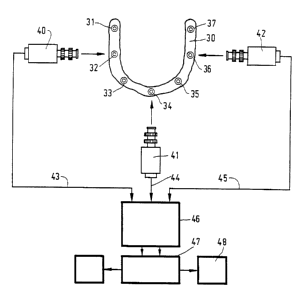
Fig. 3 shows a diagrammatic top plan view of the arch of a
jaw 30 in which seven inserts 31-37 have been fitted in the
implants tnot shown in the drawing). These inserts 31-37 will
generally have their top surfaces disposed at different levels,
while further the longitudinal axes of the implants, and hence
the longitudinal axes of inserts screwed into them, will almost
never run parallel.
As Fig. 3 shows, a number of cameras 40, 41, 42 have been
arranged around the arch 30, all disposed in the same plane,
approximately in the plane of the arch 30. They are special
cameras, such as for instance pixel cameras or ccd cameras, in
which the image obtained with an optical lens is projected on a
screen and is converted into a series of electronic signals
through electronic scanning procedures.
The number of cameras shown in Fig. 3 is three, which is
adequate to obtain a good survey of the different inserts. From a
theoretical point of view, however, two cameras are sufficient.
On the other hand, it has also been found to be possible to use a
single camera, which is then swivelled around the mouth at short
intervals into at least two accurately defined positions and
makes the pictures in succession.
Each camera is in communication with an interface 46 through
a corresponding connecting cable 43, 44, 45, which interface 46
can provide for the conversion of the signals into digital form.
It is also possible, however, to utilize a very modern camera, in
which the recorded images appear at the output terminals directly
WO 95103007 ~ ~ ~ ~ ~.
PCT/NL94/00173
in digital form. The interface :~6 is connected to a powerful
calculating unit 47, which provides an analysis of the received
signals in that the received electronic signals are processed and
combined in coordinates of the different inserts, and their axes '
5 and their top surfaces, and the calculating unit 47 transmits
these data, again in digitized form, as recording functions to a
recording device 48, in order to be recorded there on a suitable
recording medium such as a magnetic tape or possibly a diskette.
The calculating unit is provided with software which is known per
10 se, for determining coordinates defining the position and
orientation of the implants and/or inserts. In particular, the
relative orientation and position are determined, i.e., the
orientation and position of the implants relative to each other.
Because the implants and/or inserts have predetermined
dimensions, the calculating unit can process the information
about these dimensions in combination with the above-mentioned
position and orientation information for obtaining the
information f or determining the dimensions of a prosthesis which
can be fixed to the implants free of stress. If the dimensions of
the implants and/or the inserts are not known, these too can be
determined, in accordance with the invention, by photogrammetric
route, but this will generally yield less accurate results than
the preferred embodiment outlined above. If the dimensions of the
implants and/or the inserts are predetermined, this information
can also be used in known manner to recognize the inserts and/or
implants by the photogrammetric method (pattern recognition),
which makes it possible to accurately determine the above-
mentioned position and orientation information.
Preferably, recordings of implants are taken when they
comprise inserts. Preferably, the inserts are cylindrical, while
the calculating unit determines ellipse variables of the
circumf erential edge of an insert and determines the position and '
orientation information on the basis of these variables. These
variables can, for instance, be the variables of dimension, '
flattening, and angle.
WO 95/03007 216 810 4 PCT/NL94/00173
11
If an object provided with a number of calibrated optical
recognition points is introduced into a patient's mouth, these
points can function as reference. As described hereinbefore, a
number of implants are provided with at least one optical
recognition point. This is understood to include inserts which
are connected to the implants and are provided with optical
recognition points. These recognition points are used for the
photogrammetric determination of the orientation and position. In
particular, for the purpose of the photogrammetric determination
of the course of the longitudinal axes of the inserts, these are
provided with calibrated optical recognition points at their top
surface. In addition, a recognition means can be attached to an
implant or inserts, the recognition means being provided with
calibrated optical recognition points. As will be discussed
hereinafter, these optical recognition points have, for instance,
a predetermined position relative to the implant and/or insert
when the recognition means is attached to it. As a consequence,
on the basis of the photogrammetric determination of the position
of the recognition points, the orientation and position of the
associated implant and/or insert can be determined.
The above-mentioned magnetic recording medium, after the data
associated with the patient in question have been recorded, is
taken out of the device 48 in order to be utilized in a different
place and at a different time for controlling a five- or six-axis
turning and milling machine for mechanically machining a metal
part of the later prosthesis, for instance a crosspiece, on which
the superstructure is subsequently fitted. The drive of the
turning and milling machine takes place in accordance with the
values of the above-mentioned coordinates as found by the
photogrammetric route, which is known per se, in a manner so
accurate that in the product the position and the orientation of
the later fixing holes correspond with the recorded situation in
the mouth to within a few microns.
In order that a fixed reference be available when recordings
are being made, it is preferred, in the practice of the method
WO 95/03007
PCT/NL94/00173
12
according to the invention, to use a measuring scale shown in
more detail in Figs. 4a-4e.
Fig. 4a shows a small bar 50 used for this purpose, showing a
trapezoidal shape in side elevation;
Fig. ~b shows the top plan view of the bar 50;
Fig. 4c shows the right-hand end view of the bar;
Fig. 4d shows a pin 59 with a round head 58, to be screwed
into an insert; and
Fig. 4e shows the assembled measuring scale screwed on an
insert 10.
This measuring scale, as a recognition or identification
means during photogrammetric work, accordingly consists of a
'small bar 50, of substantially rectangular cross section, which
bar, on three sides thereof, viz. the two sides 51, 52 and the
top side 53, is provided with a row of optical marking points 54
of very minor dimensions, which marking points 54 have been
engraved very accurately, for instance by means of a laser beam.
The diameter of the marking points 5~ is, for instance, 100 to
200 microns. The relative distances of these points 54 are
calibrated. To increase the contrast, the bar 50 has, for
instance, been colored dark blue and the marking points 54 have
been colored white. The bar 50, at the blunt end 55 thereof, is
provided with internal thread, in which a screw knob 56 can be
manually turned by the implantologist. At the underside the bar
50 comprises a spherical recess 57 for receiving therein a round
head 58. This round head 58 forms the top end of a pin 59
provided, at the lower end thereof, with screw thread 60 for
screwing the measuring scale into an insert 10 on the jaw of the
patient (Fig. 4e) .
To make recordings with the cameras, the assembled measuring
scale according to Fig. 4e along with the insert 10 is screwed
into one of the implants placed in the arch of the jaw. At this
point it cannot be predicted whether the axis of the selected
implant is vertical. However, in order that, in the case of a
non-vertical axis, the bar 50 nevertheless extends level in the
~WO 95103007 PCT/NL94/00173
13
mouth as far as possible, the bar 50 is tilted about the round
head 58 of the pin 59 screwed into the insert 10, until it has
the desired level orientation and is then secured with the screw
knob 56.
The recordings by the cameras derive their scale of reference
from the calibrated distances of the measuring points 54.
Fig. 5 shows a recognition means 80 comprising at least two
spheres 82 which have been fixedly positioned relative to each
other and are mounted on a pin 8~. At its lower end 86, the pin
8.~ is provided with screw thread and can thereby be secured to an
implant andjor insert in the mouth of a patient. The white
spheres 82 represent the actual measuring objects. They off er a
good possibility of providing contrast and can be automatically
located and measured. In this connection it is important that a
spherical shape is imaged as a circle under any angle of view of
a camera. This will facilitate the automatic measurement of the
centres of the spheres as a representation of the insert axis.
The inclined orientation of the insert and/or implant can be
derived from the coordinates of the centre of the two spheres.
For this purpose, it is important that the pin and the spheres be
accurately in line. If the distance between the centres of the
two spheres is known as a fixed measure, this method at the same
time provides an elegant solution for the provision of scale in
the images. This can be realized by making the recognition means
in one piece. In particular, the recognition means further
comprises a plate 88 to indicate the proper height of the insert
and to cancel any play in the screw thread. Optionally, the plate
can be painted black and so serve as a contrastive background to
the white spheres. Alternatively, an elastic black backdrop can
be slid over the spheres. Also, the edge of this plate can be
ribbed, so that the indicator can be easily screwed into the
insert. In addition, from the centres of spheres associated with
different implants and/or inserts, the relative position and/or
orientation of the corresponding implants and/or inserts with
respect to each other can be determined.
WO 95/03007
PCT/NL94/00173
14
The invention, in teens of its application, is not in any way
limited to the manner of fixing a superstructure as discussed
with reference to Fig. 1, but can equally successfully be applied
to a covering prosthesis as shown in Fig. 6.
Here, for instance four implants have been placed in a
patient's mandibular arch 100. Two implants are shown in the
drawing in the front of the arch and indicated with ref erence
numerals 101 and 102. The implants 101, 102 and possibly others
are connected to each other with a crosspiece construction 103,
which is bent in this example, for instance of substantially egg-
shaped cross-section with the small end directed downwards, which
comes to lie somewhat above on top of the wall of the jaw. In the
example shown, this crosspiece 103 is fixed onto inserts in the
implants, for instance by means of small screws. The crosspiece
103 and the holes provided therein are made using the above-
described photogrammetric recording methods and the CAM methods
and can later be secured on the implants 101, 102 in the arch 100
entirely without stress.
The actual prosthesis 104 is of the clip-on type and to that
end comprises a metal base having at the underside thereof a
concavity complementary to the contour of the crosspiece
construction 103. Accordingly, this prosthesis 104 can be clipped
around the crosspiece 103 onto the jaw 100 with a close fit.
Other forms of implants and prostheses, too, lend themselves
for use of the invention.
While recording the coordinate data by means of the cameras
arranged around the opened mouth of the patient, it is possible -
for the purpose of increasing the accuracy of the measurement
inasmuch as a better spatial impression is thereby provided - to
move the entire arrangement parallel to itself in upward
direction over a slight distance, say a few centimeters.
Immediately thereafter, again recordings are made from this
slightly higher position. By comparing the signals, the software
governing the calculating unit can determine the coordinates of
the positions of the different inserts and the measuring scale
~WO 95/03007 21 b 810 4 PCT/NL94/00173
unequivocally and with great accuracy. The height and the
orientation of the top surfaces of all of the inserts can be
determined very accurately, which is indispensable in the
fabrication of the prosthesis to provide f or stress-free
5 placement in the mouth.
The recorded data coming from the calculating unit are made
available to a five- or six-axis turning and milling machine. By
means thereof, a crosspiece or connecting plate can be made which
subsequently forms a perfect close fit with the measured insert
10 surfaces and is provided with throughbores perfectly in line with
the axes of the implants and inserts as placed. By the use of
this advanced technique, the three-dimensional coordinates are
accurate to within 20-30 microns.
The photo cameras are basically achromatic electronic image
15 'recording tubes. To obtain the desired information with regard to
the minuscule recognition points, a good depth of focus is
essential, which imposes stringent optical requirements on the
optics of the lens and the diaphragm. Because the recognition
points on the inserts and on the measuring scale have been made
white, a high brightness sensitivity of the target inside of the
camera, on which the light impressions are collected via the
optical lens, is essential. Because these objects to be recorded
basically do not move, the operation of the target may otherwise
be fairly slow. The achromatic images recorded by the cameras are
transmitted to the interface as a video signal containing the
requested information, in order to be converted in the interface
into the digital form which is fed to the calculating unit.
The photogrammetric equipment is,'of course, arranged at the
implantologist~s. The data recorded on magnetic recording medium
are used in the dental laboratory in the manner analogous to that
known as computer aided manufacturing (CAM) for numerical control
of the suitable production machine.
An important advantage of the invention is that it eliminates
the occurrence of situations where superstructures have to be
made again, implants have to be re-made because they do not fit
WO 95/03007 PCT/1VL94/00173
16 '
or cause stresses in the arch of the jaw as well as the loss of
implants with all the harmful health consequences thereof. The
technique described can naturally be used as well for patients ,
with superstructures on natural elements.
Accordingly, this entails the advantages that the dental ,
laboratories can work more accurately and even in those
situations can preclude internal errors. It then prevents
products having to be remade because of the laboratory~s own
mistakes. This development will lead to a saving on labor time
and cost, also f or the dentist. From a health service point of
view, too, this aspect is not unimportant. In addition, the
method according to the present invention can be qualified as
more hygienic and patient-friendly in all respects.
A major advantage is also that stress-free superstructures
clearly prolong the life of implants. Further, a well-nigh
unlimited range of applications in the medical field is possible.
In the development of dentistry this method is a major step
f onward .