Note: Descriptions are shown in the official language in which they were submitted.
~1~3~14
FIBER OPTIC DIFFUSE LIGHT REFLECTANCE SENSOR
Field Of The Invention
The present invention generally relates to the field
of medical diagnostic equipment used in clinical chemistry.
More particularly, the present invention relates to a
sensor used in a visual imaging system that analyzes a
light reflectance change associated with one or more test
pad areas on a reagent test strip following contact thereof
with a liquid specimen containing occult blood.
Backaround Of The Invention
Reagent test strips are widely used in clinical
chemistry. A reagent test strip usually has one or more
test areas (pads), and each test area is capable of
undergoing a color or brightness change in response to
contact with a liquid specimen. An analyte is reacted with
the reagent strip in order to ascertain the presence of one
or more constituents or properties of interest in the
analyte. The presence and concentrations of these
constituents of interest in the specimen are indicated by a
change in the test strip when reacted with the analyte.
Light reflected off of the reacted reagent test strip is
analyzed to determine if the constituents of interest are
present and in what quantity. Usually, this analysis
involves a color comparison between the reacted test pad
and a color standard or scale. In this way, reagent test
MSE-1898
~~73014
- 2 -
strips assist physicians in diagnosing the existence of
diseases and other health problems.
Reflected light comparisons made with the naked eye
can lead to imprecise measurement. Today, reagent strip
reading instruments exist that employ reflectance
photometry for reading test strip changes. These
instruments determine the color change of a test strip, but
only with limited resolution. Color variations smaller
than the resolution of current instruments can be
critically important for diagnosis, yet undetectable with
current instruments. For example, such an instrument can
fail to detect traces of non-hemolyzed blood within a urine
specimen. Reagents like the occult blood pad on a
Multistix~ 10 SG reagent strip of the type sold by Miles
Inc., Diagnostics Division, of Elkhart, Indiana 46515
develop small colored spots when reacted with low
concentrations of non-hemolyzed blood in urine.
Concentrations at these levels are commonly referred to as
non-hemolyzed trace (NHT).
After a urine specimen has contacted a test pad of the
Multistix~ 10 SG reagent strip, intact blood cells appear
as tiny green blotches on the orange test area. Existing
strip readers detect the overall color or brightness of the
test pad and large green areas, but ignore the small areas
of green associated with non-hemolyzed blood. Small areas
of green create a spotted appearance indicating that non-
hemolyzed cells in the urine have hemolyzed on the reagent
MSE-1898
- 3 -
test paper. Currently the NHT condition, i.e., 5 to 15
cells/~cL(microliter), is not consistently reported, thus
producing false negative readings by automated urinalysis
instruments. Furthermore, in hospital environments, NHT
occurs in approximately one of every 15 urine samples and
is only detected about 50% of the time. Current detectors
can detect NHT only down to about 15 red blood cells/~CL,
which is not sufficiently accurate for all medical
diagnosis. This is important because presence of non-
hemolyzed blood cells in occult blood is related to the
likelihood of various renal and other disorders. For
example, a more accurate NHT detector would be useful in
detecting such renal diseases such as hematuria,
hemoglobinuria and myoglobinuria. Thus, a higher
resolution diffuse reflectance sensor is highly desirable
as a diagnostic tool.
Resolution in current reflectance sensors is
inherently limited because such sensors have only been
capable of reading one field of view (FOV) of a reagent
pad; meaning, the entire reagent pad is viewed with one
sensor. The problem with one FOV is that valuable color
reflectance information occurring in small areas is lost.
For example, if the optics have a FOV that is limited to a
pad area (typically 0.5 cm x 0.5 cm(centimeters)), then the
optics would not have the sensitivity to detect color
reflectance changes of small area (typically 0.03 cm in
diameter) within the FOV.
MSE-1898
- ~1~3~~4
- 4 -
Some prior art inventions have tried to overcome the
single FOV limitation with a variety of methods. One
approach is to use a moving aperture with a smaller FOV.
An example of a device with a moving aperture is Japanese
Kokai Patent Application No. Hei 5[1993]-80048, filed
September 20, 1991 by Suzuki et al. The Japanese
Application also discloses the use of a bundle of optical
fibers to transmit light reflected from a reagent strip.
However, the application describes using a reading device
with a moving aperture to measure light transmitted by the
optical fibers. A problem with this design is that either
the pad or aperture must be precisely moved to scan the
light reflected from the reagent pad through the optical
fibers. Difficulties with precise mechanical translation
of the pad or aperture negatively affects cost, reliability
and resolution.
Summarv of the Invention
The present invention is a method and apparatus for
providing higher resolution of diffuse light reflected from
a reagent strip. The test pads on the reagent strip are
reacted with an analyte. Light reflected off of the
reagent strip is converted into electrical signals that can
be analyzed with diagnostic equipment. More specifically,
one embodiment of the present invention employs a randomly
oriented illumination bundle of optical fibers to
illuminate the reagent strip with diffuse light from a
MSE-1898
_ 2~~3~~~
- 5 -
remote light source. A baffle and lens system focuses the
diffuse light reflected off of the reagent strip into a
detection bundle of optical fibers at a first end. The
detection bundle's first end cross-section is shaped to
generally match the reagent pad area as focused onto the
first end. The detection bundle's opposite end is arranged
linearly to couple optically with a linear array detector.
The linear array converts the reflected light transmitted
by the detection bundle of optical fibers into electrical
signals for processing. A small reference bundle of
optical fibers is optically coupled to the linear detector
to create a reference signal. The reference signal is used
to prevent significant drift in gain of the linear array
detector. The capability of splitting off some of the
light to create the reference signal is another advantage
in using optical fibers in this way. In one embodiment the
electronic signals are processed to ascertain the presence
of hemolyzed or non-hemolyzed trace blood in the analyte.
Further analysis on the electronic signals, for example to
determine the quantity of hemolyzed or non-hemolyzed blood,
is also performed.
Detection sensitivity is improved to allow NHT
detection of even 2 red blood cells/~L. This is made
possible in part by using slender optical fibers in the
detection bundle which serve to break up the reflected
light into- small representative areas for separate
detection and analysis. Furthermore, each optical fiber is
MSE-1898
CA 02173014 2004-05-25
- 6 -
coupled to multiple detectors to drastically decrease the
field of view for each detector. Because each detector has
a much smaller FOV, much smaller details can be
ascertained, thus resolution is increased. The present
invention provides improved cost, reliability and
performance advantages over current systems.
Brief Description Of The Drawincr
Other aspects and advantages of the invention will
become apparent upon reading the following detailed
description and upon reference to the accompanying drawing,
in which:
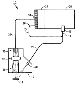
FIG. 1 is a block diagram overview of a fiber optic
diffuse light reflectance sensor according to one
embodiment of the present invention.
Detailed Description Of The Preferred Embodiments
While the invention is susceptible to various
modifications and alternative forms, a number of specific
embodiments thereof have been shown by way of example in
the drawing and will be described in detail herein. It
should be understood, however, that this is not intended to
limit the invention to the particular forms disclosed. On
the contrary, the intention is to cover all modifications,
equivalents and alternatives falling within the spirit and
scope of the invention as defined by the appended claims.
CA 02173014 2004-05-25
-
An embodiment of the present invention is used in a
medical diagnostic instrument to measure diffuse light
reflected from reagent paper that has been reacted with an
analyte, such as urine containing blood. Very small area
color reflectance patterns are capable of detection with
the present invention, particularly those of non-hemolyzed
trace blood cells that develop during a chemical reaction
between the reagent strip and the analyte.
In Figure 1, a fiber optic diffuse light reflectance
sensor 10 is designed with a readhead 12 to reflect light
off of a reagent test strip pad l4 reacted with an analyte.
The light reflected off the reagent test strip 14
originated at a light source 16. In one embodiment, the
light source 16 is a single light emitting diode (LED)
which emits light at a wavelength around 660
nm(nanometers), with a bandwidth of about ~ 13 nm and at a
narrow angle (~5 degrees). For example, the TLRA180AP LED
produced by ToshibaTM Corp. , 1-1 Shibaura, 1-Chome, Minato-
Ku, Tokyo, 105-O1, Japan, was found to be satisfactory as
the light source 16. Note that other sources of light
besides an LED may be used, such as a laser diode or a
quartz halogen lamp combined with a narrow-band filter to
provide monochromatic light. In one embodiment, the
ToshibaTM LED has a desirable high intensity output of
3mW(milliwatts)/cm2 at a 20 mA(milliamps) drive current
which is pulsed on and off using a constant-current pulsed
direct current (DC) power supply. Pulsing the LED
- g -
minimizes LED heating as well as associated LED intensity
and wavelength variation.
Light emanating from the light source 16 is directed
into a light-source optical fiber bundle 18. The light-
s source bundle 18 is comprised of hundreds of randomly oriented
very thin optical fibers each approximately 0.01 cm in
diameter. Note that the phrase "randomly oriented" does
not require the optical fibers be absolutely random in
orientation. The light-source bundle i8 is further divided
into an illumination optical fiber bundle 20 and a
reference optical fiber bundle 22. The reference bundle
Z2, which has an aggregate diameter of approximately 0.03
cm, is used to optically couple some of the light emanating
from the light source 16 to part of a linear array detector
24. A number of pixels at one end of the linear array
detector 24 are illuminated by the reference bundle 22 to
provide a reference intensity signal 26. The reference
intensity signal 26 is used to correct system drift caused
by LED output or detector response variations over time.
The reference bundle 22 therefore enables more consistent
linear array detector 24 performance, which in turn
produces greater measurement resolution of the fiber optic
diffuse light reflectance sensor 10.
The illumination bundle 20 carries light from the
light source 16 to an illumination light baffle 28 in the
readhead 12. Light emanating from the illumination bundle
20 serves to illuminate each reagent strip pad 14 that is
MSE-1898
- 9 -
analyzed. The illumination bundle 20 has an approximate
aggregate diameter of 0.28 cm and terminates within the
illumination light baffle 28. The illumination light
baffle 28 is mounted at a 30 degree angle with respect to a
perpendicular axis of the reagent test strip pad 14,
positioned adjacent the readhead 12, and serves to reduce
stray light as well as improve uniformity of illumination
over the reagent test strip.
Approximately 30 percent of the LED light is collected
and transmitted by the illumination bundle 20. This light
loss is mainly due to over filling the illumination bundle
input diameter by the LED output beam. Also the
illumination bundle 20 numerical aperture (NA) is less than
the LED output NA of 0.25. However, these optical
15 inefficiencies provide for a more fault tolerant light
source 16 to illumination bundle 20 alignment. Minor
misalignment, including tilt of the LED, has minimal effect
on illumination fiber bundle 20 illumination properties.
The reagent test pad 14 is illuminated with diffuse
20 light emanating from the randomly oriented optical fibers
of the illumination bundle 20 at an angle of 30 degrees.
It has been shown that positioning the illumination optical
fibers 20 in the readhead 12 at an angle of 30 degrees, as
opposed to the 45 degree angle used in the prior art,
improves illumination uniformity over the reagent test pad
14. Consequently, improved illumination uniformity
produces a decrease in color reflectance sensitivity to
MSE-1898
- ~l~f~Uf~
- i0 -
variations in reagent strip pad 14 height. Sensitivity to
height variations in the reagent pad 14 from the readhead
12 was reduced from 0.7% R/0.003 cm (prior art) to 0.2%
R/0.003 cm. Prior art readheads typically illuminate test
areas at an angle of 45 degrees. More consistent color
reflectance values are achieved with illumination at 30
degrees than at 45 degrees because the cone of illumination
reflected off the reagent test pad 14 expands and contracts
more rapidly at the larger angle of illumination. The 30
degree illumination is therefore less sensitive to height
and position variations in the reagent strip 14. Less
height and position sensitivity facilitates a more precise
measurement of the light reflected by the reagent strip 14.
The 30 degree illumination angle was selected because
it provided the smallest practical angle. As described
above, a small angle is more desirable in terms of
providing a more uniform reagent pad 14 illumination
intensity. The illumination baffle 28 has an aperture
diameter of 0.36 cm positioned approximately 1.37 cm from
the reagent test pad. These choices were made taking into
account the illumination fiber bundle 20 NA to provide
illumination of the entire reagent strip pad 14 including
some over-illumination to account for pad 14/readhead 12
misalignment in the plane of the reagent test strip 14.
The illumination optical fiber bundle 20 employs a
random optical fiber distribution, instead of other kinds
of distributions such as direct (coherent) distribution,
MSE-1898
23~"~301~
- 11 -
because of the more desirable features associated with
random optical fiber distribution. For example, random
fiber optic distribution, thus randomized illumination,
provides a uniformity of reagent 14 illumination that
varies by only ~ 15$. This configuration breaks up and
more evenly distributes the non-uniform light created by
the light source 16. Uniformity of illumination reduces
variations in signal to noise of each small area color
reflectance field of view detected. This improves
precision of the reflectance measurements.
Furthermore, using optical fibers to illuminate the
reagent pad 14 and carry light reflected from the reagent
pad 14 has the advantage that the illumination source 16 is
remotely located away from a sample area where reagent
strips are analyzed. The optical fibers can be remotely
illuminated with LEDs, laser diodes, or other light
sources. Another advantage of fiber optic illumination is
that the bundle 18 can be divided into a plurality of
smaller bundles as needed.
After the diffuse light has been reflected off of the
reagent strip 14, it passes through a detection light
baffle 30 to a bi-convex lens 32. The detection light
baffle 30 functions to reduce stray light entering the bi-
convex lens 32. It was discovered that using a 2.08 cm
long x 0.36 cm diameter cylindrical element to view the pad
14 and threading this element using a 0.164-32 UNC-2B
thread design provided a suitable detection light baffle
MSE-1898
~17~OI4
- 12 -
30. Multiple reflections within the threaded region
effectively absorbed unwanted light.
The lens 32 to pad 14 distance is preferably at least
0.84 cm. This displacement is necessary to prevent the
detection light baffle 30 and lens 32 from being
contaminated by sample on the pad 14. The detection light
baffle 30 forms a 0.25 cm diameter aperture in front of the
lens 32, thereby improving three performance factors. The
aperture increases the f-number of the lens 32. An
increase in f-number of the lens 32 reduces optical
aberrations versus pad 14 height variation (i.e., improves
depth of field or height sensitivity). The detection light
baffle 30 restricts the FOV of the lens 32 to within the
pad area 14. This helps to ensure that only pad 14
reflected light is imaged onto a detection optical fiber
bundle 34. The detection light baffle 30 also reduces
extreme off-axis light (stray light) from entering the bi-
convex lens 32. Off-axis light originating from the
illumination or ambient room light is trapped within the
detection light baffle 30.
The bi-convex lens 32 collects the reflected light
passing through the detection light baffle 30 and images it
onto an input end 36 of a detection fiber optic bundle 34.
In one embodiment of the present invention the bi-convex
lens 32 has a focal length of 0.64 cm, a diameter of 0.64
cm and the lens 32 is located 2.54 cm from the reagent pad
14. The bi-convex lens 32 produces a 3X magnification,
MSE-1898
- 13 -
therefore, the reagent pad 14 image is enlarged by 3 times
as it is projected onto the input end 36 of the detection
optical fiber bundle 34. The bi-convex lens 32 magnifies
and projects onto the input end 36 of the detection optical
fiber bundle fiber 34 a spot size (from the reagent pad 14
surface) of 0.02 cm. Therefore, the ratio of NFiT spot size
to magnified image spot size is 2Ø The size of a feature
detected on the reagent pad 14 is dependent on the diameter
and number of detection fibers in the detection bundle 34
and magnification of the lens 32. A 2:1 fiber to spot
ratio is desirable for reliable spot detection.
Like the light-source optical fiber bundle 18, the
detection optical fiber bundle 34 is made up of hundreds of
very thin optical fibers held together to form a bundle.
Each individual optical fiber in the detection bundle 34
receives reflected light from a small field of view (FOV)
without moving the pad 14 or detection bundle 34. This
avoids misalignment problems. Optical fibers in the
detection bundle 34 are assembled randomly in order to save
costs. However, the detection bundle 34 can also be
configured as a coherent assembly. At the input end 36 of
the detection bundle 34, the optical fibers are bundled
into a shape that matches that of the image of the reagent
pad 14 as transmitted through the bi-convex lens 32. In
one embodiment the input end 36 is square, however, the
input end 36 of the detection fiber 34 can be constructed
into various shapes, e.g., round, rectangle, etc., that are
MSE-1898
__ ~~'~3~14
- 14 -
consistent with the shape of the reagent pad 14 or pattern
being detected.
The readhead 12 mechanically holds the illumination
and detection fiber optic bundles 20, 34 and baffles 28, 30
as well as the bi-convex lens 32 in alignment. The
readhead 12 component can be molded or machined.
In one embodiment, the detection optical fiber bundle
34 uses 400 randomly oriented fibers that are each 0.01 cm
in diameter. The 400 detection bundle 34 fibers are
assembled into an approximately square (0.1 cm x 0.1 cm)
pattern at the input end 36 in order to match the square
shape of the reagent test pad 14. The given input end 36
size, in conjunction with the lens magnification factor of
3 provides a 0.3 cm x o.3 cm magnified reagent pad 14
image. At an output end 38 of the detection bundle 34,
i.e., the end in contact with the linear array detector 24,
the detection bundle fibers 34 are arranged in a linear
array 0.01 cm high x 2.03 cm long.
The light output of the detection optical fibers 34 is
averaged to determine the overall reflected intensity.
This average color reflectance value indicates the presence
of hemolyzed blood in the sample. Therefore, the sensor 10
detects both hemolyzed and non-hemolyzed blood levels in a
urine sample.
At the output end 38 (opposite the input end 36) of
the detection optical fiber bundle 34, the optical fibers
are linearized into a ribbon or line. In one embodiment of
MSE-1898
__ ~17301~
- 15 -
the present invention the optical fibers are linearized in
order to optically couple with the linear array detector
24. The fibers are mounted directly onto the face of the
linear array detector 24. In an alternative embodiment the
line of detection fibers 34 can be imaged with a lens (not
shown) onto the linear array detector 24. Each fiber must
have one or more corresponding detectors in the linear
array 24 in order to maintain the maximum spatial
resolution. Devices usable as the linear array detector 24
l0 include charge coupled devices (CCDs), photocell arrays,
color CCD arrays, or CMOS (complementary metal oxide
semiconductor) photodiode arrays.
In one embodiment of the present invention a CCD is
employed as the linear array detector 24. Each light
sensing element within the CCD has an electrical response
that is proportional to the light intensity received from
the corresponding detection bundle 34 optical fiber. The
electrical response is utilized by processing electronics
25. The processing electronics 25 serially clock out the
electrical response of the array 24 into an analog to
digital converter (not shown) which in turn converts the
electrical response into corresponding digital data. The
processing electronics 25 also include a microprocessor
(not shown) which stores and utilizes the digital data to
calculate contrast variations indicated by the individual
detection elements in the linear array detector 24. The
number and locations of contrast variations is used to
MSE-1898
CA 02173014 2004-05-25
- 16 -
determine a concentration of NHT or hemolysis in the
analyte tested.
The CCD supports a 2,048-pixel array. For example, a
commercially available CCD array from EG&G ReticonTM Inc., 35
Congress Street, Salem, Massachusetts 01970, was used in
one embodiment of the present invention. This particular
CCD uses pixels 14 ~m(micrometers) wide by 130 ~Cm high,
providing 3.5 pixels per fiber and a 2.54 cm CCD length.
With this arrangement the output intensity of each of the
400 fibers of the detection bundle 34 can be accurately
recorded. Furthermore, additional space near the edge of
the array 24 is available to record dark CCD pixel output
from uncoupled pixels and the reference intensity signal 26
from the reference fibers 22. Dark CCD pixel output is used
to correct pixel response intensity and is then combined
with the detected reference intensity signal 26 to minimize
detector 24 gain effects.
No transfer optics are required between the detection
bundle's linear face 38 and the CCD because the direct
contact method was selected. To accomplish this direct
connection, the linear end 38 of the bundle 34 is bonded
(e.g., by epoxy), directly to the CCD package face. An
index matching material may also be placed between the
output end 38 fibers and the CCD pixels to reduce light
spreading. This arrangement facilitates a smaller sensor
package. Furthermore, this arrangement produces an optimum
CCD output modulation when recording the intensity of a
2.~ "~~01~ - 1~ -
darkened fiber (one observing an NHT spot) that exists next
to a number of bright fibers (negative pad region).
One embodiment of the present invention has the
following specifications:
Non-hemolyzed trace spot size diameter detection
- < 0.03 cm
Bi-convex lens 32 diameter = 0.64 cm
Bi-convex lens 32 focal length = 0.64 cm
Bi-convex lens 32 magnification = 3
Bi-convex lens 32 field of view = 0.3 cm x 0.3 cm
Detection fiber optic bundle 34
Detection fiber 34 assembly is random
Number of ffibers = 400
Fiber diameter = 0.01 cm
Fiber numerical aperture (NA) - 0.25
Detection bundle input end 36 size = 0.1 cm x 0.1
cm
Detection bundle output end 38 size = 2.03 cm x
0.01 cm
Linear Array Detector 24 CCD array
Number of pixels = 2048
Pixel width = 14 gum
Pixel height = 130 um
Array length = 2.54 cm
Thus, there has been described herein a fiber optic
diffuse light reflectance sensor 10.
Obviously, many modifications and variations of the
invention as hereinbefore set forth can be made without
departing from the spirit and scope thereof and therefore
only such limitations should be imposed as are indicated by
the appended claims.
MSE-1898