Note: Descriptions are shown in the official language in which they were submitted.
~1~3719
WO 95/10758 PCTlUS9.t/10855
-1-
METHOD AND DEVICE FOR MEASURING ULTRASONIC ACTIVITY
IN AN ULTRASOUND DELIVERY SYSTEM
Meld of the Invention
The present invention relates generally to medical
devices and more particularly to a method and device for
measuring ultrasonic activity in an ultrasound delivery
system so as to assure proper operation of the ultrasound
delivery system during therapeutic procedures.
Background of the Invention
A number of ultrasonic devices have heretofore been
proposed for use in ablating or removing obstructive
material from anatomical structures, such as blood
vessels. Examples of devices which purportedly utilize
ultrasonic energy, alone or in conjunction with other
treatment modalities, to remove obstructions from
anatomical structures include those described in United
States Patent Nos. 3,433,226 (Boyd), 3,823,717 (Pohlman,
et al.), 4,808,153 (Parisi), 4,936,281 (Stasz), 3,565,062
(Kuris), 4,924,863 .(Sterzer), 4,870,953 (Don Michael,~Fet
al.), 4,920,954 (Alliger, et al.), and 5,100,423
(Fearnot) as well as other patent publications W087-05739
(Cooper), W089-06515 (Bernstein, et al.), W090-0130
(Sonic Needle Corp.), EP316789 (Don Michael, et al.),
DE3,821,836 (Schubert) and DE2,438,648 (Pohlman).
Ultrasound transmitting catheters have been utilized
to successfully ablate various types of obstructions from
blood vessels of humans and animals. Particular success
has been achieved in ablation of obstructions located in
peripheral blood vessels such as the femoral arteries.
Successful application of ultrasonic energy to smaller
blood vessels, such as the coronary arteries, has also
been achieved. Such applications necessitate the use of
ultrasound transmitting catheters which are sufficiently
small and flexible to undergo transluminal advancement
CA 02173719 1999-10-04
-2-
through the tortuous vasculature of the aortic arch and
coronary tree.
Additionally, ultrasound transmitting catheters may
be utilized to deliver ultrasonic energy to blood vessel
walls for the purpose of preventing or reversing
vasospasm as described in Canadian Patent 2,117,176.
Thus, it is apparent that the use of ultrasound
therapeutic procedures provides substantial benefits.
However, a problem commonly associated with the
l0 performance of -such procedures is the inability to
readily determine proper functioning of the ultrasound
delivery system. As those skilled in the art will
appreciate, the presence of the desired level of
ultrasound energy at the distal end of the ultrasound
catheter cannot be determined by visual inspection or
feel. Thus, the operator cannot easily ascertain whether
- or not the ultrasound delivery system is providing the
y desired level of ultrasound energy at the distal end of
. the ultrasound catheter.
It is necessary that the ultrasound delivery system
be functioning properly in order to provide the desired
therapeutic effect. It is common for ultrasound delivery
systems to function suboptimally or not at all due to
looaa mechanical connection of the ultrasound
transmission member, breakage of the ultrasound
transmission member, failure of tht ultrasound
transducer, and failure of the ultrasound generator and
control electronics, as well as for. various other
reasons.
- 30 Various things can happen to the different
components of the ultrasound delivery system during
shipping, handling, and use thereof so as to cause the
WO 95/10758 PCT/US94/10855
-3 -
ultrasound delivery system to function suboptimally. For
example, rough handling of the piezoelectric crystal of
the ultrasound transducer can result in damage thereto
. which causes the system to function at less than the
desired level and which is not readily apparent to the
user. The piezoelectric crystal itself may become
cracked or broken, or the electrical leads thereto may
fail so as to provide inadequate electrical conduction.
Indeed, a wide variety of different types of malfunctions
and component failures may occur so as to render
operation of the ultrasound delivery system suboptimal.
It is also possible for the operator to improperly
set up or operate the ultrasound delivery system so as
to
inadvertently cause the system to operate suboptimally.
For example, an inappropriate level of ultrasound
vibration may inadvertently be selected by the operator,
thus potentially rendering the therapeutic procedure
ineffective. For example, a level of ultrasound
vibration appropriate for coronary procedures may
inadvertently be selected when a peripheral procedure
is
to be performed. The level of ultrasound vibration
commonly associated with coronary procedures is
substantially lower than that generally desired for use
in peripheral procedures. Thus, even though a visual
indication, e. g. , status light or digital readout, of
the
selected procedure may be provided at the signal
generator, it is possible for the operator to overlook
such visual indication and to perform the procedure at
an
ultrasound energy level other than that desired.
Thus, it is possible to perform an entire
therapeutic procedure with an ultrasound delivery system
providing suboptimal or zero output and without the
operator being aware of such problem. Contemporary
methodology provides no means for assuring proper
operation of the ultrasound delivery system during
therapeutic procedures.
2173~1~
WO 95/10758
PCTlUS94/10855
-4-
The performance of such therapeutic procedures with
an ultrasound delivery system providing suboptimal or no
ultrasound energy has potentially serious consequences
for the patient. For example, rather than ablating the
material comprising a stenosis, the distal end of the
catheter may undesirably dislodge portions thereof or may
compact the stenotic material against the vessel walls.
Such breaking away of stenotic material or compaction
thereof may go unnoticed until a serious problem caused
thereby arises. Stenotic debris may potentially form an
embolism, thus impeding the flow of blood to a vital
organ, e.g., the brain. Compaction of stenotic material
may provide a base upon which further stenotic material
may subsequently accumulate.
As such, it would be beneficial to verify proper
operation of the ultrasound delivery system prior to
commencing the therapeutic procedure for which the
ultrasound delivery system is to be utilized.
summary of the Invention
The present invention specifically addresses and
r
alleviates the above mentioned deficiencies associated
with the prior art. More particularly, the present
invention comprises a method and device for measuring
ultrasonic activity in an ultrasound delivery system so
as to assure proper operation of the ultrasound delivery
system during therapeutic procedures. Thus, the
undesirable consequences of equipment malfunction and
operator error are mitigated. The device for measuring
ultrasonic activity in an ultrasound delivery system
comprises a sensor for providing an output representative
of a sensed ultrasound vibration level, an indicator for
receiving the output of the sensor and providing an
indication of the vibration level sensed thereby, and a
rigid body to which the sensor is attached. The rigid
body comprises a sensor attaching portion configured to
receive a sensor and a catheter abutting portion
2113~~~
WO 95/10758 PCTlUS9a/10855
-5-
configured to abut the distal end of an ultrasound
delivery system catheter. The catheter abutting portion,
the sensor attaching portion, and the sensor, taken
together, define a sensing head. Ultrasonic activity of
the ultrasound delivery system is measured by abutting
. the distal end of the catheter to the catheter abutting
portion of the rigid body and then noting the indication
provided by the indicator.
The sensor preferrably comprises an accelerometer
apparatus and the indicator preferably comprises a meter
apparatus. Those skilled in the art will recognize that
various other types of sensors are likewise suitable.
For example, a displacement sensor or a velocity sensor
may alternatively be utilized and the output thereof
optionally converted to acceleration so as to facilitate
use thereof in combination with a meter apparatus
configured to receive an acceleration signal.
Alternatively, the meter apparatus may directly use the
output of such a displacement sensor or velocity sensor.
The meter apparatus preferrably comprises a
voltmeter apparatus configured to provide an indication
of the ultrasound vibration level. Those skilled in the
art will recognize various types of meter apparatus and
indications are likewise suitable. For example, a
digital readout of the acceleration, velocity, and/or
displacement of the distal end of the ultrasound catheter
may be provided.
The sensor attaching portion preferrably comprises
a female threaded coupling and the sensor, e.g.,
accelerometer apparatus, preferrably comprises a male
threaded coupling engaging the female threaded coupling
so as to provide rigid attachment of the accelerometer
to
the rigid body. The catheter abutting portion
preferrably comprises a recess configured to receive the
distal end or end of the ultrasound delivery system
catheter.
WO 95/10758 2 ~ ~ 3 71 ~
PCTlUS9.1/10855
-6-
The recess may optionally be configured to conform
to the shape of the distal end of the ultrasound
catheter. Alternatively, the recess may merely be a
dimple or bore conf figured so as to receive the distal end
of the ultrasound catheter. Those skilled in the art
will appreciate that various different configurations of
the recess are likewise suitable.
The catheter abutting portion is removeably and
rigidly attachable to the sensor attaching portion so as
to facilitate placement of the sensor attaching portion
upon one side of a sterile barrier and placement of the
catheter abutting portion upon the opposite side of the
sterile barrier. The sterile barrier is thus captured
intermediate the sensor attaching portion and the
catheter abutting portion.
The catheter abutting portion is preferrably
maintained in a sterile condition, e.g., disposed within
a sterile enclosure, prior to use thereof. The catheter
abutting portion is preferrably disposable, such that a
new, sterile catheter abutting portion is utilized for
each therapeutic procedure. In the preferred embodiment
of the present invention each catheter abutting portion
is thus maintained in a sterile condition within a sealed
plastic package prior to use thereof and is disposed of
2 5 after each use .
The indicator is preferrably configured so as to
provide an indication of the condition of ultrasound
delivery systems providing various different desired
ultrasonic activity levels so as to accommodate various
different therapeutic procedures. Thus, for example, the
operator may select the particular therapeutic procedure,
e.g., coronary or peripheral, which is to be performed so
as to utilize the device for measuring ultrasonic
activity of the present invention to verify proper
operation of the ultrasound delivery system for the
particular therapeutic procedure to be performed.
CA 02173719 1999-10-04
_7_
In this regard, the device for measuring ultrasound
activity of the present invention preferrably comprises
a selector for selecting the type of procedure in which
the ultrasound delivery system is to be utilized. The
indication of the condition of the ultrasound delivery
system is responsive to the selector. The selector
causes the attenuation or amplification of the sensor
signal so as to provide an accurate indication of the
acceptability thereof.
According to an aspect of the present invention
there is provided a device for measuring ultrasonic
activity in an ultrasound delivery system, said device
comprising: a) a sensor for providing an output
representative of a sensed ultrasound vibration level;
b) an indicator receiving the output of said sensor and
providing an indication of the vibration level sensed
thereby; c) a rigid body to which said sensor is
attached, said rigid body comprising: i) a sensor
attaching portion; ii) a, catheter abutting portion
configured to abut the distal end of an ultrasound
delivery system catheter; and d) wherein ultrasonic
activity of the ultrasound delivery system is measured by
abutting the distal end of the catheter to the catheter
abutting portion of the rigid body and noting the
indication provided by the indicator.
According to an aspect of the present invention
there is provided a device for measuring ultrasonic
activity in an ultrasound delivery system, said device
comprising: a) an accelerometer apparatus for providing
an electrical output representative of a sensed
ultrasound vibration level; b) a meter apparatus
receiving the output of said accelerometer apparatus and
providing an indication of the vibration level sensed
CA 02173719 1999-10-04
7a
thereby, said meter apparatus configured to provide an
indication of the condition of ultrasound delivery
systems providing various different desired ultrasonic
activity levels so as to accommodate various different
S therapeutic procedures, said meter apparatus comprising a
selector for selecting the type of procedure that the
ultrasound delivery system is to be utilized in, the
indication of the condition of the ultrasound delivery
system being responsive to the said selector; c) a rigid
body comprising: i) an accelerometer apparatus attaching
portion to which said accelerometer apparatus is
attached; ii) a disposable catheter abutting portion
comprising a recess configured to receive the distal end
of an ultrasound delivery system catheter, said catheter
abutting portion being removably and rigidly attachable
to the accelerometer apparatus attaching portion wherein
the accelerometer apparatus attaching portion is
disposable upon one side of a sterile barrier and the
catheter abutting portion is disposable upon the opposite
side of the sterile barrier such that the sterile barrier
is captured intermediate the accelerometer apparatus
attaching portion and the catheter abutting portion.
According to an aspect of the present invention
there is provided an improved ultrasound delivery system
comprising: a) an ultrasound signal generator for
providing an ultrasound drive signal; b) an ultrasound
transducer receiving the ultrasound drive signal for
converting the ultrasound drive signal into ultrasound
vibration; c) an ultrasound catheter, having proximal and
distal ends, attached at the proximal end thereon to said
ultrasound transducer, for transmitting ultrasound
vibration from the ultrasound transducer to a desired
anatomical site; d) a device for measuring ultrasonic
activity at the distal end of said catheter; and e)
wherein verification of proper operation of said
CA 02173719 1999-10-04
7b
ultrasound generator, said ultrasound transducer, and
said ultrasound catheter. is provided by measuring
ultrasonic activity at the distal end of said catheter.
According to an aspect of the present invention
there is provided an improved ultrasound delivery system
comprising: a) an ultrasound signal generator for
providing an ultrasound drive signal; b) an ultrasound
transducer receiving the ultrasound drive signal for
converting the electrical ultrasound drive signal into
ultrasound vibration; c) an ultrasound catheter, having
proximal and distal ends, attached at the proximal end
thereon to said ultrasound transducer, for transmitting
ultrasound vibration from the ultrasound transducer to a
desired anatomical site; d) a device for measuring
ultrasonic activity of the distal end of said catheter
said device comprising: i) an accelerometer apparatus for
providing an output representative of sensed vibration
level; ii) a meter apparatus receiving the output of said
accelerometer apparatus and providing an indication of
the vibration level sensed thereby; iii) rigid body
comprising an accelerometer apparatus attaching portion
to which said accelerometer apparatus is attached and a
disposable catheter abutting portion comprising a recess
configured to receive the distal end of an ultrasound
delivery system catheter, said catheter abutting portion
being removably and rigidly attachable to the
accelerometer apparatus attaching portion and disposed
upon one side of a sterile barrier and the catheter
abutting portion disposed upon the opposite side of the
sterile barrier, the sterile barrier being captured
intermediate the accelerometer apparatus attaching
portion and the catheter abutting portion; e) wherein
CA 02173719 1999-10-04
verification of proper operation of said ultrasound
generator said ultrasound transducer, and said ultrasound
catheter is provided by measuring ultrasonic activity at
the distal end of said catheter.
A method for performing therapeutic ultrasound
procedures according to the present invention generally
comprises measuring ultrasound activity in the ultrasound
delivery system prior to commencing ultrasound therapy
and preferrably repeating measurement of the ultrasound
activity subsequent to the therapeutic procedure so as to
verif continued
Y proper operation of the ultrasound
delivery system during the therapeutic procedure.
The method more particularly comprises the steps of
activating the ultrasound delivery system, abutting a
distal end of an ultrasound catheter of the ultrasound
delivery system to a device for measuring ultrasonic
activity, and noting the level of ultrasonic activity as
indicated by an indicator responsive to the device for
measuring ultrasonic activity. The method preferrably
further comprises the step of selecting the desired
procedure to be perfor~aed, the indicator being responsive
to such selection so as to indicate whether the measured
level of ultrasonic activity is sufficient for
performance of the particular selected procedure or,
alternatively, is insufficient for performance of the
particular selected procedure. The level of ultrasonic
activity preferrably is indicated as a fail/pass
indication.
The step of abutting a distal end of an ultrasound
catheter to a device~for measuring ultrasonic activity
preferrably comprises abutting the distal end of the ~
2173719
WO 95/10758
PCT/US9-1/ 10851
_8_
catheter to a catheter abutting portion formed upon a
rigid body, the rigid body having a sensor attached
thereto for providing an output to the indicator
representative of the level of ultrasound vibration
sensed thereby.
The method preferrably further comprises the step of
attaching the catheter abutting portion to the sensor
attaching portion of the rigid body so as to capture a
sterile barrier, e.g., a bag, therebetween. Thus, the
sensor attaching portion of the rigid body need not be
maintained in a sterile condition. The sensor attaching
portion of the rigid body is disposed within a sterile
bag so as to isolate it from the sterile environment in
which the ultrasound procedure is being performed. The
ultrasound transducer is likewise disposed within the
sterile bag and attached to the ultrasound catheter which
extends therefrom. Only the catheter abutting portion of
the rigid body needs to be maintained in a sterile
condition, since the catheter abutting portion is
disposed outside of the sterile bag during performance of
the ultrasound therapeutic procedure. Similarly, the
ultrasound catheter is typically maintained in a sterile
condition prior to use and is likewise typically
disposable.
Thus, according to the methodology of the present
invention, an ultrasound transducer is disposed within a
sterile bag such that a sterile ultrasound catheter
extends from the bag and a rigid body having an
ultrasound vibration sensor attached thereto is likewise
disposed within the sterile bag. A catheter abutting
portion of the rigid body is rigidly attached thereto
such that the catheter abutting portion is disposed
outside of the bag. The ultrasound delivery system is
then activated and the distal end of the ultrasound
catheter abutted to the catheter abutment end member of
the rigid body so as to transmit ultrasound vibration
CA 02173719 2000-02-28
9
thereto. The level of the ultrasonic activity as
indicated by the indicator is then noted and preferrably
provides a fail/pass indication.
The particular procedure to be performed is
preferrably selected at the indicator such that the
indication provided by the indicator is specific to the
level of ultrasound vibration required for the selected
procedure.
Measurement of the ultrasound activity at the distal
end of the ultrasound catheter is preferrably repeated
after performance of the ultrasound therapeutic procedure
so as to verify continued proper operation of the
ultrasound delivery system throughout the procedure.
According to an aspect of the present invention
there is provided a method for measuring ultrasonic
activity in an ultrasound delivery system, said method
comprising the steps of: a) activating the ultrasound
delivery system; b) abutting a distal end of an
ultrasound catheter of the ultrasound delivery system to
a device for measuring ultrasonic activity; and c) noting
the level of ultrasonic activity as indicated by an
indicator responsive to the device for measuring
ultrasonic activity.
According to an aspect of the present invention
there is provided the use of an ultrasound delivery
device for providing ultrasonic activity, said use
comprising the steps of a) disposing an ultrasound
transducer within a sterile bag such that a sterile
ultrasound catheter extends from the bag; b) disposing a
rigid body having an ultrasound vibration sensor attached
thereto within the bag; c) attaching a catheter abutting
portion of the rigid body thereto such that the catheter
abutting portion is disposed outside the bag; d)
. CA 02173719 2000-02-28
9a
activating the ultrasound delivery system; e) abutting a
distal end of an ultrasound catheter of the ultrasound
delivery system to a device for measuring ultrasonic
activity; f) noting the level of ultrasonic activity as
indicated by an indicator responsive to the device for
measuring ultrasonic activity; and g) using the
ultrasonic activity.
According to an aspect of the present invention
there is provided a method of using an ultrasound
delivery device for providing ultrasonic activity, the
method comprising a) disposing an ultrasound transducer
within a sterile bag such that a sterile ultrasound
catheter extends from the bag; b) disposing a rigid body
having an ultrasound vibration sensor attached thereto
within the bag; c) attaching a catheter abutting portion
of the rigid body thereto such that the catheter abutting
portion is disposed outside of the bag; d) activating the
ultrasound delivery system; e) abutting a distal end of
an ultrasound catheter of the ultrasound delivery system
to a device for measuring ultrasonic activity; f) noting
the level of ultrasonic activity as indicated by an
indicator responsive to the device for measuring
ultrasonic activity; and g) using the ultrasonic
activity.
These, as well as other advantages of the present
invention will be more apparent from the following
description and drawings. It is understood that changes
in the specific structure or methodology described herein
may be made within the scope of the claims without
departing from the spirit of the invention.
Brief Description of the Drawin9~s
Figure 1 is a perspective view of the device for
measuring ultrasonic activity in an ultrasound delivery
CA 02173719 2000-02-28
9b .
system of the present invention being utilized within a
contemporary ultrasound delivery system;
Figure 2 is an enlarged perspective view of the device
for measuring ultrasound activity of Figure 1;
S Figure 3 is a cross-sectional side view of the sensing
head of Figure 2, shown in cross-section;
Figure 4 is an exploded perspective view of the sensing
head of Figure 3; and
Figure 5 is a flow chart illustrating the steps for
performing a therapeutic procedure utilizing the method
for measuring ultrasound activity in an ultrasound
delivery system according to the present invention.
Dpta i l ad Da~r~ri nfi nn of rhea Drofcrreri Ti!",l,nra;.nn.,+~
WO 95/10758 ~ 1 ~ ~ PCTlUS9.t/10855
-10-
The following detailed description and the
accompanying drawings are intended to describe and show
presently preferred embodiments) of the invention only
and are not intended to limit the scope of the invention
in any way.
a. A Preferred Vibration Measuring Device ,
As shown in Figures 1 and 2, the device of the
present invention may be utilized to measure the
vibrational output of a medical ultrasound catheter prior
to insertion of the catheter into a mammalian body.
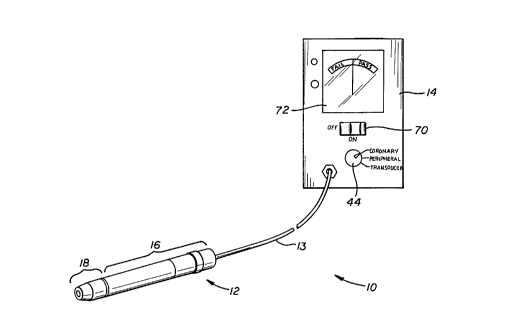
As shown, the device 10 of the present invention
generally comprises a vibration sensing head 12 connected
to a meter apparatus 14.
The vibration sensing head 12, described in more
detail herebelow and shown in Figures 3-4, generally
consists of an elongate rigid housing 16 connectable to
a disposable abutment end member 18.
The rigid housing 16, as well as the abutment end
member 18 and the sleeve 56, are preferrably comprised of
a polymer, preferrably acetal resin (e. g., Delrin'~
manufactured by Du Pont De Nemours , E . I . , and Co . , Inc f ) .
The rigid body member 52 is preferrably approximately
3.150 inches long from the deepest portion of the recess
42 to the accelerometer 50 (dimension A of Figure 3).
The diameter of the rigid body member 52 is preferrably
approximately 0.600 inch (Dimension B of Figure 3).
Those skilled in the art will recognize that various
other substantially rigid materials and various other
dimensions and configurations of the rigid body member 52
are likewise suitable.
The meter apparatus 14 of the device 10 preferably
comprises a voltmeter apparatus having a readout or
display 72 which is calibrated or delineated to indicate
either acceptable (i.e., "pass") or non-acceptable (i.e.,
"fail" ) levels of vibrational energy sensed by the device
10.
WO 95/10758
PCTlUS94/ 10855
-11-
As shown in Figure 1, the device 10 of the present
invention may be utilized in conjunction with a medical
ultrasound catheter system. The typical medical
ultrasound catheter system comprises an elongate catheter
20 having a proximal end and a distal end. A proximal
end connector assembly 22 is positioned on the
roxi
l
p
ma
end of the catheter 20 and is coupleable to an ultrasound
transducer 24. The ultrasound transducer 24 is connected
by way of cable 26 to signal generator 28. Signal
generator 28 is provided with an on/off foot pedal 30.
Depression of on/off foot pedal 30 causes signal
generator 28 to emit an electrical signal through cable
26 to ultrasound transducer 24. Ultrasound transducer 24
converts the electrical signal received thereby to
ultrasonic vibration. An ultrasound transmission member
or wire (not shown) extends longitudinally through the
length of the catheter 20 so as to transmit the
ultrasonic vibration from transducer 24 to the distal end
DE of the catheter 20.
Various untoward circumstances may result in
disruption or mutation of the ultrasonic vibration
transmitted to the distal end DE of the catheter 20. For
example, if the ultrasound transmission member or wire
(not shown) should become fractured or broken, such may
significantly diminish the quantum of ultrasonic
vibration transmitted to the distal end DE of the
catheter 20. Similarly, if the connection between the
proximal connector assembly 22 and the ultrasound
transducer 24 has been disrupted, there will be a
resultant diminution or interruption of the ultrasonic
energy transmitted to the distal end DE of the catheter
20. Also, if the signal generator 20 or transducer 24
were to have been improperly set, or malfunctioning, such
may also result in an incorrect amount of ultrasound
vibration reaching the distal end DE of the catheter 20.
WO 95/10758 ~ PCT/US9-1!10855
-12-
If, in fact, the desired level of vibrational energy
is not being transmitted to the distal end DE of the
catheter 20, it is desirable to determine such fact
before the catheter 20 has been inserted into the
patient. Thus, the device 10 of the present invention
may be utilized to test_,the ultrasonic vibration at the
distal end DE of the catheter 20 prior to insertion of
the catheter so that adjustments or remedial measures may
be undertaken before the catheter 20 is inserted into the
patient.
As shown in Figure 1, it is preferrable that the
catheter 20 be maintained in a sterile condition during
the testing procedure. Accordingly, the accelerometer
apparatus housing portion 16 of the sensing head 12 is
initially inserted into a sterile barrier bag 40 or
sheath defining a sterile barrier, along with the non-
sterile ultrasound transducer 24. One commercially
sterile barrier bag which may be utilized for this
purpose is the Baxter "' Arthroscopy Camera Drape
(Sterile) available from Baxter Healthcare Corporation,
Hospital Supply Division, Deerfield, Illinois 60015.
Thereafter, the sterile disposable catheter abutting end
member 18 is screwed onto the distal end of the
accelerometer apparatus housing portion 16 of the sensing
head 12, outside of the sterile barrier bag 40 such that
a portion of the material of the sterile barrier bag 40
is trapped or clamped between the non-sterile
accelerometer apparatus housing portion 16 and the
sterile disposable catheter-abutting end member 18.
Similarly, the proximal connector assembly 22 of the
catheter 20 is threaded onto and coupled to the
ultrasound transducer 24 with the sterile barrier bag 40
being tightly closed therearound so as to maintain the
proximal connector assembly 22 of the catheter within the
sterile field.
2i 7719
WO 95/10758 PCTJUS9-t/10855
-13-
After the catheter 20 has been operatively connected
to the ultrasound transducer 24, the distal end DE of the
catheter 20 is inserted into the catheter receiving
recess or well formed in the distal end of the catheter
abutting end member 18. The distal end DE of the
catheter 20 is held in firm abutting contact with the
floor of the recess 42 and the on/off foot pedal 30 of
the signal generator 28 is utilized to activate signal
generator 28. Signal generator 28 is typically preset at
a desired output level expected to provide the acceptable
ultrasonic vibration at the distal end DE of the catheter
20.
As the signal passes from signal generator 28
through cable 26, the ultrasound transducer 24 will
convert the signal to ultrasonic vibration. The
ultrasonic vibration then will be transmitted through
catheter 20 to the distal end DE thereof.
Abutment of the distal end DE of the catheter 20
with the floor of the catheter receiving recess 42 of the
probe member 12 causes the vibration of the distal end DE
to be sensed by accelerometer apparatus 50 and converted
thereby into an electrical signal. The electrical signal
is then transmitted through cable 13 to monitor 14 and a
corresponding acceptable/unacceptable indication is
displayed by monitor 14 as a result of the vibrational
energy sensed by the accelerometer apparatus 50 of the
sensing head 12.
Depending on the intended therapeutic application of
the ultrasound system, the signal generator 28 and
monitor 14 may be specifically set to desired ranges or
energy levels pre-determined to be suitable for the
intended therapeutic application. For example, in
' clinical settings wherein the catheter 20 is to be
inserted into a blood vessel for purposes of ablating or
ultrasonically treating vaso-obstructive matter within
the blood vessel, the setting of the signal generator 28
WO 95/10758 2 PCTNS9.1/10855
-14-
may differ depending on whether the obstruction to be
treated is within the coronary or peripheral vasculature.
Accordingly, the mode setting apparatus 44 of monitor 14
may be appropriately set on "coronary" or °'peripheral°'
settings such that the monitor 14 will be thereby
adjusted to seek the appropriate vibrational levels for
the intended "coronary" or "peripheral" use.
Additionally, the device 10 of the present invention
may be utilized to test the vibrational output of the
ultrasound transducer 24 itself, without the attachment
of the catheter 20. When utilized for such purpose, the
mode setting apparatus 44 of monitor 14 will be switched
to its "transducer'° setting and the distal end of the
transducer horn will be inserted into the recess 42 of
the sensing head 12, in firm abutment therewith. As
such, the vibrational energy emanating from the horn of
the transducer 24 will be sent by the accelerometer
apparatus 50 of the probe and, provided that the mode
setting apparatus 44 of the monitor 14 is appropriately
set on the "transducer" setting, the monitor will display
r
an indication as to whether the vibrational energy sensed
by the accelerometer apparatus is within the desirable
range defined for the transducer test.
1i. Preferred Construction of the
Vibration Sensing Head
The vibration sensing head 12 of the device 10 may
constructed and configured in various ways. One
presently preferred mode of constructing the vibration
sensing head 12 as shown in Figures 3 and 4.
As shown, the presently preferred sensing head 12
comprises a detachable abutment end member 18, a rigid
body 52, an accelerometer apparatus 50, an accelerometer
apparatus cable connector 54 and a guide sleeve 56.
A threaded male projection 58 is formed on the
proximal side of abutment end member 18. A corresponding
threaded female bore is formed in the distal end of rigid
2173719
WO 95/10758 PCT/US9-t/10855
-15-
body member 52. By such construction, the threaded male
projection 58 of the abutment end member 18 may be
screwed into the threaded female bore 60 of the rigid
body member 52, thereby pinching or trapping the sterile
barrier 40 therebetween, as shown in Figure 3.
A proximal accelerometer apparatus housing member 62
is mounted on the proximal end of rigid body member 52.
The proximal accelerometer apparatus housing member 62
has an inner bore 66 which is sized and configured to
receive accelerometer apparatus 50 therewithin. A
threaded accelerometer apparatus receiving bore_ 66 is
formed in the proximal end of rigid body member 52. A
corresponding threaded male projection 64 is formed on
the distal face of accelerometer apparatus 50 such that
accelerometer apparatus 50, when inserted into the inner
bore 66 of accelerometer apparatus housing member 62, may
be firmly threaded into bore 66, thereby causing
accelerometer apparatus 50 to be firmly and rigidly
mounted in abutting contact with the rigid body member
52. As such, vibrational energy received by the distal
end member 18 will be transmitted through the rigid body
member 52 and will be sensed by accelerometer apparatus
50.
One commercially available accelerometer which may
be incorporated into the device of the present invention
is the PCB Piezotronics, Inc. , model number 353B18 quartz
shear mode accelerometer apparatus, such as that
available from PCB Electronics, 3425 Walden Avenue,
Depew, NY 14043-2495 having a sensitivity of 10 Mv/g and
a frequency range of 0.35 to 30,000 Hz (+/- 3 Db). Those
skilled in the art will recognize that various other
accelerometer apparatus and/or vibration sensors are
likewise suitable.
An accelerometer apparatus-cable connector 54 is
mounted on the proximal end of accelerometer apparatus 50
WO 95/10758 2 ~ ~ ~ ~ 19 PCT/US9.l/10855
-16-
so as to couple accelerometer apparatus 50 to cable 13.
A male threaded projection 68 is formed on the
proximal end of accelerometer apparatus housing 62. A
corresponding female threaded bore 70 is formed in the
distal end of guide sleeve 56. Optional guide sleeve 56
may then be threaded onto projection 68 so as to surround
and restrain the lateral movement of connector 54, while
allowing cable 13 to pass outwardly from the proximal end
of the sensing head 12.
By the above-described preferred construction of the
sensing head 12, such sensing head 12 may be utilized to
conveniently sense vibrationally energy emanating from
the distal end DE of catheter 20 or from the distal end
of the ultrasound horn of transducer 24.
c. Preferred Monitor Apparatus
In the preferred embodiment of the present
invention, the monitor 14 comprises a vibration meter
apparatus such as PCB series 291 available from PCB
Electronics, 3425 Walden Avenue, Depew, NY 14043-2495.
The monitor 14 may optionally have custom indicia formed
thereon to indicate a Fail/Pass condition. Additionally,
the range selector switch may optionally comprise indicia
indicative of the particular ultrasound therapeutic
procedure to be performed. Thus, according to the
setting of the range selector switch, the vibration
signal received by the meter is attenuated or amplified,
as necessary, so as to provide an indication of
acceptability thereof according to the particular
ultrasound therapeutic procedure to be performed.
d. Preferred Methods of Using_ the ,
Vibration Measuring Device of the Present Invention
The above-described device 10 may be utilized in
various clinical applications for testing the operability
and efficiency of an ultrasound transmitting member or
ultrasound catheter 20.
2173719
WO 95/10758 PCTlUS9-t/10855
-17-
A method for testing an ultrasound catheter prior to
(and preferrably also after) therapeutic use is shown in
the block diagram of Figure 5.
More specifically, with reference to Figures 1 and
2, the ultrasound catheter 20 shown may be tested prior
to use in a coronary artery ablation procedure by the
following steps:
Step 1. Connect ultrasound transducer 24 to signal
generator 28 by cable 26.
Step 2. Set signal generator 28 at desired output level
for coronary ablation procedure.
Step 3. Connect accelerometer apparatus housing portion
16 of sensing head 12 to monitor 14 by way of cable
13. Set monitor 14 on °'coronary" setting and turn
on/off switch 70 to "on" position.
Step 4. Insert accelerometer apparatus housing portion
16 of sensing head 12 into sterile barrier bag 40.
5. Position disposable end member 18 on outside of
sterile barrier bag 40 adjacent distal end of
accelerometer apparatus housing portion 16 and
threadably mount abutment end member 18 onto
accelerometer apparatus housing portion 16 thereby
pinching the surrounding portion of sterile barrier
sack 14 therebetween. The male threads 58 of the
abutment end member 18 may penetrate the bag 40 or
the bag 40 may alternatively remain intact, captured
intermediate the abutment end member 18 and the
accelerometer housing portion 16.
6. Operatively connect the proximal connector
assembly 22 of catheter 20 to the ultrasound
transducer 24.
7. Insert the distal end DE of catheter 20 into
the receiving recess 42 of sensing head 12 such that
the distal end DE of the catheter 20 is in firm
abutment with the floor of the receiving recess 42.
WO 95/10758 ~ . PCTNS9.l/10855
-18-
8. Depress foot on/off pedal 30 thereby actuating
signal generator 28 so as to cause ultrasound
transducer 24 to send ultrasonic vibration through
catheter 20.
9. Observe the display 72 of monitor 14 to
determine whether the sensing head 12 has sensed
ultrasonic vibration at the distal end DE of the
catheter 20 which is within the acceptable or "pass"
range.
10. If the monitor 14 indicates that the ultrasound
vibration at the distal end DE of catheter 20 is
within the acceptable or °'pass" range, the catheter
may then be inserted into the vasculature and
advanced to the desired coronary location for
15 purposes of effecting the therapeutic application.
11. If, however, the monitor 14 indicates that the
ultrasound vibration sensed at the distal end DE of
the catheter 20 is within the unacceptable or "fail'°
range, appropriate steps may then be taken to
20 troubleshoot the system and/or to change the
catheter 20 prior to proceeding with the therapeutic
procedure.
Although the invention has been described herein
with specific reference to presently preferred
embodiments thereof, it will be appreciated by those
skilled in the art that various additions, modifications,
deletions and alterations may be made to such preferred
embodiments without departing from the spirit and scope
of the invention. Accordingly, it is intended that all
reasonably foreseeable additions, deletions, alterations
and modifications be included within the scope of the
invention as defined in the following claims.