Note: Descriptions are shown in the official language in which they were submitted.
,.~.. 21 l ~ 5 2 3
- 1 - TY052
8llCfCGROVND Op T~ INVENT=Orl
1. Field of the Invention:
. ~, The present invention relates to a plasma separa-
tion filter, a plasma separation method usir~ the filter
and a plasma separation apparatus comprising the lilter.~
More speoilioslly, the present invention relates to s
plasma separation filter capable of easily, speedily and
safely oolleot3nQ s ~onll quantity o! plasma necessary
!or blood tests or the like. The plasma contains sub-
stantially no blood cells and/or hemoglobin and retains
vubstantiaily the same composition of protein end eleo-
trolytea as in the blood. The present invention also
relates to s plasma separation method using the filter
and a plasma separation apparatus comprising the filter.
2. Description of the Related Arts
Hioohemioal tests, which measure components in
blood, are widely utilised !or diagnosis and observation
of pxoprsss for a variety of diseases, and occupy'an
important position as a clinical test. Analysis tech-
niques !or the biochemical tests have been significantly
~ewloped in recent years. For Esample, the development
of a variety of automatic analyzers has enabled a number
of specimens to be accurately and speedily analyzed.
Howwer, in some biochemical taste, the contami-
nation o! erythrocytes or the like interferes with~the
analysis of a targeted substance. Thus, plasma or serum
which is prwiousiy separated frors blood is used. The
plasma or serum for a teat is obtained by collecting
r., 21 ? ~ ~ ~ ~.
TY032
- Z -
blood f's~ra a patient, followed by coagulation and cen-
trifugation of the blood cell components. The operation
of the coagulation and the aantrifuQstion takes a long
period of time, and thus not only prevents the psriod~of
time .for the clinical test to ba short~nad, but also
requires a lns~s sanled oantrifugal machine, xccording~
ly, excopt for large hospitals, the alisriaal test is _
generally only performed by external laboratories at the
present. As n result of such outsourcir~ of the tests,
several days are required to acquire the test results.
' Thus, despite the automation of a number of pro-
asssas for aliniaal taste, the separation of plasma is
still mostly manually performed at the present. Thare~.
fore, the operation of saparatin9 plasma disadvantageous-
ly makes not only the aliniaal tests ineffiaiant, but
also puts a person involve4 at risk such as infection.
caused by oontaCting an infected blood.
ZO A tsohniquw generally celled dry chemistry ie
known as a means for solving the above-mentioned problem.
According to this technique, when a trace amount of blood
is dropped onto a sonail plate consisting of a serum
separation leyer~lorn~ed of a micro fiber filter roads of
glass fibers or the like and a reeation lnyar located
beneath the~serurn separation layer, the serum is separat-
ed in the strum separation layer. Then; the serum is
rsaeted in the underlyir~ reaction layer and colored,
then measured by a spectrophotometer. In such dzy
chemistry, a liquid type coloring reagent is not used,
nor is troublesome serum aoliaction by aentsifuQatioa
reqcsired. Alth~ouQh the dry ahemistsg is such a simple
method, it has the following disadvantages: the number of
CA 02178523 2000-11-24
- 3 _ TYCO 52
measurable items is limited when compared with general biochemical analysis
and immunological analysis using the liquid type reagent; a number of plates
are required in order to test a plurality of items because one plate is used
for
one test item, thus impairing the advantage of shortening the operation time;
and the dry chemistry is expensive. Accordingly, the dry chemistry is not
widely used.
One example of means for speedily obtaining plasma is a
separation method using membranes. Japanese Laid-Open (Kokai) Publication
No. 53-72691 (Laid-Open Publication Date: June 28,1978) discloses a method
for separating plasma from blood by using a fme tube-like filter device (pore
diameter: 0.05 to 0.5 Vim) having one closed end as a filter medium. In this
method, however, blood cells deposit on the surface of the membrane.
Accordingly, a long period of time for filtering plasma is not only required,
but
also permeability of components such as protein contained in the plasma is
poor. On the other hand, when filtering pressure is raised in order to raise a
filtering rate, hemolysis (a phenomenon that erythrocyte membranes are
ruptured and hemoglobin inside the erythrocyte is released) adversely occurs.
Furthermore, Japanese Laid-Open (Kokai) Publication No. 60-
11166 (Laid-Open Publication Date: January 21, 1985) proposes a method in
which a filtering cartridge (pore diameter: 0.05 to 1 Vim) employing hollow
fiber membranes is used so as to separate plasma from the blood. However,
this method requires a priming (wetting the hollow fibers with saline) before
separation. Thus, problems arise in that not only the preparing operation
before
the separation takes more time than the plasma separation itself, but also
since
CA 02178523 2000-11-24
_4_ TYC052
the obtained plasma is diluted by the saline, accurate analysis data cannot be
obtained.
In the aforementioned separation methods using membranes, a
permeability of a relatively large molecular weight substance such as protein
in blood is low because the separation is performed based on the molecular
size. Thus, a composition of protein contained in plasma does not accurately
reflect the original composition of protein in the blood. In addition, when
the
pore size of the membrane is too large, hemolysis adversely occurs due to
erythrocytes clogged.
Other proposed techniques for separating serum or plasma for
clinical tests using a fiber filter are as follows. Japanese Lai-Open (Kokai)
Publication No. 61-38608 (Laid-Open Publication Date: February 24, 1986)
discloses a solid-liquid separation instrument formed of fibers using a volume
filtering effect. In the solid-liquid separation instrument, plasma can be
obtained by allowing blood to flow through the fibers while applying pressure.
However, since pressure loss is large and thus resistance of a filter medium
is
large, several minutes are required to obtain plasma. In addition, since
protein
in the blood is adversely adsorbed in the fibers, a concentration of protein
in
the plasma obtained at an early stage is reduced. Thus, the solid-liquid
separation instrument has not been practically used yet.
Japanese Laid-Open (Kokai) Publication Nos. 4-208856 (Laid-Open
Publication Date: July 30, 1992) and 5-196620 (Laid-Open Publication Date:
August 6, 1993) disclose a separation filter including glass fibers containing
polyacrylate derivatives and polyethylene glycol, and lectin impregnated
layer, a
2 ~ T~
TY052
- 5 -
method for separating and aollaating saru~n or gleams
oomponsats using the filter end a device for separating
serum or plasma using the separation filter. Although
those methods and devices can OOlleot serum yr plasma for
clinical tests without performing Centrifugation, the
serum or the plasma is obtained in amounts as amali ao
about 100 iii, and in addition, the period of time' re-
quired for t-,he separation is about 2 a~nutvs. This is
not so different from s period of time reauired when
aentrifuQntion is performed. Furthermore, since these
techniques use pleas fibers as a separating medium,
electrolytes era eluted from glass fibers and blood
components are adsorbed to the fibers. As a result,
conoentrstiona of electrolytes, phosphorus and lipid iri
obtained plasma or serum ere significantly different from
those is the original blood. For this reason, those
tsahniquss are sot widely used.
As deaaribed above, no lifter provides satisfac-
tort' performance to efficiently and safely separate
piasma_or serum components for use in clinical tests from
a small amount of blood for a short period of time
presents a sufficient performnnee at the present.
~ 81J~11RY OF T83 IHVEHTIOtt
The purpose of this invention includes
(1) providing a plasma eepnration filter capable of
easily, speedily and snfely separating plasma from blood
and exaelleat in assamblabilityf (2) providing a method
using such n filter; and (3) providing an apparatus
comprising such a filter.
217853
TY0.52
- 6 -
AccosdinQ to the present imrvntion, play
components having the same component oomposition as in
the blood~can be obtained without damnging blood culls in
the blood. The present invention provides a method for
svpar~tting blood oomponents.uainQ a micro fiber medium.
The msahnnism of separating plasma using the micro fiber
medium aopprdin~ to the present irnrantion is. to generate
a differenos in the moving rates between erythrocyte and
plasma moving in the micro fiber medium. The difference
in the movir~ rates is attained by optimising faatorv
such as materials of the micro fibers, the average fiber
diameter and the size of dap between fibers, the length
of the blood oell separation layer, the form of the mioro
fibers, the direction of blood flow, the improwmvnt in
the surface of the fibers. As a result, plasma 3n the
blood is separated fr~n the blood componvnto such as
erythrocytes and collected. Furthermore, since the
prvvsure los~ of the filter o! the present invention is
low, the blood id speedily treated, end the oonoantra-
tione of electrolytes and protein in the obtained piasrma
are substantially the same as that of the blood without
separation. ACCOrdingly, the pr~sent invention makes it
possible to obtain plasma eQuivslent to the plasma
obtained by en ordinary aentrifuQation. I;erainaft~r, the
present invention will by dvsaribvd in detail.
The present invention relates to a plasma separa-
tion filter. Thv filter according to the prevent inven-
tion comprises a micro fiber medium 1n a container having
an inlet and en outlet, whorein
(1) a ratio (L/D) of a blood flow passage length (L) to
a blood flog passage diameter (D) o! the micro fiber
217~~~~
TY052
medium iv 0.15 to 6s and
(Z) an sveraQe hydraulic radius of the micro fiber
medium is 0..5 to 3.0 pm.
~..isi ono preferred embodiment, the plasma separa-
tion filter comprises the eharaateristio that when fresh
bovine blood having an erythrocyte concentration of 6 to
8 x 10'/ml is separated at a pressure of 0.2 to 0.4 kQ/cml
and plasma filtrate iv collected in an amount equivalent
to 10~ of s pore volume of the micro fiber medium,
(a) a ratio of an erythrocyte concentration in the
plasma to the erythrocyte concentration in the blood
begore the separation iv 0.1~ or lesss and
(b) erythrocytes are not substantially hemolyzed.
Furthermore, the~~piasma separation filter com-
prises the characteri~tio that when plaaaa is collected
' under the above-mentioned conditions, a difference
between an electrolyte concentration in the separated
plasma and that in the plasma obtained by centrifugation
iv lass than 10~, or a difference between n protein
concentration in the plasma obtained at the start of
filtration, that in the plasma obtained at the and of
filtration and that in the plasma obtained by centrifuQa-
tion is less than 10~r.
tn one preferred embodiment, the micro fiber
mediws is made of polyester, polypropylene, polyamide, or '
polyethylene. c
~rosa
- 8 -
In another prelerred embodiment, the micro fiber
medium is a form of a nonwovsn fnbria.
In still another preferred embodiment, the single
or multilayered nonwovsa fabric is placed is the contain-
es, and blood flows substantially in parallel to a faoa
of the single or multilayered nonwoven fabric of the
mioro fiber medium.
In yet another preferred embodiment, the ~ntain-
er is a disk-like container, and blood flown from s
ai~cuaferant3al portion toward the centrnl portion of the
micro fiber medium plnaed in the coatairur.
Furthermore, the present invention relates to a
filter is which a hydrophilla agent is immobili$ed to the
micro fibers.
~In one preferred embodiment, the hydrophilic
substance is immobilized to the surface o! the micro
fibers.
In another preferred embodiment, the hydrophilic
substance is polyvinyl pyrolidone, and the filter com-
prises the following characteristic that:
(1) a rstio (L/D) of s blood flow passage length (Lj to
a blood flow passage diameter (n) of the micro fiber
medium is 0.13 to 6s end
(2) sn average hydraulic radius of the miarv fiber
medium is 0.5 to 3.0 arm. .
~1T~~~~
TY051
- 9 -
In one preferred wnbodia~ent, the filter in which
the hydraphiliv substance is immobilized to the micro
fibers caaaprises the follo~oing oharaoteristio that:
. when fresh bovine blood having an erythrocyte
oonosntrntion of 6 to 8 x 10~/ml is aeperatad at n
prssaure of 0.2 to 0.' kg/am' and plaems filtrate is
collected in as amount equivalent to 10~ o! a pore volume
of the micro fiber medium,
(a) a ratio of an erythrocyte voncentration in
the plasma to the erythrocyte concentration in the blood
before the separation is 0.1; or less; and
is (b) erythrocytes are not substantially hemolyzed.
Furthermore, the pla8ma separation filter oom-
psises the oharaateristio that ~hsn plaame ie collected
under ths_ nbovs-mentioned conditions, a difference
betoveen an aleatrolyte eonaentration in the separated
plasma and that in thd plasma obtained by centrifugation
is 1~se than 10~, or a difference bet~rean a protein
aono8ntration in th~ pla9ma obtained at the start of
filtration, that~in the plnama obtained et the end of
filtration and that in the plasma obtained by centrifugs-
tion is leas than 10~.
In one praferred.embodimant, the micro fiber
madiuaa is made of polyester, polypropylene, polyamide, or
polyethylene.
xn another preferr~! ombodimerrt, the micro fiber
medium is a form of a nonwoven fabric.
21 %8~,~~3
r-,
TY052
- 10 -
In still another preferred embodiment, the single
or multilayered nonwoven fabric is placed in the oontein
ar, and blood flows substantially in parallel to a plane
face of the single or multilayered nonwoven fabric of th~
micro. fiber medium.
In yet another preferred embodiment, the eontain-
er and the nonwoven fabric are shaped like a disk, end
the inlet is lc~rawd auah that blood is supplied across
the entire aide lace of the perimeter of the disk shaped
nonwoven fabric, and the outlet is formed such that
separated piasmt ie diacha~rged from the aentrnl portion
of the disk shaped nornaoven fabric.
Furthermore, the present invention relates to a
method for eeparatinQ plasma using the piasyna separation
lifter comprising the above-mentioned characteristics, or
the plasma. separation filter in which the hydrophilic
substance ie immobilized to the micro fibers.
In one preferred embodiment, a filter obtained by
placing the single or muitilayered nonwoven fabric in the
container is used.
, In another preferred embodiment, a filter wherein
blood flows substantially in parallel to a face of the
single or muitilayered nonwoven fabric o! the micro fiber
medium is used. Furthermore, a filter wherein the
container is a disk-like container, and blood flows from
a aireumf~rantial portion toward the central portion o!
the micro fiber medium placed in the container is used
for the method of tho present invention.
!~,
~ro5a
- 11 .-
in still another preferred embodiment, a linear
velocity of blood to by treated is 0.05 to 50 cm/min.
Furthermore, the present invention relntas to an
apparatus comprising the plasma separation filter having
the above-mentioned characteristics, or the plasma
separation filter in which the hydrophilic substance is
immobilized to the micro fibers.
In cnv preferred embodiment, the apparatus of the
present invention further comprises blood supplying means
for supplying blood to the filter, pressurizing means for
pressurizing the blood supplied to the liltvr and/or
depressurizing means for reducing pressure at the fil-
trate aids in order to separate plasma from the supplied
blood, and plasma draining means for draining the sspa-
ratvd plasma.
~n another preferred embodiment, the apparatus of
the prosent invention further comprises blood and/or
hemoglobin detecting means for detecting blood cells
and/or hemoglobin in the separated plasma, switching
means for fractionating plasma contaminated by blood
cells and/or hemoglobin, and blood cell and/or hemoglobin
contaminated plasma draining means for draining the
fractionated plasma contaminated by blood cells and/or
hemoglobin.
In still anofihar prelerred embodiment, in the
appsratu, of the present invention, the filter is
rvlsasably provided between the blood supplying means and
the plasma collecting means.
217~~23
TY052
- 12 -
=n yet another preferred embodiment, the appara-
tus of the present invention comprises blood supplying
means for supplying blood in a pradatarmiasd amount,
plasma Collt~otinQ atvan~ for collecting plasma in a
predetermined amount, or both of the means.
Theaa and other advantngu of the present irrvan-
tion will baooam< apparent to thoaa skilled in the art
upon rasding and under4tanding the following detailed .
description with reference to the aooompanyinQ figuros.
HRIE1 DESCRIPTION Og T83 DR71WIh1Q8
F3gure 1 is a view illustrating en ~emplery
filter used in a plasma separation apparatus according to
the present invention.
Figure Z iv a view illustrating an axamplsry
lilter used im a plasma separation npparatua accordiap to
the present invention.
Figure 3 is a view illustrating the filter in
Figure Z with a base medium filling a space.
, Figure 4 is a view illustrating nn exemplary
filter used in a plasma separation apparatus aocording to
the present invention.
Figures Ss and bb are views illustrating esewpia-
ry filtors used in s plasma separation apptratua accord-
ing to the present invention.
F3guras 6a and 6b are viawa illustrating the
/~'~
TY052
- 13 -
filters is Figure 4 further inoludinp pressurizing means.
Figure ~ is a view illustrating an apparatus
including the filter in Fi~ura 1 connected to blood
supplying and pressurising means.
Fi~ura 8 is a schematio view illustrating an
euamplary plasma separation apparatus according to the
present invention.
Figure 9 is a aohmatia view illustratirsg
wcsatplary struoture automating the plasma separation
apparatus according to the present invention.
DSBCRiPT=OH.OF T8: pR~p~Oa=fig
The blood used in the present invention generally
includes components o! blood cells, plasma and the like.
The blood can be derived lrom .any origin including a
20' human, n bovine, n goat, a canidae and n rabbit. The
blood can be used as it is, or the blood containing an
additive such as as anticoagulant and an erythrocyte-
agglutinant ears be used. Ptsrthermore, in the cage where
the blood is kept without an anticoagulant, or in the
Z5 base where a coagulant is added to the blood, fibrinogen
in the blood is changed to fibrin, and the coagulation o!
the blood proceeds. This type of coagulated blood can be
also used as it is. Furthermore, the blood which has
been subjected to a chemical treatment after centrifuga-
30 tion or the like can be used.
The plasma used in the present invention refers
to plasma which contains substantially no blood cell.
217823
TY052
- i4 -
fhu=. the present invention is not limitad to plasma
which contains strictly no blood call at ail. Further-
more, in the case where the blood is coagulated and then
solid components are separated and removed, serum con-
s taininq no fibrinogen can be obtained. Thus, in the
pruent invention, a term "plasma" inaludas saran unless
it speoifiaally refers to plasma only,
Hereinafter, s plasma separation filter, a plasma
separation method and a plnsme separation apparatus will
be described in this order.
(Plasma separation filer)
~1 plasma separation filter of the present inven
ts tion includes a micro fiber medium and a container having
an inlet and as outlet. The micro fiber medium is placed
in the container.. An average hydraulic radius of the
micro fiber medium is preferably 0.5 ~n.to 3.0 pm, nacre
-prelerabiy 0.S pCn to 2.5 pm, and most prelerabiy 0.5 pCa
to 2.0 pen. Herein, an average hydraulic radius refers to
a concept in piece of a diameter in the case where a pore
O! the micro fiber medium does not have a shape of a
circle. . The average hydraulic radius is defined as
followsa
,
Average hydraulic radius
~ Cross-sectional area of tube /
circumterential length of tha tubs
~,Volume o! liquid in tubs / inns= surface
area o! tube in contact pith the liquid
~ Volume o! pore of porous member / surfaw
~17~~2
TY05Z
- 15 -
area of pore of the porous member
According to the present invention, the average
hydraulic radius is cnlculatad with the following formula
(1): .
DR ~ R x (p - rm) / 4 rm (ly
wherein DH indicates an awrsQe. hpdrnulio radius o! a
micro fiber madium in a container (pm), R indicates as
average fiber diameter of a micro fiber ( pm ) , p indicates
s density of the micro fiber ( Q j~a~ ) , and rat indicates an
avereQe bulk density of the micro fiber medivu~t in a
container (Q/cm~)
1'~
As~ shows in Formula ( 1 ) , the awrnge hydraulic
radius DH of a micro fiber medium is a container is
determined by R and rm in' the cans vrhere micro fibers
made of :one type o! mnterial are usod (i.e., p is con
stunt).
zn the case o:here the average hydraulic radius
azCeeda 3.0 pm, blood cells pass through fiber gaps more
easily. Thus, the filter having an average hydraulic
radius more than 3.0 ptn cannot sepnrntn plasma from
blood.
In the Case where the average hydraulic radius is
below 0.5 ~, the fiber gap o! the filter, i.e., a flow
paasaQa of blood is too nnrrov~, so that blood cells nra
easily olo9ged in the flow passages. Furthermore, When
the filter is pressurized in order to increase the
quantity of the blood passing throvQh, pressure loss is
1 l'8~23
TY052
- 16 -
increased and hemolyaia easily occurs.
In the awrape hydraulic radius range of 0.5 pm
to 3.0 pie, a smaller awraQe hydraulic radius affects
less p~rmeabiiity of components having s small particle
diameter such as plasma, at the same Lima, components
having a large particle diamatsr sueh as blood cells are
difficult to pass through the filter. Thus, the evaraga
hydraulic radius is preferabiyØ5 pm to 2.6 yua, and most
l0 prefsrebly Ø5 1sm to 2.0 ym.
Furthermore, the averapa hydraulic rsdiu, o! the
micro fiber medium of the present invention can be
vonstant in the axis dissation from the supply side o!
blood to they outlet side of plasma, or can be varied
depending on the portion of the micro fiber medium.
Moreavar, the awraga hydraulic radius can become gradu-
ally smaller from the inlet toward the outlet. With rush
a~ structure, separation sf~iciency between blood cell
components and plasma ooaaponents in the vicinity o! the
outlet can ba enhanced.
In the present invention, the average hydraulic
radius re!~rs to an average hydraulic radius of the micro
fiber medium when the micro fiber medium is placed in a
container having an inlet and an outlet, and can be
substantially involwed in plasma separation. Thus, in
the case wham a ' micro fiber mediuaa is used as a base
medium lY for filliaQ a space 16 in Figure Z, the micro
fiber medium used as the base medium 19 (as shown in
Figure 3) is not involved in plasma separation. Accord-
ingly, the awraga hydraulic. radius referred.to in the
present inwntion is an average hydrauliv radius of the
''1 21 %8523
TYOSa
mioro fiber medium excluding that used for the base msdi-
wa i9.
In other words, when all the micro fiber mediums
placed in the filter are considered, some mioro fiber
mediums have average hydraulic radii beyond the prefera-
ble range. However, the loot that plasma can bs sepa-
rated even in this case indicates that nt least part of
the micro fiber mediuaes placed in the oontainar hoe sn
average hydraulic radius in the preferable range.
=n the present invention, s prefiltsr can be
provided to remove contaminants in the blood before the
micro fibor medium for plasma separation. The nvwrage
oeeulum diameter and the average hydraulio radius of the
prefilter is naturally lerper than the average hydraulic
radius of the micro fiber medium. xowswr, when an
average hydraulic radius as the entire filter ie deter-
mined, .the average diameter o! the preliiter is not
a0 ooneidered, but the average hydraulic radius of the main
filter should be used.
In the Dresent invention, the micro fiber medium
refers to the state where micro fibers aro irregularly
a5 e~Qregated. Such a state can 1» obtained, for example,
by compressing, for example, mess, nonwovsn, woven,
knitted mioro fibers independently or in combination.
Ths micro fiber medium ie preferably nonwovsn fabric or
mass of m~,aro fibers in view of moldability,
30 proeessability, easiness of handling and difficulty of
ahennaling alter packed in a container. Particularly,
nonwovsn fabric is preferable. When the nonwoven fabric
is placed in a filts~ case, uniformity is easily main-
2 0 ~~
TY052
- 18 -
twined, end sparse portions are unlikely to be gansreted,
whereby blood flow is uniformalized.
A materiai.for the micro fibers is not limited,
but .exemples of the matsrinl include polyester,
polypropylene, polyamide or polyethylene and the like.
The material. is preferably hydrophobio polypropylene sad
polyesters (e.Q., polyethylene terephthelata). The
nbowe-mentioned materials are preferable beonuse when the
materials oontaot blood or plasma components are not
adsorbed to the materials, or a part of the materials is
not eluted in the plasma. Jas described in the section of
Prior Art, when plasma or asrum separation filter o!
glass fibers is used, electrolytes are eluted from the
plnss fibers, or phosphorus or lipid is sdaorbsd to the
glass fibers, so that the resultant substances cannot
provide aoourate measurement results.
The length of the blood osll separation layer in
the present invention is preferably 5 mm or more. The
length of the blood oaii separation layer refers to the
length from the point where the micro fiber medium
contacts blood to the point where the blood (plss~ma)
leaves the micro fiber medium. As desoribed above, the
present invention utilizes a difference in the mcvi~ag
rates between the bioo4 oomponents in the micro fiber
medium to separate plasma from the blood. Pressure is
applied fr~n the inlet,of the blood separation layer, or
prusure isreduced from the outlet thereof, or both of
the operations era simultenaously performed so that blood
is allowed to flow in the micro fiber, medium. Then,
blood call oomponents-repeatediy collide with the micro
fibers while flowing through the qap of the mioro fibers,
~1~~~~3
TY03Z
- 19 =
Adhesive leukocytes and platelets are adsorbed to the
micro fibers, and orythrooytss, ~rh,ioh i4 not aQhesive,
are repeatedly transformed while moving. On the other
hand, since plasma is a liquid component, the plasma more
rapidly moves through the micro fibers than erythrocytes,
and reach the outiet'sariier than the erythrocytes, When
the length of the blood~cell separation layer is 5 mm or
less, a sufficient diffarenCe in the moving distance
between the blood cells and the plasma is not generated.
Therefore, the separation between the blood cells and the
plasma is insufficient. Thus, the. length of 5 mm or lass
is not preferable. As the length of the blood cell
separation 7.ayer becomes larger, the efficiency of
separation between the blood cells and the plasma becomes
higher. On the other hand, problems arise in that
pressure loss is raised, or a required amount of micro
fiber medium or a~required amount of blood is increased.
Therefore, the leaQth of the blood separation layer is
determined by o required amount of plasma. a blood amount
to be used, the limitations of the size of the filters or
the like, but the upper limit does not theoretically
exist.
In th~ case where nonwoven fabrics are used, it
~,g preferable that blood floors in parallel to a plane
face of the nonwovsn fabric (plane face of the stacked
nonwoven fabrics). In general, when the nonwoven fabric
is used, the direction to which liquid to be treated
flows is vertical to the plans face of the nonwoven
fabric (platur face of the stacked nonvvovan fabrics ) .
However, in t~u present invention, by allowing blood to
flow in parallel to the plans face of the nonwoven
fabric, the efficiency of the separation between blood
TY052
- 20 -
cells and plasma components is enhanced, When blood is
allowed to flour in parallel to the face o! the nonwoven
fabrics, it is bolieved that the uniformity of the blood
floor is ia~pxovod, because the micro fibers are preoent
without intermittence over the entire flow passage length
when the blood flows from the inlet to the outlet.
However, the reason why the psrallel flotv is 'preferable
is not limited to the above-mentioned reason.
The aviaro fibers to which a hydrophilic substance
it immobilized osn be preferably used as a filter of the
present invention.
The immobilisation o! the hydrophilic substance
can be phyoicaily or Chemically performed. 8y immobilis-
ing hydrophilic subotanoa to the eurfnce o! the micro
fibers, the affinity betwun the micro fibers and blood
is enhanced. Thus, when plasma is separated from the
blood, pressure loss can be reduoed,_"and the eeparatioa
rate can be raised, l~ny hydrophilic substance can bs
uead, as long as it Boas not interfere with anslysis when
it is contaminated into plnsma. Polyvinyl pyrolidone is
preferable. Although polyvinyl pyrolidone is eluted to
the blood with a relatively low rate because o~ a rela-
tively lnrQa molecular weight, the elution o! polyvinyl
pyrolidone does not affect the enalysie of the blood
components. Ths method for immobilizing polyvinyl
pyrolidone is not particularly limited, but any known
method can be used. For.auample, polyvinyl pyrolidone is
easily immobilized to the surface of the fibers in such
a physical mnnnar that the micro fiber medium is dipped
in a solution of polyvinyl pyrolidone, and dried.
Furthermore, the micro fiber medium with such polyvinyl
2178523
TY052
- 21 -
pyrolidone physiaaliy immobilized on the surface thereof
is aubjsatad to a heating treatment, and/or n radiation
treatment, so that polyvinyl pyrolidonos oan be easily
cxosslinkad. The crosslinking can further suppress the
elutipn of polyvinyl pyrolidons to the blood.
The method for the heating treatment is not
limited. 8samples of the haetinQ method inolude a method
for heating under pressure suoh as an autoclave treat-
meat, a method for keeping in a tank at a .constant
. temperature and the like. Moreover, the temperature of
the heating treatment is not particularly limited, but
preferably 70'C or more, and more preferably 100'C or
mora. As the heating temperature is higher, the
orosslinking effiaianoy is improved. The upper limit of
the temperature is not simply determined because it
depends on the property of the micro fibers to bo used or
the heat resistance of polyvinyl pyrolidona, but prefera-
bly 200'C or less and suors pr~farabiy 150'C or lass. A
period of times for heating is preferably long so that
crosslinking is sufficiently formed, but restricted by
the.property of tho micro fibers to be used or the
denaturalization of the polyvinyl pyrolidone. In. gener-
al, the period of time is preferably in the range of 20
spin. to 2 hours. Furthermore, the crosslinking can be
formed by heating in both cases where the micro fibers
are immersed is the hydrophilic substance solution (wET
state), or where the micro fibers are dried after the
immersion (DRY state). xn either case, polyvinyl
pyrolidone oan be immobilizes!! to the micro fiberer.
Unreacted polyvinyl pyrolidone is removed by washing pith
water.
178~2~3
TY052
- 22 ..
The method for immobilizing the hydrophilic
substance using radiation ig riot particularly limited.
Examples of the ~nathod include Y ray irrndiation, elec-
tron beam irradiation, corona discharge and the like.
The x ray irradiation is preferable in terms of the
thickness, to be treated and its operstability. Arr
irradiation amount is not particularly limited~either, as
long ns polyvinyl .pyrolidone can be sufficiently
crosslinked. ~~owevar, the irradiation amount is prefera-
bly in~tha rangs,o! 10 KQy to SO KOy, because the micro
fiber materia7.s end polyvinyl pyrpiidons are not dena
tured by such a radiation in the range. Moreover, the
irradiation can be performed in the WET state or DRY
state. Unresated polyvinyl pyrolidone caa be removed by
is wnshing with water.
A variety of .polyvinyl pyrolidone with vnrious
molecular weights are available. In order to prevent
polyvinyl pyrolidone fraan being eluted to the blood,
polyvinyl pyrolidone having a large molecular weight is
particularly preferable.
A filter is produced using a micro fiber medium
with the hydrophilic substance immobilized.
,
The filter is produced by stacking and aompreea-
ing mass, nonwoven, woven, Or knitted miCxo fibers inde-
pendently or air combination.
=n the prea~nt invantioa, the shape of the
container o! filters is not particularly limited,
examples of the shape include reotanaular psralieiopipsd,
disk, cylinder, truncated cone, fan shape end the like.
,~1 21 X8323
TY052
- 23
In the case where s rectangular paralielopiped, disk or
fan shaped filter is used to allow the blood to flow in
psraliel to the faae~of the micro fiber medium, sopara_
tion perforeaance can be improved. In the case of a
r~CtanQular parallelopiped shaped filter, blood is
allowed to flow froea one end to the other end of the
rectangular parnllelopipad. 1~11.tsrnatively, in the case
of a disk shaped filter and a fan shaped filter, blood is
allowed to flow fraaa the perimeter portion to the central
portion. Hy pressurizing nonwoven fabriCa using Contaia-
ars of such shapes, the filter Can be sealed. Thus, such
shapes are particularly prefarsble because it to unnecsa-
sary to use an adhesive. Particularly, s fan or disk
shaped container is more preferable for the following
reason: ns the blood is moved, the cross-sectional area
of the flow passage of the blood becomes gradually
smaller. !~a a result, unevenness of the lateral movement
of the blood components is decreased. Especially, the
disk shaped aontniner is moat preferable in that its
operatability is excellent. In the case o! the disk
shaped container, the nonwoven fabric plaasd in the
container is also shaped like a disk. It is preferable
that the inlet for introducing the blood is formed in
such a manner that the blood can be supplied across the
entire perimeter of the disk shaped nonwoven fabric. For
example, a apace is provided between the perimeter of the
disk shaped nonwoven fabric and the perimeter of the 4isk
~Ded Gonteiner s0 that One Or. a plurality Of inlets
Communicating with the apaea can be provided in th~ aide
face, top feae or bottom face of the container.
An average fiber diameter of mi.CrO fiber~ used in
tho filter of the present invention is preferably 0.5 ~
2178523
TY052
- 24 -
to 3 . S pm, more preferably 0 . 5 dun to 2. 8 pm, and most
preferably 0.5 pin to 2.0 um.
The micro libers having the above-m~ntion~d
overage fiber diameter can be. obtained by an ordinary
spinning method suoh as Meltblow.
Herein, an average fiber diameter of the micro
fibers refers to an average value obtninad by anlculating
diameters of 50 micro fibers randomly selected from a
photographed micro fiber medium enlarged to a 2000-fold
'size by a scanning electron microscope, using calipers or
n magnifier.
When the average fiber diameter of the micro
fibers ssceads 3.5 pm, a length per unit volume of the
roioro fibers o! the micro fiber medium becomes shorter.
As n result, the number of intermingled portion~ in the
fibers is reduced, and a fiber gap is enlarged. Aocord-
ingly, components having a larger particle diameter such
as blood cells are likily to pass through the micro fiber
medium, resulting in insufficient separation between
blood cells end plasma.
23 ~ In the ease where the average fiber diameter of
the micro fibers is less than~0.5 pm, n length per unit
volume of the micro libers becomes longer. As n result,
the number of intermingled portions in the fibers is in-
crensed, and~a fiber gap is reduced. Accordingly, blood
cells are likely to be clogged. Furthermore, since
pressure loss of the micro fiber medium is increased,
hemolysis of erythrocytes is likely to occur:
-. 2178523
TY052
25 -
An nv~srape bulk density of the micro fiber medium
used in the present invention is preferably 0.15 to 0.60
Q/cm', more preferably 0.18 to 0.50 q/cm', and moat
preferably 0.25 to 0.60 g/cm'. .
Herein, the average bulk density refers t0 a
value obtained by dividing a weight of the micro fiber
medium by a volume of the micro fiber medium.
In the case where the average bulk density is
smaller than 0.15 Q/cm', s difference from an as spun
average bulk density (e.Q., 0.08 Q/cm' to 0.10 Q/cm' in
the Case of the Meltblow spinning method ) pf the micro
fiber medium is amnll. As s result, a compression ratio
of the micro fiber medium becomes small, l~ccordingly,
dense portions and sparse portions are likely to b~
generated fn 'the micro fiber medium, rssultinQ in uneven-
ness in moving rate4 of the blood. in addition, since
the fiber gap is everaqely large, the separstion between
blood cells and plasma is insuffiaisnt.
In th~a case where th~ average bulk density of the
micro fiber medium is more than 0.60 Q/cm', a special
process such as heating compression is required for
producing the micro fiber medium, thus.compliaating the
compression process. Moreover, since the fiber gap of
the micro fiber medium bsaomes small, blood cell compo-
nents are likely to be clopQed in the filter. In addi-
tion, since pressure loss o! the micro fiber medium is
inareaead, hemolysis is likely to occur.
xn the average bulk density rsn~ of 0.15 to
0.60 Q/cm', by inaeasinQ the average bulk density, the
2~ ~~~;~~
TY05z
- 26 -
uniformity of the mioro fiber medium ie fyimD~d,
whereaa proasssability is deteriorated. Thus, tho
awrage bulk density is preferably 0.18 to 0.50 g/cm', and
most preferably 0.25 to 0.40 g/cm'.
The awrape bulk density of the micro fiber
autdium placed irt the filter of the present invention can .
be varied depending on the portion of the filter. For
exampl~, the average bulk density van by gradually
incr0aead from the inlet to the outlet of the oontainar
of the filter. With such a structure, separation effi-
oienoy between blood cells and plasma can ba higher as
the blood components maw toward the outlet.
He for a low passage for blood comments is the
micro fiber medium placed in the filter of the apparatus
o! the present invention, the ratio (L/D) o! a flow
passage length ( L ) to a flox passage diameter ( D ) is 0.15
to 6, preferably 0.25 to 4, and most preferably 0.5 to 2.
Rerein, the flow passage length (L) of th~ blood
components refers to a straight distance in the inside of
the micro fiber .medium from the point where the blood
contacts the micro fibers to the point where plasma
leaves the micro fibers (generally, length of the micro
fiber medium). The flow passage diameter (D) of the
blood oomponent9 r8fers to a circle-equivalent diameter
of the arose-sectional area on the ~urfaoe, of the micro
fiber medium to the inlet portion of the blood extending
is the dirsation perpendicular to the flow passage
length.
The cirale~~squivalent diameter is obtained by
2178523
TY052
- 27 -
using the cross-sectional area (A) in the following
fos~aule ( 2 )
(A / s)ii' (2)
8trietly speaking, the micro fiber medium surface
has oonaavss and oonvsxev due to the curve of the micro
fibers. However, the cross-seationai area is calculated
by ignoring the concave: and aonvexev and avsuming the
surface as a plane. Ia the case where the micro fiber
median: has large concave: and oonvexes formed by vurlace
procevsing other than those due to the curve of the micro
fibers, the arose-sectional area is calculated by averag-
ing the ooncaves and eonvexes so ae to obtain a plane.
In the ease where L/D is smaller then 0,15, the
components o! the blood move in a short distance because
the flow passage is too short. Moreover, since the
cross-sectional area iv large, moving rates of the
oo~mponents are not uniform in a lateral direction. Thus,
the separation between blood cells and plasma is insuffi-
cient.
=n the case where L/D is larger than 6, separa-
xion efficiency is improved. However, since the moving
distance is longer, pressure loss is increased. Accord-
ingly, hemolysis is likely to occur.
The filter of the present invention hat the
following chsraateriatio*:
Whori frevh bovine blood hsvl,nQ an erythrocyte
concentration of 6 to 8 x 10' /ml is separated under a
,.. 21 l ~~2~
TY052
- 28 -
pressure of 0.2 to 0.4 KO/cm=, and at the point where
plasma filtrate is oolleoted in as amount equivalent to
10% of a pore volume of the micro fiber medium,
(a~ the erythrocyte concentration is the plnamn
is 0.1% or less with respect to the erythrocyte oonaen-
tration in the blood without separation; and
(b) the erythrocytes ore not substantially
hamolyzed.~
Difference betwun nn electrolyte ooncentraticn
in the separated plasma and that in the plnsma obtained
by oentriluQation is less than 10%.
I5
=n the prsaant invention, it is profarable that
90% or morn o! a conaentratioa oi~ electrolytes in the
plascaa seper~eted by the plasma separation liltar is
retained when oompared with a conoentration of eleotro-
7.ytes in ordiz~ry plasma or serum obtained by centrifugn~
tioa. Namwly, the dilfereno0 in the eiootrolyte concen-
tration after tho separation by the filter is less than
10% when Compared with the separation by oentrilugation.
Five % or less is more preferable. When the difFerence
i,n the electrolyte oo»centration in the plasma axouds
10~t, reliability of-a bioahemioal diaQ»osis becomes low.
Thus, the diflerencs of 10% or more is not prstsrable.
=n view of measurement accuracy of biochemical tests,
when the difference betwesa the eleotrolyte concentration
in the piasraa separated by the filtor and that obtained
by csntrifuQatioa is leas than 10%, no problem virtually
occurs. The difference og 5%. or less presmnts substan-
tially r~o probles. Herein, the plasma refers to a
'~ 2 ~ ~'$ ~~.3
TY052
- 29 -
supernatant obtained by centrifugation eftar an anticoag-
ulant is added to the aolleoted blood. Usually, the
Centrifugation is performed nt I000 0 for IO min.
. In the present invention, it is preferable that
difference of a protein concentration in the pls~
obtained at the start of filtration, at the end of
filtration and obtained by centrifugation is less than
10i. Namely, the difference in the protein aonoentra-
tions niter the separation by the filter is less. than 10~
when compared with the separation by aentrifuQatioa.
Five ~C or less is more preferable. When the difference
in the protein aonoentration in the plassit esp~s 10~,
reliability of a biochemical diagnosis becomes low. =n
addition, a value at the surly stags of collection may be
different from that at the time of co~mpietion of the
aolleotion, whereby an accurate diagnosis cannot bs con-
ducted. Moreover, when the difference in the protein
Concentrations sxaeeds 10~, the composition of plasma
protein is likely to be significantly changed, thus
making it impoasibia-to uav in diagnosis of disease.
Thus, the difference of less than I0~ is preferable, be-
Cause, generally, the difference of less than 10~ does
not cause a serious problem in CliniCai diagnosis, and
the difference of 5% or less is in the rungs of measure-
ment error.
A material of the container of the filter of the
present invention is not limited. Bsamples of the
material include metal, glass, plastics such as poiyvth-
y~.~. polypropylene, nylon, polycnrbonate, polystyrene,
polyester and an AHS resin. in the case phere observa~
tiOn of the inside is desired, a transparent or svmi-
~~~'~~~3
TY052
- 30 -
transparent material may be selected. Plastics arc
preferable in view of prooessability, anti~breakability,
and light weight.
S . Hereinafter, the !liter used is the plasms
s~parntion ~apparetus of the present invention will be
described with reference to the accompanying drawinQa.
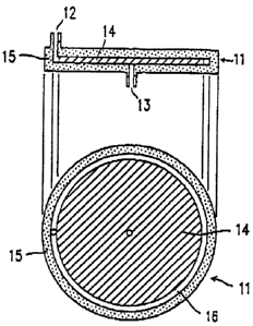
The filter of the present invention is most
simply shown is Figure 1, in Figure 1, a filter 11
inaludss a container la having an inlet iZ and an out-
let 13, end a disk-like micro fiber mediu~a 14. The micro
fiber mediuat 14 is pieced in the ooatainsr 15. in this
filter 11, the micro fibers era compressed in the c:on-
tainar 16 so that a suitable space 16 is formed between
the inner circumfarantinl aurfeca of the container 15 and
the outer circumferential surface of the micro fiber
~ediuca id. The outer circumferential portion of the
micro fiber medium i4 can be formed of, for ~arnp~.o, a
plastic plate having ~uitebla holes capable of pnaa3ng
blood through. When blood is auppiiad from the inlet is
of the contairu~r located above the oiroumferential
portion of the disk-like micro fiber medium 14, the blood
passes through the spas ib so as to be uniformly sup-
2s plied to the micro fiber medium 14. Then, the blood
flows from the outer circumferential portion to the
central portion of the micro fiber medium 14. Duri~ the
course of the flow, pla:ma,and blood cells are separated,
and the plasma is collected after being discharged from
the outlet Z3 in the centre! portion of the container 15.
Hereinafter, the "micro fiber medium ~,4" refers to the
micro fiber medium which is placed in the Biter...
TY052
- 31 -
Figure 2 shows s filter ii as another embodiment.
The filter 11 includes a Cylindrical micro fiber msdiu-
m 14 piaoed in a oyliadrical container i5. Blood is
supplied from an inlet is of the container 15, and plasma
separated by the mioro fiber medium if is collected after
being discharged frog an outlet 13 of the container 18.
The inlet 12 and the outlet I3 can be loaeted in arbi-
trary positions.
The filter in Figure 2 inoludes a space 16 in the
vicinity of the inlet 12, as in the Biter ire Figure 1.
The spaoe 16 can be provided in order to uniformly
perform the supply of blood, pressurizntion or the like.
A filter Z1 in Figure 3 is a filter inaludtng a
suitable base medium 1~ planed in the apace 16 of the
filter ii in Figure Z. Far the boss medium i~, any
materinls oan be used, as long as it has larger fiber
gaps-than the micro fiber medium 14, and all coaaponents
of the blood can flow therethrough. The base medium can
be, for example, a plats-like, paper-like, tabor-like
filter medium. In the filter in Figure I, the base
medium 19 oan bs placed in the space 16.
, A filter Il in Figure 4 includes a micro fiber
medium 14 in a container 15 having an opening portion i8
and an outlet 13.
The filter 11 can be releasnbiy provided in blood
34 supplying means and/or plasma aolleoting mesns described
later. As shown is Figures sa and 5b illustrating
another embodiment, the container 13 can: consist o! three
portions, i.e., a portion 15a including en inlet 12 or an
TY052
- 32 -
opeais~ portion 18, a portion including a micro fiber
medium 14, and a portion idb including as outlet 13.
Each of the portions can releasably form the contnin-
ar is. With such a structure, the micro fiber medium
portion can be independently exchanged. The exchangeable
micro fiber medium can be coated with a blood permeable
membrane or the like, and take a form of a rsiensnbla
cassette capable of being engaged. Examples of the
releasable attaching means inolu4e engagement, thread and
l0 a magnet. The release can be automatically performed.
As described above, in the case whore the filter
of the present invention ie produced by stacking micro
fibers, the direction of blood flow can be substantially
vartiaal to the stacked face, or substantially parallel
to the stacked face. Suitable shapes for the filter are
column or cone-like shape in order to alloy blood to flow
vertical to the stacked face. By using filters of such
shapes,., for example, the blood is allowed to flow from
the upper portion to the lower portion of the cylindrical
filter (i~iguree Z to 4). Alternatively, the blood is
allowed to flow from the bottom to the apex of the cons
of the truncated cone. The truncated cone is obtained by
vetting off the apex portion in a cone shape. On the
Other hand, when the filer obtained by stacking the micro
fibers to form a disk (e.g., Figure 1) is used, blood is
allowed to flow in parallel to the stacked face from the
periphery to the canter of the filter. Alternatively,
the filter obtained by stacking the micro fibers to form
a plans is used,~blood ie allowed to flow in parallel to
tho stacked face from one side to the other side o! the
filter. Zn the truncated oonianl or disk~lfke fiites,
since the cross-sectional area of the blood flow passage
TYOS2
- 33 -
is gradually reduced, moving rates of blood oo~aponente
are raised as the blood moves, and unevenness of movement
of the blood components in a interal direction is re
duced. 1~ a result, separation efficiency of the blood
is improved.
The plurality of filter of the present invention
can be connected. When the filters are connected ~in
series, the separation performance is improved. When the
filters are connected in parallel, s quantity of the
blood which can b~ treated is increased.
Furthermore, the filter can be coupled with a
sampling container. Such a filter coupled with the
sampling container can be used in nn automatic analysis
aDDaratus ineludir~ moans for collecting s predetermined
amount of blood and supplying the blood to the filter and
means for automatically exohnnQing~the used filter with
a new~ono.
(plasma separation method)
In order to eepnrnte plasma, a pressure loss
batwsen as inlet and an outlet is preferably in the range
of 0.03 to 5 kg/em=. The pressure loss depends on the
shape of filters end the treating rate of blood.
In the Case where the pressure lose is smaller
than 0.03 kg,/~=, load to the blood is the micro fiber
medium is small. Thus, when highly hydrophobic micro
fibers are used, blood asnnot be supplied to the inside
of the micro fiber medium. Alternatively, the processing
adversely takes too much tiara. In ndditiocr, no diffez>
ence in the moving rates is generated between blood cell
-~ 2118523
TY052
- 34 -
aomponenta and plasma components in the micro fiber
mediurrt, resulting in insuffioient separation of plasma.
. In the case where the pressure loss is larger
than B kQ/cmi, the flow rate o! the blood is too large.
Thus, ainee plasma and blood cells pans through the mioro
fiber medium with a small time difference, plasma may be
unable to be separated. Moreover, since pressure is
applied, arythrOCytea may ba hemOlyzed, or the filter or
epparatus may be damaged.
T~hea pressure is raised in the aforen~entioaed
range. Dlaome can be obtained in s larger amount, thus
obtaining plasma within a short time period. On the
other hand, since hemolysis possibly occurs, the pressure
lvss is preferably in the range of 0.05 to 3 k~/cm~, and
more preferably 0.1 to I kg/cm'.
In the plasma separation method according to the
present invention, thayratio of the total area of micro
fibers to the blood amount to be treated is not limited,
but preferably 0.1 to 3 m' per 1 ml of blood.
23 ~ In the case where the ratio of the total area of
micro fibers to the blood amount to be treated is less
than 0.1 m' par 1 ml of blood, separation efficiency of
plasma is deteriorated.
In the.aaee where the ratio of the total area of
micro fibers to the blood amount to be treated is greater
than 3 m' psr 1 ml Ol blood, the aa~unt O! blood is top
small, thus causing a diffiauity of supplying the blood
TYas2
- 33 -
acrd aollectir~ plasma. Moreover, plasma protein is
adsorbed to the micro fibers. 7~s a result, the protein
components is the plasme may not noourately reflect the
protein components in the blood without separation. In
addition, pressure loss is easily caused.
Whaa the surface area of the micro fibers is
increased, plasma can ba obtained in a larger amount. on
the other hand, the amount of protein adsorbed to the
m3oro fibers iv increased. Thus, the ratio of the total
area of micro fibers to the blood amount to be treated is
preferably in the range of 0.2 to 2 m', and more prefera-
bly 0.3 to 1.5 ma.
In the plasma separation method of the present
invention, linear velocity of treatment of blood is not
particularly limited, but typically about 0.05 to
50 cm/min. When the linear velocity 3s less than
0.05 cm/min., n period of time during which blood oompo-
vents pans through the micro fiber medium is prolonged.
As a result, separated plasma and blood cells are dif
fused during the movement, leading to inmuffiCient
separation. when the linear velocity of treatment is
greater than 50 cm/min., pressure loss is increased, thus
CnusinQ hemolysis.
For example, in the case where blood is delivered
from the outer ciraumferentisl portion of a disk-like
container (e.Q., Figure 1)~ to the central portion, or
blood is delivered to tho apex of a Cone of a conical
aontaiaer, the linear valoaity for treatment is variod
depend3n0 on the portions. In this case, the ~linear
velodty" refers to an average linear velocity durir~ a
,.-, 2178523
TYOSa
- 36 -
period of time from the point when the blood oontaots the
mioro tabors to the point when the plasma (blood) leaves
the mioro fibers,
. =n the plasma separation method of tho present
invention, the ratio (8/A) or the blood amount for
treatment (H) to the pore volume of the micro fiber
medium (11) ie preferably 0.2 or more. In the case where
the ratio is smaller than 0.2 (i.e., the volume of the
blood is a0~ or smaller than the pots volume), the blood
amount is too small, thus causing diffioulty in supplying
the blood and collecting plasma. Moreover, plasma
protein is adsorbed to the micro fibers. As a result,
the protein ca~aponents in the plasma may not aoaurately
rell6ot the protein components in the blood without
separation. The ratio is preferably 0.5 or more, and
most preferably 0.9 or more.
in the plasma separation method of the present
invention, examples of the method for supplying and
pressurizing blood include a piston and pressurized air.
In addition, liquid which is not reactive with blood
components, for example, liquid with high viscosity for
preventing mixing, paraffin, glycerin or the like, is
Z5 uped to push out the blood. The method using pressurized
air or paraffin are suitable in the case where the blood
amount to be treated is smaller than the pore volume of
the macro fiber medicos. How~wr,~ 3n the case of pressur-
ized air, the blood in the micro fiber medium may be
unable to move due to surface tension generated in pores
in the micro fiber medium. On the other hand, the method
using paraffine or glycerin is suitable is that the
problem o! the surface tensiva is not caused.
~178~23
TY052
- 37 -
(Plasma separation appnrntus)
The plasma separation apparatus according to the
present invention inaludea a plasma separation filter,
preferably blood supplying means for supplying blood to
the filter, pressurizing manna for pressurizing the
supplied blood and/or dapresaurizing means for reducing
pressure at the filtrate aids, and plasma draining means
for draining the separnted plasma. Furthes~oore, the
apparatus oan include detecting means for detvoting blood
cells and/or hemoglobin in the svparatvd~plasma, sWiteh-
ing means for fractionating plasma contaminated by blood
culls and /or hemoglobin, blood cell and/or hemoglobin-
containing-plasma draining means for draining plasma
contaminated by blood culls and /or hemoglobin, and
is plasma collecting means for collecting the plnamn which
has been confirmed not to by contaminated by blood cells
and/or hemoglobin. Furth~rmore, the blood suppiy3ng
means may supply blood in a predetermined amount, end the
plasma colivating means may oolleat plasma in a pradeter-
mined amount. Bach of the means will by described below.
<elood supplying maans>
In the apparatus of the present invention, known
2S ~penns can be used as the means for supplying bleed to n
filter. For esample, a pump den be used to supply blood:
a container containing blood is pressurized to utilize
the pressure for supplying th8 blood: and the prvs~ure in
a filter is reduosd to utilize a pressure diffvrsncv !or
supplying blood from a blood storage container (s.g., a
method using a vsauum pump, or a method in which a piston
is moved upo~ard to,reduoe the pressure in a cylinder and
blood is guided from en inlet iZ, as shown in glgure db ) .
~.. ~ ~ %~ ~2~
TY052
- 38 -
Needless to say, blood can be manually supplied using a
pipet or tho like. The means for supplying blood to a
filter oan be connected to nn inlet of the filter. any
methods can be used to connect the moans to the filter,
as long as it doss not allow the blood to leak. For
example, the mans and the filter is connected by having
the outlet of blood of the blood supplying means being
engaged, fitted or threaded with the.inlat of the filter.
Furthermore. the outlet of the blood supplying
means oen ba tightly closed, and punching means can be
provided in the inlet Of the filter. On the Contrary,
the inlet of the filter is tightly closed, punching means
oan be provided in the blood supplying means. 8xemples
. of the punching means include a hollow and tube-like
element such as an infection needle. in say cases,
punching enables blood to be supplied to the filter.
8uoh connection bstwearr the filter and the blood supply-
ing means with punching means is particularly effective
to automatically supply blood and avoid the contact with
blood. in the case where plasma is to separated fro~
blood collected using a vacuum collecting blood tuba
which is widely used in recent years, the airtight inlet
of the filter is punched and the vacuum collecting blood
tube contained in blood is inserted in the punched inlet.
By reduoiag the pressure of the container of the filter,
the blood can be supplied to the micro fiber medium in
tho filter. When the supply of the blood is completed,
the vacumablood collecting tube is extracted, and
pressure can be applied from the inlet of the filter.
The blood supplying means ce~bbs supplying mars: .
for supplying blood in a predetermined amount. For
~ ~~~3
- 39 - X052
example, an apparatus for supplying blood in n predeter-
mined amount suoh as a roller pump and a cylinder pump
can be used. When blood is supplied in n predetermined
amount, the supplying means can be stopped. For example,
b n sensor oar be provided in a blood storage apparatus to
measure a reduction in the amount of the blood in blood
storage tank and then stop the supplying means.
A volume of blood cv~aponents to be treated in the
apparatus o! the present invsation ( an amount of blood to
be supplied by the supplying mews) is preferably 20~ or
more of the pons volume of the mioro fiber medium placed
in the filter, more preferably 50~ or more, and most
preferably 70~r or more. In the case where the volume of
the blood components is less than 20~ of the pore volume
o! the micro fiber medium, it may be difficult to trans-
port the blood into the mioro fiber medium, and piasn~a
components are likely to partially remain in the epaoes
without being collected. Herein, the pore voluate (V) of
the micro fiber medium placed in the filter is defined as
lolloars:
(V) ~ Volume of the macro fiber me4ium - (Total
aright of the micro fiber medium / Density of the ~nioro
;iber medium)
<Pressurizinq aveans>
Pressure i~ applied to the blood side (inlet) of
the micro fiber medium, and/or pressure is reduced in the
permeated liquid side (outlet) so that the blood retained
in or supplied to the micro fiber medium is movrd to the
permeated liquid side, thus causing separation between
plasma and blood cells due to a difference in.the moving
X178523
TY032
- 40 -
rates between the plasma anti the blood cello. ~It this
point, the blood toll components i.a., laukoaytes and
platelets ors adsorbed to the micro fiber medium. A
raathod for applying. and reducing pressure is not limited.
For example, a pump oan be use4 for pressurization: gas
such as pressurised air can be used for pressurization:
liquid which is not reaoted.with the blood aomponenta
(s. g.; pnraffine, glycerin or the like) can be pressur-
ized for pushing out the bloodt and a vacuu~a
cylinder oan be used to sunk the blood. The method using
parnffine incompatible with the blood aomppnento is
effective whoa the amount of blood is small, and prefera-
ble b4causs the problem of fluidity due to the surface
tension of the blood components is not caused.
Figures 6a and 6b show an exemplary plasma
separation ~pparatuv including pressurizing means. =n
the apparatus shown in Figure 6a, slidable pressurizing
moans 24 including contact moans ZZ !or tightly oontsat-
a0 ing an inner wall 19 of a container 15 is inserted fraao
an opening portion~l8 of a titter 11. The pressurizing
means Z4 pressurises the blood supplied from the opening
portion 1B. The, pressurized blood is separated while
passing through o micro fiber medium l4 and plasma is
qollected from an outlet 13.
Ttu3 apparatus shown in Figure 6b inolud~s alid-
abl~ pressurising means Z4 including'a contsot means Z3
for tightly contacting an inner wall ig of a container la
of a filter li. The apparatus also inaludas a velw Z0
in an inlet i3. When the pressurizing means Z4 is moves
' upward, the pressure in the filter is reduced to open. the
valve ~0. As a result, blood is supplied from the
~178~23
TY05Z
- 41 -
inlet i2. Furthermore, when the pressurizing means 24 is
moved downward, the vnlve 20 in the inlet is is closed.
Then, blood is pressurized and thus plasma is separated.
ey repeating this procedure, plasma can be continuously
separated. Xnown means Can be used for pressurizing the
piston.
In the apparatuv o! the present invention, the
blood vuppiying mearu oan ba used as the pressurizing
mans. Figure 7 shows ores example o! the ease where the
blood supplying means is also used as the pressurizing
means. Zn the apparatus shown in Figure 9, an inlet is
o: a filter ii iv oonnaoted to blood supplying masns 2s.
The blood supplying means 25 irioludes a member 24 having
is contacting means 22 !or tightly contacting an inner
wall 23. Hlood.26 is stored in the blood supplying
means 28. When the member 24 is pushed down, the
blood 26 is supplied to th~ :filter 11 and pressurized.
Thus, plasma is separated. In this manner, the blood
supplying means 25 asn also ir~lud~ the pressurizing
means 24, and can be used as the pressurizing means. A
specific example i~ a syringe for in~oct3on.
In the ~ case where thv pressurizing or
c~epressurizirig msana is used, pressure controlling mearu
fvr controlling levels of pressurization and
depressurization aan be inoluded in order to control the
moving ratev of plasma and blood cells. Pumps ere
provided before and behind the container p! th0 micro
fiber medium to control pressure ire order not to cause
hemolysis. ~n this ease, the presvures to the blood
~D~~ts before and after passing through the, miorv
fiber medium oan be independently controlled. =n this
TYasa
- 42 -
oasa, th0 levels of pressurization and dapressurisstion
aan be automatically oontroilad by a computer or the
like.
. The pressure lose (a pressure differenos between
et an inlet and et an outlet of the filter) due to the
pressurization and/or dapressurization depends on shapes
of the micro fiber medium ~d t~ treatirrQ rata of blood,
but prefersbiy 0.03 to 5 kg/orai, morn preferably 0.05 to
i0 3 kQ/om=, and most preferably 0.1 to 1 kQ/c~a=. In the
space where the pressure loss is less than 0.03 kQ/c~n',
treatment possibly takes more time, or it possibly
results in i;nsuffioiont separation of plasma. When
highly hydrophobic micro fibers are used, it may De
is diffiCUlt to trar~port blood into the micro fiber medium.
Ia the case where the pressure ions is greater than 5
kp/cmi. the time difference for passim: through the micro
fiber medium between the plasma and blood cells betames
smaller. Aa a result, plasma can be contaminated by
20 blood oella, thus oausir~g difficulty in colleatinp the
plasma. Moreover, erythrocytes aan be hemolyzed, and the
apparatus and the micro fiber medium aan be damaged.
<CollectinQ mean~>
23' , The plasma, which is sepnrated by the micro fiber
medium and troves to the perm~ated liquid side, is then
collected for use in tests. Known moans aaa bs used ns
the colleatinQ means. For examplo, a differential
pressure nppiied to the blood in the filter can be
30 utilized to collect separated plasma in a sample contain-
er. l~lternat~ively, a pump can be used to suck the
permeated liquid, or push out the permeated liquid to
collect the separated plasma in a sampl~ container. The
217$23
TYOS2
- d3 -
separated ple,en~e can by collsatsd in the sample container
immediately below the outlet of the filter. Alternative-
ly, the separated plasma is introduced into the sample
container by conneoting a hollow tuba to the outlet. In
the ogee where the outlet of the filter ie connected to
the oollsoting means, the same maethod~ ae those for
oonnvotinp the blood supplying means to the filter can be
used herein.
The plasma etoreQ is the ssnple container or the
.like is provided as a test sample. 8xnmplee of the
sample container inolu4e a tube and a vial. Any mntsri-
als can bo used for the oontainor, as long as the matori..
al of the aontainvr does not affaot the storage of the
obtained plasma. For example, the same material as used
for the oontainar !or the filter can be used.
In separating plasma by the micro fiber medium,
sinco blood cells are permeated later, the separated
plasma can bas contaminated by the blood cells. In
another case, erythrpoytva are destroyed in tho micro
fiber medium, and the eluted hemoglobin can contaminate
the permeated liquid. In separating plasma for clinical
teats, the contamination of obtained plasma with blood
galls or hemoglobin should be avoided. when plasma is
contaminated by blood cells or hemoglobin in a predeter-
minsd amount o:r more, the obtained plasma cannot be used
as a clinical test ssmpl~.
In order to solve the above-mentioned problem,
early permeated liquid (plasma) not containing blood
cells can be collected in a preselected amount. For
e:ampiv, plasma is collected in an amount equivalent to
TY052
- 44 -
10~ o: tho pore volume of the micro fiber medium under
the conditions described in the specilioation ao that
plasma ooataining substantially no blood cells can be
obtained. Such an amount can be datarmlned by providing
6 a liquid quantity meter in the plasma collecting contain-
er, as in measuring the supplying amount of blood de-
scribed above. The liquid amount can be measured by
detecting with a sensor. Alter plasma is collected in a
predetormined amount, the plasma colloetinp container is
10moved to obtain several samples with the same plasma.
8uah oovem4nt can be manually or automatically oonduated.
In'this case, all the plasma can be pooled in one con-
tainsr, and than divided into several other containers
with s predetermined amount. Furthermore, the apparatus
15 ann be constructed in such a manner that the blood
supplying an~aris and the pressurizing means can be stopped
whoa plasma is obtained in a predetermined amount. These
means can be controlled by a computer. Alternatively,
after plasma is collect~d in a predetermined amount,
20 further permeated plasma can be disposed of or drained in
another oontainor by switching means described later.
mien blood sell and/or hemoglobin detecting means is
provided, the apparatus for deterlaining plasma amount is
preferably provided in the vicinity o! the' outlet leading
25 to a container to drain a blood cell and/or hemoglobin-
containing samples, or in a plasma sample containor.
<Hlood cell and/or hemoglobin detecting meana>
ors described abovi, the plasma contamtinatsd by
30 blood cells or the like cannot be used as a test sample.
Thus, the apparatus of the present invention can prslera
bly include blood cell and/or~hemoQlobin detesting scans
for detecting blood cells and/or hemoglobin in the perms
TY052
- 45 -
aced liquid side. when blood cells and/or hemoglobin are
detested, prwasurization and thus blood supply are
stoppod, and the lines are switched eo that the draining
means can work to drain or dispose of the .plasma contami~
hated. by blood soils snd/or hemoglobin. With such ~n
atruoturo, the plasma not contaminated by blood coils or
hemoglobin and thus useful as a clinical teat sample can
~ o~n~y obtained. The plasma with blood cells
detected or blood cell components permeated van be
returned back to a blood supplying port.
Examples of tho means for detecting blood cells
and/or hemoglobin in the plasma permeated liquid include
optical means and coloring means by ohem3aal reaction.
gaeily, the optical means can directly detest blood veils
and/or hsmoglobia. For example, when plasma is contami-
nated by blood calls, light transmittance is decreased.
~llternntivaly; why plasma is oontamiaated by hemoglobin,
the plasma is colored red. In either case, the detection
i4 performed by the optical means. For example, a leaked
blood sensor for detecting leaked blood n predetermined
value or more by ors optical system van be used. The
leaked blood sensor is a aersaor for detecting blood cells
or hemoglobin :Ln a solution. For e~camplo, the absorbance
qr the traasmittanoa o! a predetermined wevelenQth of the
solution is measured by a spectrophotometer. More
specifically, by measuring turbidity (s.g., nbaorbnnce of
a wsvele~th of 630 nm) due to blood coils in the~solu=.,
tion, or the nbsorbanos of hemoglobin (e. g., absorbanos
of s wavelength of 430 nm), the leakago of blood cello
and hemoglobin to the solution can bs detected.
1~ method for coloring by chemical reaction with
- 46 - ~os2
blood oolla and/or hemoaiobin oan be used ns the ohromo-
gsnic manna. The coloring can b~ detected by the optical
means. For oxa~apia, a method utilizing a psro~cidaee like
function of hemoglobin can ba used. Sensitivity oan be
oontrolled by selecting suitable peroxide end chrompQ,dn,
In view of the objectives of the present inven-
tion, any method can be used in addition to the optiael
method, as long as it~ can dstsot blood cells and/or hemo-
glob3n. For exa~npie, s cell counter, i.e., a counter fot
eleotriaally counting the number of oelie can be used to
detect blood cells.
~on~ the blood cell and/or hemoglobin detecting
means, the optical means oen be located at any position
betsvean the outlet through which plasma flows and the
collaetinp means. For example, a sensor to externally
inserted to directly contact plasma, whereby blood cells
and/or hemoglobin can be detected. l~lternstively, a
sensor to located so that a tube through which plasma
flows is interposed between the sensor. Then, light or
laser light is emtttad at one side of the sensor, end tho
emitted light is receiv~d at the other, thus detecting
blood cells and/or hemoglobin, xn this ease, the tube is
preferably transparent so that light can be trnnsmitted.
J~ tube having a low transmissibility can be used, as long
as the transmittanc~ of the tubs alone is previously mea-
sured. Moreover, detecting means by sampling providod
in the tube.aan detect whether the oolleated plasma ii
contaminated by blood cells and/or hemoglobin using the
~~g~.a reagent such as a test paper. When: the blood
cells and/or lobin era detected, the plasma contain-
ing blood cells and/or hemoglobin can b~ disposed of or
~~~~~~3
- 4~ - TY052
drained by switching m~ans. Furthermore, the blood
and/or hens~lobin dsteating means can be connected to the
blood supplying means, the pressurizing means, or the
~switchinQ means described later, and operated in connoo-
tion with the propesses o! th~ stop o! supply or pressur-
ization, disposal or ooliection by the switching maan~,
Generally, plasma slightly contains hemoglobin
even when hemolysis dons not ooour, and t~ concentration
is varied with individunls. tt is known that when blood
is colleoted using a vacuum blood cplleoting tub w~,~
is widely used in recent years, hamolysis is slightly
caused in the collected blood. However, it is known that
hemoglobin contained in plasma in such a loan coneentra-
is tion does not substantially affect clinical test data.
Thus, there exists an acceptably range in which blood
aelis and/or hemoglobin can be contninsd in plasma.
Accordingly, no serious problems arise, as long as blood
Celia and/or hemoglobin contained in the plasma are in
the acceptable range. For example, an acceptable eryth-
rocyte aoncvntration in obtained plasma is D.i~ or lees
with respect to the erythrocyte coneentrntion in the
blood without separation.
, Thus, in the appnratus of the present invention,
the blood cell and/or hemoglobin detecting means can by
connected to the switching means so that when the blood
cell and/or hemoglobin detecting means dvtvotv blood
cells and/or hemoglobin exceeding a predetermined concen-
tration, the switching amians is allowed to operate to
dispose of the plasma containing blood calls and/or hemo-
globin in a large amount or stop the supply or pressur-
ization of blood.
~ 7~~~23
TYOS2
- 48 -
<8witching mans>
Switching means for freotionetinp gleam contemt-
nated by blood cells and/or hemoglobin is located down-
stream of the outlet. Examples of the switching means
include n switching oock and a switching valve. The
switching mans can b0 operated either automatioally or
manually. =x~ the ease where it is automatically operat-
ed, the blood cell and/or hemoglobin dets~tir~~means is
provided at a position before the switching mans. The
switching means can be coupled with the blood culls
and/or hemoglobin dsteating means. When's concentration
of blood cells and/or hemoglobin exceeds a predetermined
value, the switching means is operated by a signal from
the blood cell and/or hemoqiobin detecting means to
switch the line for aollecti~ plawaa to the line for
draining or disposing of plasma oonteminated by blood
coils and/or hemoglobin. The plasma collacting.method
described above can be used sa the draining and disposal
method.
2p
A separation mechanism of blood cells and plasma
in the present invention utilizes the difference in the
moving rats betw~an the blood coils and the plasma in the
micro fiber medium. The moving rats of plasma in the
picro fiber medium is larger than that of blood calls.
Thus, by supplying blood to the micro fiber medium and
applying pressure, plasma is moved faster than blood
calls. ~coordingly, after the plasma and the blood dells
move irc n predetermined distance, they are completely
separated. =n this manner, sinus the plasma is first
permeated. end then the blood cells permeated; the pia~ns
can be collected utilizing this time differor~ov.
TY052
- 49 -
~s described above, the prevent invention is
fundamentally different from n centrifugation method
utilizing diffarena0 in specific gravity betwun blood
oomponents, or n ma~rnne seperntion method utilizing N
ditfvin size between the blood cc~apo~ts. Fur-
thermorv, the present. invention is different from a
separation mwthod for leukoaytvs utilizing only adsorp-
tion o! the blood components to the micro fibers.
the gleams separation apparatus of the present
invention utilizes n difference in the moving rates
between plnsms and blood cells. Thus, the obtained
plasma components will not have been diluted, or the
components not changed. ~s a result, the plasma is the
=ame se the plasma obtained by oentrifugstion. Furthvr-
more, means for detecting blood cell and/or hemoglobin
and a soak operated in Connection therewith are provided
in the permeated aids so that the oontaminatioa of n
plasma sample with blood cells and/or hemoglobin can be
prevented. According to the apparatus of the present
invention, a plasma sample not contaminated by blood
culls and/or hemoglobin can by easily, speedily and
safely obtained. Thus, the apparatus of the present
invention is uselul in the field where plasma should be
separated from a small amount of blood in a short period
of time for use in a clinical test. Furthermore, the
apparatus of the present invention can realize automation.
of collectit~ a plasma sample for a clinical test, thus
significantly contributing to promote the automation,
sp~dinvss and safety.
In Ssamplvs, a nonarovsa fabric formed of micro
~ ~ is~z3
TY052
- 50 -
fibers (duuityt 1.38 Q/cm') made of polypthylsne
terephthalnte obtained by spinning by an ordinarp~
Maltblow method was used as a micro fiber medium. ~s a
boviru blood, fresh bovine blood within 8 hours after
oollepted pas used. urn anticoaQtiiant (ACD: citric acid
dextrose) wns added immediately alter Colleotinp the
blood is order. to present the blood from ooapulatiaQ. 7~r~
erythrocyte concentration in the bovine blood wad ~.2 x
~ /ml,, a leukocyte Concentration was 6.8 x 10' /m1, end
10 n platelet concentration was 2.i x 10' /ml.
The performance of a plasma separation filter was
evaluated by analyzing concentrations of blood culls
(erythrocyte,, leukooytev and platelets) contained in
is plasma, the presence or absence o! hsmolyate and blood
oompoaente (serum amylase, total cholesterol, neutral
fat, urea nitroQsn, .blood sugar, total protein concentra- .
tion and albumin). The plasma obtained by centrifuging
the above-mentioned bovine blood wes used for comparison.
The conoentratioa of blood cells contained in
plasma was measured with n Coultsr counter method. The
upper limit conoentratioa of detectir~ erythrocytes wad
~ x 10' /ml, that for leukocytes was 1 x 10' /ml, end that
for platelets was 1 x 10~ /ml.
Hemolysis was detected by measuring s coaaeatra
tio~c of hemoglobin conteinsd in plasma usiuQ as O
toluidine method.
analysis of each blood components wss performed
in the following method:
~ ~ ;~.:~:5
TYosa
. 51
1. Serum amylase: GSCNP a~thod
2. Total cholesterols cholesterol
oxidase~peroxidase coloring method
3. Neutral fat: LPL~glycerol-3-phosphate
. o:ddase~peroxidase eolorin~ method
4. Urea nitrogen: urease~indophenol method
5. eiood sugar; gluaosa oxidase~peroxidase
coloring method'
6. Total protein oonventration: 8uret method
. 7. Al.bumins eCG (Hrom Cresol Green) method
8saeaple 1
A plasma separation filter as shown in gigure 1
was produced in the ioiiowing manner; A disk-like oon
tamer with a diameter of 52.0 mm and a thickness o! 2.0
mm was made from acrylic polymer. The container had s
hole with a diameter of.l.0,mm on the top end portion for
working as nn inlet, and. a hole witty a diamatar o! 1.0 mer
on the aentrai 'portion of the bottom for working ns an
outlet. As a micro fiber medium, 14 :ihssts of nonwoven
fabrics ( 50 g/m', thiotsness of about 2 mm ) formed of micro
fibers made of Dolyethylene terephthnlate o! an average
fiber diametex o! 1.8 y:m were stacked in th~ container so
that a resultant multilayer has a weight of i.4 Q, a
, diameter of 50.0 enm and a thickness of 2.0 men. At the
same time, a space having a width of 1.0 erme was provided
between the outer surface of the miorv fiber erredium and
the inner surface of the Container. In the produced
filter, a volume of the micro fiber medium was about.3.9
cm', and a volueew of the space was about 0.32 cta'. ~ The
total surface area of the micro fibsra in the plasaa
eeparetion !liter, the average bulk density, the average
hydraulic radius and the pore volume and L/D of the micro
TYoSa
- 52 -
fiber medium nre shown in Table 1. 8aah value was
aaiculated in the following lormulao. Tho doru,ity of
polyethylene terephthalate was 1.38 Q/cm~ described above.
. Total surface area
~ d x weight of micro fiber. medium / ( deity
of micro fibers x average fiber diameter)
d x i.d g/(1.38 Q/Cm~ x1.8 Wn) ~ 2.3 m~
10~ average bulk density
~ weight of micro fiber medium / volume of
micro fiber medium
~ 1. s g/3. 9 cxn' ~ 0.36 g/c~'
1$ ~lveraQe hydrsulio radius
~ average fiber diameter x (density of micro
,fibers - average bulk density) /
(d x average bulk density)
1.8 Wn x (1.38 0/om~ - D.36 g/cm') /
20 (4 x 0.36 g/cm~) . i.28 pm
Pore volume
~ Volume of micro fiber awdium -
(weight~of mioro fiber medium /
25 , density .af mioro fibers)
~3.9Cmi- (1.4g+1.38Q/cm~) =2.9am~
L/D ( flow passage length of blood campossents /
flog pnssaQ~ diameter of blood components)
30 ~ L/2 (cross sectional area o! micro fiber
aggrsgste surface st blood islet portion / ".
n~o.s .
~ (2.5cm) /2((5.0cmxr~ x0.2 cm) +~c
'~' 217823
TYOS2
- 53 -
~ 1.25
The bovine blood was inJaoted in an smount 4 ml
from the inlet of the contninar o! the plasma separation
filter to file the space between the outer surface cf the
micro fiber medium and the inner surfnos of the contain-
er. and then a pressure of O.Z 1cp/om~ wee applied to push
out the bovine blood. Ths bovine blood entered from the
outer surface o! the micro fiber medium and moved frown
the outer portion to the center portion of the inside.ot
the micro fiber medium in a horizontal dirsotion. Attar
seconds passed from the start of the pressurizati~,
permeation of plasma from the outlet of the container wan
started. Then, 4 seconds later, the oontnmination o!
15 esythrooytes ;in the liquid occurred. Thus, the plasma
was collected for about 2 ooconds from the start of the
permeation, thus obtaining 0.29 ml of plasma. An average
linear velocity for treatment was 75 mm/min. (. 25 mm/20
aec.). .
The oanoentrations of erythrocytes, leukocytes,
and platelets in the plaame of 0.29 ml obtained above
were below the detection limit. Thus, the ratio of the
erythrocyte concentration in the obtained plasma to
~rythroeyte concentration in the bovine blood without the
separation (hereinafter, referred to as an erythrocyte
contamination ratio) was 0.003 (~ (2 x 10 ' t 7.Z x 10')
x 100) or less. The hemo~iobin conceatratian in ttu
obtained plasma was 5 mg/dl, and the analysis results of
the blood comp,onenta are shown in Table 1.
sxa~aple Z
A plssme separation filter was produced i.n the
_ 54 - . TY052
same manner as in Example 1, except that a filling amount '
of the noawaven fabrics was 0. 8 Q ( a muitilayror of 8
fabrics).
. The total surface area of the micro fibers in the
plasma separation filter, the average bulk density, the
average hydraulio radius and the pore volume and L/D of
the micro fiber medium are shown in Table 1.
The plasma separation was performed using the
plasma separation filter in th~ came manner ss in Example
i. After 15 seconds passed from the start of the pres-
surization, permeation of plasma was started. Then, 2
seconds later, the contamination of erythrocytes in the
liquid occurred. Thus, the plasma was colieoted !or
about 1.5 seconds from the start of the permeation, thus
obtaining 0.33 ml of.plasma. ~ average linear velocity
for treatment was 100 mm/min. '
The conoentrntions.Of leukocytes and platelets in
the,plasma of 0.33 ml obtained above wars below the
detection limit. The concentration of erythrocytes was
3 x 10' /ml. Thus, the erythrocyte contamination ratio
was 0.004. The hemoglobin concentration in the obtained
plasma was below the detection limit, and the analysis
results of the blood components are shown in Tablie 1.
Example 3
A plasma separation fiites as shown in Figure 3
was produced in the following manner; A column-likes oon
tainer with a diamete~t of 29.0 mm ~ a t~,~es of 6.5
mm was made from polypropylene. The.container had a hole
with a'diameter of I.0 aim on the top end portion !or
~ ) 78 ~~~
- 55 - TYOS2
working as an inl~t, and a bolo with a diameter of 1.0 mm
on the oentral portion of the bottom for working as en
outlet. As s micro fiber medium, 12 sheets of nonwoven
fabrics ( SO g/cal, thickness of about 3 ma ) formed of mioro
fibers made of polyethylene tsrsphthalate of an average
. Tiber diameter of 1.8 yua wars stacked ire the oontair~ar so
that a resultant multilaysr has a.wsiQht of 1.2 g, a
diameter o! 29.0 mm and. a thickness of 6.0 mm, and a
apace having s width of 0.5 mm was provided between the
top surface of the micro fiber medium and the inner top
surface of the container. The apace wan filled with 2
nonwowri fabrics ( 50 g/m', thicknsaa of about 1 mm ) having
a woight of 0.33 Q~form~d of micro fibers mado o! poly-
ethylens terephthalate of en average fiber diameter of 10
pea. Thus, a plasma separation filtor was produced (a
volume of the micro fiber medium: about 4.0 om't a volume
of the space: about 0.33 cm').
The total surfaes area of the micro fibers in the
plasma soparation filter, tht average bulk duuity, the
average hydraulic rndiua and the pore volume and L/D of
the micro fiber medium are shown in Table 1.
The bovine blood was injsct~d in as amount 4 ml,
from the inlet o! the container of the plasma separation
filter to fill the space between the tap surface of the
micro fiber medium and the inner top surface of the
container, and than a prassus~a of 0.4 kg/om= wan applied
to push out the boviru~ blood. Ths bovine blood was
entered from the top surface of the micro fiber medium
and anowd downward through the. inside, of the ~,orq.,fiber
median. After 30 seconds passed from the otart of-the
preasvrisetion, permeation of plasma from the outlet of
217823
- 56 - X052
the container was started. Then, 5 seconds later, the
contamination of erythrocytes in the permeated liQuid
occurred. Thus, the plasma was collected for about 3
seconds from the start of the permeation, thus obtaining
0.31 .ml of plasma. An average linear velocity for
treatment ryas 12 mm/min.
The concentretiono of blood cell components
o~tainad in the plasma of 0.31 ml obtained above were
all below the detention limit. Thus, the erythrocyte
o~taaination ratio was 0.003 or less. The hemoglobin
conoeatration in the obtained plasma was 8 mp/dl, and the
analysis results of the blood composts are shown is
Table 1.
Example 4
A plasma separation filter ns sin Fipura 1
was produced in the following wanner: A divk-like con-
tainer with n diameter of 134 mm and a thickness of 0.30
mm was made from acrylic polymer. The oontainor had a
hole ,with a diameter of 1.0 mm on the top end portion for
working as err inlet, and a hole with a diameter of 1.0 mm
on the central portion of the bottom for working ns an
outlet. As a micro fiber medium, 3 nonwoven fabrics (50
~/m~, thickness of about Z mm ) formed of micro fibers made
of polyethylene tgraph~alats of an average fiber diame-
ter of 3.0 pm were stacked in the container so that a
resultant multilayer has a a»iQht of 2.0 Q, n diameter of
130 mm and a thickness of 0.30 mm. At the same time, a
space having a width of 2.0 aim was provided between the
outer surface of the micro fiber medium and tho inner
surface of the contniaer. In this maruser. the plasma
separation filter shown in Figure 1 was produced (a
z a r$~z3
TYoSa
- 57 -
volume of the micro fiber mediums about 4.0 cm', a~ a
volume of the space: about 0.25 cm').
Tha total surface area of the micro fibers in the
plasma separation filter, the avera~~ bulk density, the
average hydsnulio radius and the pore volume and L/D of
the micro fiber medium are shown is Table 1.
The plsama separation was performed uainQ t~
l0 piasme separation filter in the .same ma>snsr ns in Exempla
1, except that a pressure of 0.4 kg/cm= was applied to
push out the bovine blood. After 120 sacoads passed from
the start of the pressurization, permeation of plasma was
atartad. Then, 18 seconds later, the contamination of
erythrocytes in the permeated liquid occurred. Thin, thp
plasma was callactad for 12 seconds from the start of
persaeation, thus obtaining O.Z6 ml of plasma. An average
linear velocity for treatment was 32.5 mi~/min,
The concentrations of leukocytes and platelets in
the plasma of 0.28 ml obtained above wars below the
detection limit. The Concentration Of erythrocytes was
2.9 x 10' /ml. Thug, the orythroayta contamination ratio
was 0.04~t. The hemoglobin concentration in the obtained
Z5 plasma was 4 mg/dl, and the analysis results of the blood
components are shown in Table 1.
Example 5
A plavma separation filtor a. shown in Figure Z
was prepared in the following manner; J1 column-like con
tainer with a diameter of 15.Z mm and a~thickn~as of Zd
mm was made fram polypropylene. The container had a hole
with a diameter of 1.0 mas on tho top and portion for
,~-. ~ ~ 7 .~ ~
TYosa
-se-
working as an inlot, and a hole with a diameter o: 1.0 aam
on the oeatrai portion of the botto:rt for working as an
outlet. As n micro fiber medium, mans of micro fibers
made o! polyethylene terephthalate with an average fiber
diamiter o! i . 8 pm was plaoed in the oontainer so that
the mioro Tiber medium has a weight of 1.4 g, s diameter
of 15.3 mr~ and a thickness of as aAa:, end a spays having
a width o! 2.0 mm was provided between the top surface of
the micro liter mediuu and the truer top surlnoe O! the
oontainer. Thus, a plasma separation filter was produa~
(a volume o! the micro fiber medium: about d.0 om'~ a
volume of the speoe: about 0.36 om').
The tvtai surface area of the micro fibers in the
plasma separation filter, the average bulk density, the
average hydraulio radius and the pore volume and L/D o!
the micro fiber medium era shown in Table 1.
The bovine blood was in~eoted in an mount 4 n1
from the inlet of the container of the plasma separation
filter to fill the space between the tvpr surface o! the
mioro fiber medium and the inner top surface of the
oontniner, and then a pressure of p.4 kg/crr= was applied
to push out the~bovine blood. The bovine blood entered
,from the top surface of the micro fiber medium acrd moved
downward through the inside o! the micro fiber medium.
After 300 seconds passed from the start of the presaur-
ization, permeation o! plasma. was started. Then, 70
eecond~s later, the aonteminstion of erythrocytes in the
permeated liquid occurred. Thus, the plasma was collect-
ed for shout 30 seconds from the start of permeation,
thus obtaining 0.30 ml ol.plasma. An average linear
velocity for 'treatment was 4.d mm/min.
~ ~'~~~3
TY032
_ ~g _
The coneentrations of blood oall oomppnente
contnined im the phasme of 0.30 ml obtained above were
all below the detection limit, Thus, the erythrocyte,
aontamirrntion ratio wss 0.003 or less. Th~ hemoglobin
conoantratiox~ in the obtained plasma was 6 m0/dl, and the
nnnlysis results of the blood oomponents are shown in
Table 1.
A plasma separation filter wan produced in the
same manner as in 8xampls 3, except that mnss of micro
fibers made oaf Dolyvthylvnv tvrvphthalate of an avsrapv
fiber diamat~ar of 0.8 pm was used as the micro fiber
medium.
13
The total surface area of the micro fibers in the
plasma separation filter, the av4raQv bulk density, the
average hydraulio radius end the pore volume and L/D o!
the micro fiber medium' wag the seine as in example 3, as
shown is Table 1.
The plasma separation was performed usio~ the
plasma separation filter in the same manner as in Example
3. After 810 sseo:~ds passed from the start of the
23 pressurization, permeation of plasma was started. Than,
115 seconds later, the contamination of erythrocytes in
the permeated liquid occurred. , Thus, the plasma was
oolleoted for about g0 svbonds from the start of the
permsation,.thus obtaining 0.31 ml of plasma. 1~ average.
linear velocity for treatment was p.41 mm/min.
The oonoentrations of blood colt conpo~ts is
the plasma of 0.31 ml obtained above were slI below the
~ ~;~~~3
TYOSz
-
detection limit. Thus, the erythrocyte contamination
ratio was 0.003 or less. The hemoglobin aonoeatration
in the obtained plasma was 10 mQ/dl, and the analysis
results of the blood aomponenta nre shown in Table 1.
3
Bxampl-
A plasma separation filter was produced in the
same manner as in Example 5, except that: n container
with a diameter of 10.0 mm and a thickness of 55.0 mm was
used= as micro fiber medium, mess of micro fibers of as
average fiber diameter of 3.2 pm was placed is an amount .
of 2.O.Q in the container so that the micro fiber medium
had a diameter of 10.0 mm and a thickness of 51.0 mm~ and
a space having a thickness of 4.0 mm was provided between
15 the ton surface of the micro fiber medium and the inner
top surface of the container. Thus, a plasma separation
filter was produced (a volume of the micro fiber medium:
about 4.O cm'; a volume of the space: about 0.3I dn').
Z0 Th~ total surface area of the micro fibers in the
gleams separation lifter, th~ average bulk density, the
awraQe hydraulic radius and the pore volume end L/D of
the micro fiber medium are shown in Table 1.
25 ~ The plasma soparation~ was performed using the.
plasma separation filter in the same manner as in Exempla
5. After 264 seconds psssed from the start of the
pressurization, permeation of plasma eras started. Then,
25 seconds later, the contamination of erythrocytes in
30 the permeated liquid ooaurred. Thus, the plasma Was
cvileeted for about Z~ ae~nde from the start ot. the
permeation, thus obtairsir~ 0.26 ml of glasses. 1~1n avera~
linear wlacity for treatment was 11.6 mm/min.
~178~23
TYOSz
- 61 -
The concentrations o! leukocytes and platelets in
the plasma of 0.26 ml obtained above veers all below the
dateotion limit. The oonoentration of erythrocytes was
4.3 x 10~ /ml. Thug, the erythrocyte Contamination ratio
was 0.06. The hemoglobin concentration in the Obtained
Dia~ was below the detection limit, and the analysis
results of t1x~ bioo4 oomponeats are shown in Table 1.
..
A plasma separation filter was made is the same
manner as is gsample 5, esoept that the lillis~'amount of
the mass o! the micro fibers was 0.8 g. The total
surface area of the micro fibers in the plasma separation
filter, the average bulk density, tile average hydraulic
radius and the pore volume and L/D of the micro fiber
m~di~ are shown is Table 1.
. The plasma separation was performed using the
piasma~ separation lifter in the saa~ mann~r as in 8xampie .
20~ 5. After 150 seconds passed frown the start of the pros-
surization, permeation o! plasma was started. Then', 20
' seconds later, the ooastamination of erythrocytes is the '
permeated liquid,occurred. Thus, the plasma was collect-
ed for about 17 seconds lr~a the start of the, permeation,
thus obtainin0 0.34 ml of plasma. An nvernQe linear
velocity for treatment was 8.8 mm/min.
The concentrations o! leukoaytu and platelets in
th~ plasma of 0.34'ml obtained above were all below the
3d ~ detection lim~,t. The concentration v! erythrocytes was
2.2 x l0a /ml. Thus, the erythrocyte contamination ratio
was 0.03. Ths hemoglobin co~oentratioa is the obtained
plasma was below the detection limit, and the analysis
~~%~
TY052
- 62 -
results o! the blood components ores shorn in Tables 1.
Cvmpnrative- Example i
A plasma separation liltsr rns produced in the
same manner ns in Esample 1, except that 8 shoats of
nonroven tabrics (50 g/m': a thickness: 2 mm) formed of
micro fibers of an average fiber diamatsr of Z.S pm was
used as the micro fiber medium to be plncsd in an amc»t
o! 0.8 Q in the container. The total surfaea area of the
t0 micro fibers in the pissms separation filter, the average
bulk density, the average hydraulic radius and the pore
volume and L/D of the mior0 fiber medium ass sham in
Table 1.
is The piasnia separation was performed using the
plaa~ea separation filter in the same manner as in E~eam-
ple 1.. after~l0 seconds paaaed from the start of the
prsssuriaation, the permeation of liquid was started.
The permeated liquid was oontaminsted by bloo4 ~o011s from
20 the beginning. Thus, the permeated liquid Wee oollsotad
for about 1.0 aaoond from the start of permeation, thus
obtaining 0.33 ml of tha~permsatad liqyid.
The oonoentrations of leukocytes and platelets in
25 the permeated liquid of 0.33 ml obtained above were beloar
the detection limit. However, the concentration of
erYthrocytea rns 3.8 x l0i /ml. Thus,~the arythrooyte
contamination ratio was 5:3 ~. The hemoglobin coneentra_
tioo in tha~ nevrmeated .liauid obtained wee belop the
30 detection liml.t. The analy$ia raeults of the blood
components era omitted.
Comparativ~ 8'ac~nla 2
2 ~ ?~5~3
- 63 - X052
h plasma separation lifter was produced in the
same manner as in Example 1, except that 20 sheets of
nonwoven fabrios (50 g/m'J a thickness: 2 mm) formed of
micro fibers of an average fiber diameter of 1.0 Nm were
used as the micro fiber medium to be placed in nn amoy~nt
of 2.0 g in the container. The total surface area of the
micro fibers in the plasma separation filter, the average
bulk density, the avera0e hydraulic radius and the pore
volume and L/D of the micro fiber medium arm shown in
Table 1.
The plasma separation was performed using the
plasma separation filter in the same manner as sa Example
1, except n pressure of 0.4 kg/cm= was applied. ~lftar
1365 seconds passed from the start of the pressurization,
the permeatian of liquid was started. The permeated
liduid was colored red from the beginning. Thue~ t~
permeated~liqui4~ gas collected for about 1600 eeoonds
from the start of the permeation, thus obtaining 0.26 oaf
20_. of permeated liquid.
The concentrations of blood cell oomponeats in
the permeated liquid of 0.26 ml obtained above were all
below the detection limit. The hemoglobin conaantration
~n the plasma in the obtain0d pereaeated liquid wns 95
mg/di. The analysis results of the blood co:aponents are
omitted.
Comparative.~xample 3
'
~ plasma sepas~ttion filter was produced in the
.same manner as in Eaampi~ 3, except that:.a,column-like '
container s~ith n diameter of 50.0 mm and a thickness of .
2.3 mm was made from acrylic polymsrt as a micro fiber
TY052
- 64 -
medium, 14 sheets of nonwoven fabrics ( 50 Q/m=, thickness
of about 1 mm) formed of micro fibers made of polyethyl-
ene terephthalate with an avsraQa fiber diameter of 1.8
Wn were stacked in the container so that a resultant
multilayer has a weight of 1.4 Q, a diameter of 50.0 mro
and n thickness of 2.0 nun, and a space having a width of
0.3 mm was provided between the top surface of the micro
fiber medium and the inner top surface of the container;
the specs was fiilod with s nonwoven fabric (50 g/m',
thiolcnass of about i mm) with a weight of 0.17 p formed
of micro fibers made o! polyethylene tvrephthalate with
an average fiber diameter of 10 dun. Thus, a plasma
asparntion filter was produced (a volume of the micro
fiber medium: about 3.9 cx~~ a volume of the space: about
is 0.59 cm~).
The total surface area of the micro fibers in the
. plasma separation filter, the avernQe bulk density, the
avarnQs hydraulic radius and the pore volume and L/D of
the micro fiber medium are shown in Table 1.
The plasma separation was performed using the
plasma separation filter in the same manner as in Example
3. After 15 saccade passed from the start of the pree-
,surization, the permeation o! liquid was started. The
permeated liquid was contaminated Dy blood culls from the
beginning. Thus; the permeated liquid was collsatad for
about 1.5 seconds from the start of the permeation, thus
obtaining 0~30 ml of permeated liquid.
Tho concentrations o! leukocytes and platelets in
the permeated liquid of 0.30 ml obtained ~ ~1~
the detection limit. However, the oonoentratioa of
~' i ;~~~~.~
65 _ TY052
erythroaytos was 6.~ x 10' /mi. Thus, the erythrooyti
oonteiminatioa rntio.wns 55 d. The hemoglobin conceritra~
tion in the plasma is the obtained permeated liquid was
below the deteati~ limit, The analysis raaulta of the
blood, oompoaents ere oanittod.
' Comparative 3xampie d
~1 plasma separation filter was produaad in the
seas menuer as in 8xample 5, axoept that: a container
with a diameter of 8.0 mm and a thiekness of 86.0 mss was
made from polypropylene: the micro fiber medium was
placed in thw container to haw a diameter o! $:0 mm, a
thickness of 80.0 mm and a apace with a thiaknsas of
6.0 mm provided betwesn the top surface of the micro
fiber medium and the inner top surface of the container.
Thus, a plasyas separation filter was produced (s volume
of.the micro fiber medium: abort d.0 em': a volume of the
space: about 0. 30 oa~~ ) . The total surface area of the
micro fibers in the plasma separation filter, the averagt
bulk_denoity,.the average hydraulic radius and the pore
volume and L/D of the micro fiber medium are shown in
Table 1.
The plasma separation was performed using the
plasma separation filter is the same manner as ire Bxample
5. After 1920 seconds passed from the start of the
pressurization, the permeation of liquid was started.
The permeated liquid was colored red from the beginning.
Thus, the permeated liquid was collected for about 3600
seconds fra~o the stem o! th4 per~aeatioa, but es little
as 0.25 ml.of permeated liQuid was obtained.,
The aonoentratio»s of blood cell oomppnente is
TY05Z
- 66 -
the permeated liquid of 0.25 mi obtained above ware all
below the detection limit. The hemoglobin concentration
in the plasma in the obtained permeated liquid was 75
mg/dl. Ths analysis results of the blood components era
omitted.
Comparative Examplo 5
_.
Tha same plasma a~parstion filter as obtained in
8xampl~ 1 was produced, and the plasma separation wss
1,0 performed irt the same manner as in Exempla 1, sucapt that
the bovine blood was used in an amount of O.W nl.
8ven when all the blood was supplied into the
micro fibers, liquid was not pas7meatad. Thus, pressur-
13 izsd sir wns further supplied from the inlet of the
container. However, the preesuriZSd air passed through
the micro fibers faster than the blood. A~coordingiy,
only bubblos of bioo4 Containing air were obtained nt the
outlet. It is believed that this was because the blood
amount was too snail for the 3.0 ml of pore volume of the
micro fiber medium so that tha blood was not uniformly
distributed.
Comparative 8xampie 6
..,...._._..
plasma separation filter was produced in the
soma manner as in Saampla 3, except that: a container
with a diameter of 35.0 mra and a thickness of 4.6 mm was
made from polypropylenet~ as a micro fiber medium, 18
nonwovsn fsbrics ( 50 g/as, thickness of about 1 mm ) formed
30 of micro fibers made of polyethylene terephthalate with
an averegm fiber dia~ter of 1.8 pm wars stacked in the
container ao that a resultant multilayer has a right of
0. 87 ~, a diameter of 35.0 mm arrd a thickness of 4. Z mat,
21~5~23
TY052
- 57
and a space having a thickness of 0.4 ma was provided
between the top surface of the macro fiber medium and the
inner top surface of the oontainerf the epees was tilled
with a nonwoven fabric (50 Q/m', thickness of about 1 mca)
with a weight of 0.89 g formed of micro fibers made of
polyethylene terephthalate with an average fiber diameter
of 10 Wn. Thus, a plasma separation filter was produced
(a volume of the aicro fiber mediums 4.0 cm'f a volume of
the epees s 0 . 38 caa' ) .
l0
The total surface area of the micro fibers in the
plasma separation filter, the average bulk density, the
average hydraulic radius and the pore volume and L/D of
the micro fiber medium are shown in Table 1.
IS
~ plasma separation was performed using the
plasma separation filter in the same manner as is Buample
3. lifter 15 seconds passed from the start of the pros-
surization, the permeation of liquid was started. The
20 permeate4 liquid was contaminated by blood cells front the
beginning. Thus, the permeated liquid was collected for
'about 1.5 seconds from th0 start of th~ permeation, thus
obtaining 0.34 mi of permeated liquid. The average
linear velvoity was 16.8 mm/min.
25 ,
The oonoentratiosss of leukocytes and platelets in
the permeated liquid of 0.34 ml obtained above wars below
the detection limit. .- However, the concentration of
erythrocytes ~a~ea 1.1 x l0' /ml. Thus, the erythrocyte
30 remaining amount was i5 ~. The hemoglobin concentration
in the plasma obtained was below the deteetioa limit.
The analysis results o! the blood components are omitted.
r~'~ ,
-68-
i 1 I t 1 I 1 I 1 I 1 I a I I 1 1 I I
1 I ~I I I s I I I I 1 1 I I I I 1 ! IMINI,~~ppl~i~
1 1 I I t 1 I I I I I I 1 I I I 1 _
li li li li li ti li II li li li li I Ii li li li li ! ~i i~i~i~i I~i
i 1 1 1 ; 1 1 I 1 1 I 1 I I I I ~; i 1 i . 1 1 1
I I 1 I ~ i i i i i i I I i i i
°° i i °°.I,'~~~ s 1 ~y
._ 1°°,'y I~yN,i~i i~ ~'i°.i i~r~1 r~ .r ~, .r~ !~
1 I I C~ ~ ~ u~ . ~ C~I I I Ir-I
1 i i i i~i~i~: ~.-i.- d;"'' i~; i~ a; ;Hj~~-: ~ L.
~~ i 1 1 1 I I I 1 I I ~ 1 t I I 1 1 I 1~ I
i I t 1 1 1 t 'I 1 I 1 I I I I I 1 1 I ! I ;
i ~~I~;~~N~~I t"r:l~i~t~;~ ~I~I~t I 1 Irfl 1_NIaDW ~lfr~!
1~f t IG~i ~i~iNI ~ I~ o~~I~i~i~i~ ~I i~l~l'~I I~Is
I 1 1 1 1 ~ .1 I 1 a I 1 I I I 1 ~ i = 1 1 i
I ; j 1 I 1 I ~ ; i 1 1 I I ! 1 I f I ~ ~ ~ i
1 I~pi~l~l~! I~I~I~I~1'~ ''~IC~I, tl I I"'~ O h. N7 fr~
li 1 1~t 1 I~;a;~l I i~ o ~i°i~i~i~ ~i ~~-~~i~i i"-i~,
f I i ; t 1 t ! I ~ i I i I H I ~ i ~ i i i
i i ~ I I I 1 I I I 1 f I I 1 I ~ i
~r~ iNi~i01 I 1 I ,~ 1 ,~IQ1~1 I 1 1 I I 1....1 1
I~1- ! I I ~ I1 ~' ~ 1 1 I I 1 I 1 1
1 i i ;
I- i 1 ~;. 1 1 1 I 1 1 I I I 1 I ~ I 1
i 1 1 I ~I 1 I 1 1 1 1 t 1 1 I t
OI~I~ 0101 INI~I 1~1~ ~IO~N ~p
ixil...i~i~]i.wit~;~l I~1~1 pl~lt~ 1~1'"1~'~l 1 I~1~I"~1~I~I~
I 1 1 1 I 1~1 I 10 C31 1 1~1'-I I 1
. ~ ~i ~ ~ i 1 ~ i i ; ~ i ~ ~ i i i i ~ i i 1 i ~ i
1 ,,..I.. I I 1 I 1 . I. 1. I I I I. 1 I I
a ~ ! 1 1 _ ,~
1 ~ol~l~l'~I~l~i~l"~IOl~la ol~i~l~Ml~ ~01~1~I
1 r I i 1 i i I I i i i ~i i 1 I 1 1 I
I I 1 1 I
i ~ i = ~~ ~ _ ~ i i i ~i i i i i i i
N I I~I~I~i~~~l~:l~i~l I I pl~l~l~~~It~ ~i ~"'
1 1 ~ ~ IOI~IaI I""31 ! 1... 1 1~1 1 10 I 1~1~1'"'1 i 1
1 ~ i ~ I, 1 ~ ~ ~. i i ; ~ ~ di i i ! ~ I i
I I I i I 1 I 1 1 I i I I ~ i ; ~ i ~ ~ ~ i
~~'~~~i~i~i'~i~I~iNi~i~c~, O~.O~''~i~icri~ ~i i~~~i'~~"i~i'~I~
. ~ '-'i 1 i0~ I I I ~ i 1 1 t i J I QI 1 1 I I 1
1 1 ~ i ~ i ~ i 1 . 1 . 1 ~i ~ i i ~ ~ i
I I I ! I
I 1 1 i ,,"~ , 1 1 I. 1 1 . 1 t I
. . ~ ~ ~ t
i si~ i ~ I''' w ~I Ei ~i ~i i j;~~~ ~i ~i~ ~i ;'sl~$i~ ~ ~ ~
1 1 ~ ~I I ~ ~~ °K ~~ 1~p i ~1 I
i i ~ i i i i = ~. I~''"'i"i ~11 ~ i i' i ; ~i i i I i I i
1 1 I 1 1 ! I I I I 1 . t I 1 1 ( : ~ ; i ~ i ~ i
1 I 1 I t I 1 t yl 1 I 1 1 y 1
I 1 I 1 1 I I 1 I I. I 1 I I 1 1
I i ~ 1 I i ~1 1 i I I 1~~1 I
y I I I I I t 1 1 I I I I 1 1 1 1 1 I 1 I
I I 1 t I 1 I I~ 1 ~I I ~ I I I I
~, ,~ i i ~ i i i~~ ~' i ~ ~ ~ ~ I
I~1~1 1~1 1 I~i I I 1 I ; 1 ~1 I 1 1 1 1 I
1 ! f 1 ~1 I 1 1 1 I I 1' 1 1 Ibl 1
i 1~y 1 ~1 I 1 1 I I
1 1 I I I v' v 1 I 1 1 1 I ~; ~ I 1 I I I
1 ~ j~j ~ I I t I ~I I 1 1 1 I "'1 I I I t 1 I 1
-. ~1~11~1~1 I 1 I I 1
d ~ ~I l~jl~l1 1 I I 1 1
,~., 2178523
IIUJL
-69-
.rf~ni~ioi iii i i~i_N .,.io; i~nil~i ,~ni
~I~1°r~!~!'.s!~!'"! ~'~~of~ri''~°i~-i..'io~~~~
f f f ~ i ~ ~ ~ f ~ i i i i i i~i iliiilil;l
j i f ~ i I
I I I I i ~ f f i i
i f i i i i i .i i i
I 1 1 I . I 1 I I 1 h I
...L--~ i c
I f ~ I .,r.l l l l l IO I 1 1 1 1 1 I l l l l i
~l~~~l..ll,~~r~~~l~l~l~l~ ~IQI 1 ~ I I ~ 1 1 t 1
1 1 I Qt~I 1 1 ~ 1 ( 1 1 1 f I _I 1 I f
I IIIIIIII IIIII (n1111111
1 I I ~ I I I 1 ~ I 1 I 1 1 1 1
f Ififf fIl iii
f ~ ~ 1 1 f
r I I I I ~ I I 1 I I i i iv ~ i i i i i i
~=, ~ 1 ' 1 I I 1 1 i' i s i i
t I~l~j~l~j~j"'~jMj~=~f~~~ ~~r~'.I~je~j~j~ ~i INI 1 I t I
I I 1 ° I 1 t~l I 1 I~ 1 ! I~= C3I
f ~ ~ ~ ' 1 f ~ ~ ; . ! 1 ~ 1 ~f f ~ ~ f f f
f I r I I 1 I 1111111111
1 l 1 1 1 f r 1 I I I I I r 1 r ; 1 1 r ~ j i
1 I ! I 1 ~ 1 1 I 1 1 f 1 1 f I
1 ( ( 1 ( I I I 1 I I I I I ~ ~ ~ ~ ~ I I
I y~
I Ipe;l f h.flpfl lepl INI I ~IOIOfI~e>f~ j 1 1 I I I I
1 1,...l~l~l,-I~~~I~"rl~l I I 1 Ih~'~I I I 1
pl.,rl~l~"'1~1~
I 1 1 1°I !'-'~ ~ I Icd ~ I f I 1° ~ ~~I I I 1
f I 1 ~ 1 ~ ~ ! 1 I ~ ~ 1 I t j f 1 1 f 1 ~ I ~ I ~ ~ I 1
r 1 ~ 1 '1 1 1 t 1 I 1 1 j I 1 _I I 1 1
1 f .. 1 I ' J. ~ I I I I 1 I E ~ I ~ I 1 I
i I 1 I
1
1 I 1 1'~I I 1 1 1 I I 1 r I~-1~1 pr I ~ 1 ~ I I
~I I I r I I I ~ I I I I I 1 V11 1 I I I I I I
I I ~ f I 1 1 I I 1 I - 1 1 I ' n ( f r ~ f f ~ 1 I
I , I 1 I I 1 1 I I I t 1 1 1 1 I 1 r
1 I 1-~ I I I 1. 1 1 1 1 1 1 1 I 1 1 1 I I I 1
=iW ffl~f~f If~~~ I.....'
i i i i - 1 1 I I, 1 I I 1 1 I 1 . I I I 1 -Itj ~~
I 1u71e~lr'11a010>tl'~l~f~f~7f~~~ al~l~l~l~l~ ~1 IVIf f f
I I ~JI O ( N I o l r~ l H ~t I'll
I 1 IQI 1 1 I I 1 1~ I I I 1 f~ 1 I 1 1 I I 1
I I I ~ I 1 I 1 1 I I 1 1 I ~ I ! I ( I ( I
I I I I 1 1 1 I 1 I 1 I 1 ~ I ~ 1 I I I
1 ~ 1 I 1 I ~ 1 1 1 I 1 I~ 1 I 1 r
I r 1 1 1 I I 1 I 1 i 1 I I I~
I I 1 1 I I I I 1 I I I I
1 . 1 1 r 1 I I ~1 1 1 ~I ~1 ~1 ~ ~; ~i~ ?!I 1°$1~1~1'~1~1$I~
I I a~~ ~ 1 ~ ''"1 ~ ~ ij ~'a 1 I 1 I'~ 1~ 1 1
1 t 1 I '~I 1 t 1 I I 1
I~I 1 1 1 I B I 1 I~ 1 I 1 I ~ i I 1 ; . I 1
lei f f I I 1 ~~ I I 1 I 1 I I ~ ~ I I
I I I 1 ~ I I 1 1 I I 1 I 1
I~I 1'1 1 1 a I 1 11 1 1 1 1 I~I i I' ~ I 1 i f
i i i_~~ i i i '1 I i i i i,#i I~ 1 ~ ,~ ~ I 1 1 a I
' ' 1 1 I
~~~~~ ~I 1 t I t,~~ ~ I I ; 1~ ,I n~ ~ 1 ~ ~ I I I
~' ~i I I f 1 ' I f f iii i~ 1 I I 1 t i i
-~~ I I I I I I
t ~I~I ~f-Ei ~ I~i~ j 1 I i a i~i I = I I 1 r
~~ I~1~i i~ i I ri~i~l 1 r~l I I~ ~~ i~~. i
f 1 ii I t I I I f i i 1 1 f i i ~,r~ vI
I~ I~i ~ ~f I 1 1 I 1 ! I 1 I 1 I I ISr i
1- ~ yl 1 1 I I 1 I I I o
d 1 I 1 yl I 1
. . . .
'~T'' ~ ~ . 2't78523
TY052
- 70 -
7l nonxoven febrio formed of aiaro fibers msde of
polyethylene taraphthalata xith an swraQe fiber diameter
of 1.5 yam was out to have a weight of 2 Q, a diameter of
50 ~ and a thioknass o! 2.0 mm, and xas planed in the
same disk-like oontsinax as used in Buample 1 xith a
space of i.0 ymr between the inner surface of the oontain-
ar and the outer ourfaoe of the micro Tiber medismi. 71t
this point, en average hydraulio radiw xae :p..64 Wn~
i0 . '
~ovina blood having a heaistoarit of t5~ xith .an
ACD liquid added as an anticoagulant was supplied lro~n an
outer oiroumfarantial portion to an inside portion st a
pressure of O.S kg/om'. The blood passed through the
is oicro fiber iosdium in the horizontal direetion.xith
respevt to the nonxown fabrio face in the container, and
then plee~a was permeated Iron the outlet about 30
sands later. The plsana xas vontiauously permeated for
shout ~5 seconds, and thereafter blood cells began to
20 contaminate the plasma. Thus, plasma xithout contamina-
tion by blood cells was aollsctsd for 45 seconds after 30
s~xnds lroa the start of the pressurization. The total
soount was 0.7 ml. The hemoglobin concentration in the
obtained plasma Kse 3 n~/ai or less, and hemolysis was
2S dot observed: Tha ooncentrstione o! erythrocytes,
leukoCytu and platelets measured by a blood cell analyz-
er were all below the detection limit. Bioahemiaai
analysis values of the obtained plasma are shown in Table
2. No significant difference was observed between the
30 permeated liquid at the early stage of oolleotion, the
permeate liquid at the time of completion o! the ooileo-
tion, t sample of 0.25 mi xhich is equivalent to a lOt of
the pore volume .of the micro fiber mediums, aryd gleans
A
.-
r.. 217853
TYOS2
- 71 -
obtained by oentrifugation of the saaw blood, when a
f~.brinopen in the obtained plasma was measured by oeilu-
loss aoetatv electrophoresis, the result showed that the
oonoentration was reduced to about 30~ of the conoentra-
tion 3.n the plQ~, obtained by centrifugation. Although
fibrinogen was not completely removed, alter the obtained
plasma was kept for 24 hours, the eoapulation of fibrin
was not observed.
Example 10
The same giitar as used in Example 9 and human
blood with a hsaatoarit of d7~ immediately after being
colieoted were used. a1n anticoagulant was not added to
. , the bhd, The blood was supplied to the filter at a
iS pressure of 0.3 kg/cm'. The blood passed through the
mioro fiber modium sa tho horisontal diroofiion with
respect to the nonwoven labric face in the container, and
thus.about 35 seconds later liquid was started to be
permeated.., Then, ~ths contamination by blood cells in
permeated liquid ooCUrred after 53 aeppnds from the start
of the pressurisation. Thus, the permeated liquid
without contamination by blood cells was oolleatsd for 20
seconds after 35 seconds passed from the start of the
pressurization. The total amount obtained was 1.0 ml.
the hemoglobin concentration in the obtained permeated
liquid was 3 mq/dl or lees, and hemolysis was not ob-
served. The concentrations of erythrocytes, leukocytes
and platelets measured by a blood pall analyser wars all
below the detention limit. 8iochemioal analysis values
of the obtained permeated liquid are shown in Table 2.
No significant difference was observed between the
permeated liquid at the early stage.. of coileotion, the
permeated liquid at the time of completion of the aollea~
218523
fY05S
- 72 -
tion, a sample o! 0.25 ml which is equivalent to a 10~ o!
the pore volume of the micro fiber mediums, and serum
components obtained by asntrifugation of the same blood
after coagulation. Furthermore, fibrinopAn was not
detected in the obtained permeated liquid. The obtained
liquid was not pinsms, but serum. 7~ fibrinogen in the
plasms obtained by centrifugation oi~ the blood added with
heparin ao as antiCOaOulant collected from tha same
subject was 220 mQ/dl. ~
l~lthouQh an experiment was conducted in the aa~se
manner as in the s:ample 1 eacspt that the human blood
was used, a dilferenoe in the amounts of trio permeated
liquid collected was obearvnd. It is believed that this
iS is beaauss the size o! erythrocytes is di!lsrent betap~
bovine and a human. Furthermore, the reason why fibrino-
gen was c~apietoly removed was that an anticoagulant was
L~l..
not added so that tibrino0en was adsorbed to the !liter.
Example ii
Nonwoven fabrics formed of micro fibers made of
polyethylene terephthalate used in Bxample 9 wars im-
mersed in a solution of 0.1% polyvinyl pyrolidone k-90
(trnda names r~Olidon K 90 manufactured by HA9F, MW 360
~d), and irradiated with y rays of 50 Kciy whil~ main-
taining a wet state. The Y ray irrndiated nonwown
fabrics wars washed with pure water to remove
unorossiinlced polyvinyl pyrolidons and dried, thus
obtaining nonwavsn fabrics with polyvinyl pyrolidans
immobilized to the surface. Ths nonwovezz fabrics were
placed in the container so as to separate plasma in the
same~manner sa in 8xampls 10. The blood movsd~in paral-
lel to the stacked face of the nonwovsn fnbries in the
''' 2118523
TYos2
- 73 -
container, and plasma was permeated from the outlet about
15 seconds later. The blood cells began to contaminate
tho plasma after about 25 eeoon4s passed sine. the start
of thn pressurization. Thus, plasma without contamina-
tion by blood cells was aolleated for 10 seconds. The
total amount obtained was 1.2 ml. The hemoglobin oonoen-
tration iti the, obtaina4~ plasma was 3 mQ/di or less, and
hemolyeis was not observed. The concentrations of
erythrocytes, Isukooptae and platelets measured by, a
blood cell aaslyzer were all below the detection limit.
9iochemical analysis values in the obtained plasma are
shown in Table 2. No significant difference wss obserwQ
betweexs the permeated liquid at the early stags of
aolleotion, the permeated liquid at the time oi' cxaaple-
tion of the collection, a sample of 0.25 m1 which is
equivalent to s 10~ ol~the pore volume of the micro fiber
~~~. ~d serum components obtained by centrifugation
of the same bxood after coagulation.
Whets a fibrinogen in the obtstned plasma was
measured by cellulose acetate electrophoresis, the result
showed that the concentration was roduced to about 20t of
the concentration in the plasma obtained by centrifu-
gation, and fibrinogen was not Complotely removed.
I~owewr, after the obtained plasma wee kept for 24 hours,
the coagulation of fibrirs was sot observed. 8y fixing
polyvinyl pyrolidone to the micro fibers, the affinity of
the blood with respect to the micro fibers is improved,
thus reducing t resistance when the blood passed through
the micro fibers. Thus, a Deriod of time during which
the plasma pas separated and permeated was shortened.
Comparative Example 7
~~ ~ 17853
TYoSa
- 94 -
A Qiaas fiber filter paper ( s fiber diameter: 0. 8
to a.5 ~) was out to obtain a square of about a by 2 mm,
and wss sdded with water. Thereafter, the resultant
paper was stirred by a mixer and dehydrated in order to
be placed in a syringe container of 5 ml as shown in
Figure 4 to a density of 0.5 g/am=. At this point, an
average hydraulic radius was 1. 4b pm ( epeoific gravity o!
the glass fiber: 2.2). eovins blood having a hematvcrit
of 45~ with an ACD liquid added as an anticoagulant in
the . same manner as in 8xample 9 was suppliid from the top
of the container at a pressure of 0.5 kQ/cm=. The blood
passed through the micro fiber medium from the upper
Dortion to the lower'portion in the container, and then
plasma was perawated from the outlet about 60 seconds
later. The pi.asma was continuously permeated, for about
30 ascends, and thereafter the plasma was contaminated by
blood ells. Thus, plasma without contamination by blood
cells was collected for 30 seconds after 60 seoon4s from
the start of prusurizntion. The total amount obtained
wa4 0.2 ml. Ths hemoglobin eonoentration in the obtained
plasma was 3 mg/dl . or leas, ~ and hemolysis was not ob~~
sewed. The concentrations of orythroaytes, leukocytes
and platelets aaeasured by a blood cell analyzer were all
below the detection limit. Hioahemical analysis values
ip the obtained plasma are shown in Table 2. The pera~e-
ated liquid nt the early stage of collection, the perme-
ated liquid at the time of completion of the collection,
0.2 ml sample as obtained, and plasma obtained by csn-
trifugation of this same blood were co~apared. The teased
why the 0.2 ml sample ns obtained wad used is that a
sample of 0.a5 ml which is equivalent to a 10~r of the
pore volume of the micro fiber mediuw was net able to be
obtained. The total protein oonoentration in the perms-
TY052
- 75 -
ated liquid at the early stagy of colieation wss sipnifi-
cantiy lower than the others, and vnluea o! electrolytes
and lipid were different fsc~a those o! the plasava ob-
tained by centrifugation. It 18 believed that this is
beoswe protein, electrolytes and lipid were adsorbed to
the glass fibers, and electrolytes were pertially eluted
frees the Qlaee fibers to the plasma. When a fibrinogen
in the obtained piaama wss messured~by cellulose acetate
eleatrophoruis, the results showed that the oonoentra-
tion was reduced to about 30~ of the aoneentration in the
plasma obtained by centrifugation. l~l,thouQh fibrinogaa
was not ooeapletely removed, aftir the obtained plasma was
allowed to stand for 24 hours, the ooapulatioa o! fibrin
was not observed.
Compsratiw ~campie 8
Nonwoven fabrics formed of a~i.cro fibers made of
polyethylene terephthalate having a fiber diameter of
3.5 yua pare placed in a quantity of 2.4 Q in the saws
container as in' 8sa~opie 1. At this point, an average
hydraulic radius was 1.13 pm. Hlood wns supplied its the
same manner as in gacampie 1. The liquid was permeated
from about 45 sevonds later, but the permeated liquid
oontsined erythrocytes from the b~oinnir~. Thus, plasma
o~as unable to be separated. It is believes that this is
because the fiber diameter was as large as 3.5 pm,
whereby thm transforiaation degree of azythrooytss was
small v~hen erythroaytas passed through the fiber gap, and
thus praventinQ a difference in the moving retie betaeea
arythroaytes and plasma.
n ~ V W
~t
"~~~~~~y~~
~~a~~=~~~~~~
~ j
~~~'a~~~~o~tq
~aa~a~: ~~~x~ ~~~oR~ H =a ~~~~~a
=~ ~~~~~a
~~a~g~~~~~
=~~~~~~,'ec
~~~g~~=~~°~~~~
~~a~'a~ ~X~R~~~~~~~~"
x * *****'*s*
c a E E E
. ~
,~
'IS
~ ~a
A
~ ~ 7 ~ ~.~ 3
TY052
" -
Eaampl~ 12
Figure 8 is a sohematio view illustrating an
exemplary struature~ o! an apparatus aoeording to the
present invention. A container 31 accommodates s blood
sample. The container 31 is connected to a plasma
separation filter ll.vis a line (or tube) 3Z for supply-
ing blood. Th~ container 3i is also connected to a
supplying line 33 far eunpiying Pressured air.
The containes 3i irraludes a airtight, releasabi~
lid 34. The oontsiner 3i can further include a stirring
a~eens or shsking mesas. The container 3i or the line 3Z
can include a line for *upplying an sntiaoagulaat (not
shown in Figure 8).
l3
. . .. glue 28 ~ the container 31 is pushed out by the
pressured air transported through the line 33 from a oom~
prusor 38, arid is supplied to the filter ii through the
line 32. The pressure of the pressured air is controlled
by the compressor 33.
The blood supplied to the filter ii passes
through a micro fiber medium 14 by the pressure caused by
the pressure air~supniie4 to the container 31. During
the course of passing through the mi.oro fiber mediu~a i4,
th4 blood is separated into plasma end blood cells.
Plasma emitted from the aids of a permeated liquid in the
filter i1 passes through a line ( or tube ) 39 for colleet-
ing plnaaa via a three way cock 36, and is collected in
a container 38 for aooo~aod~ting a sample piavma.
~ blood cell and/or hamoplobin detector 40 !or
4ataotin~ tht oontaaination by blood cells and/or heno-
~roaz
globin is provided 3a the aide of the permeated liquid in
the filter 11. In addition to the blood cell and/or
hemoglobin detector 40, a sampling opening fo; detesting
the contamination by blood calls and/or hemoglobin by a
method uair~ oheaticsl reaction or by s cell counter can
be further provided. Wham blob cells and/or hemoglobin
are detected iii plasma, the three way cook 36 (e. g.,
possibly a three port valve operable by an eisotromagnet-
ic function) is.switohed so that the permeated liquid is
transported to a disposal liquid (or draining) tank 4Z
via a disposal, liquid (or drainis~) line 11. At the saws
time, the supply of the pressured air to the container 31
i~ stoppeQ, thus completing the plasma separating opers-
tion.
13
These means can be connected to the blood cell
and/or hemoglobin detector, so that, !or example, when
blood cells and/or hemoglobin are detected, the plasma
separation operation can be automatically stopped.
ZO
Furthsr~ra, the blood transporting lira 3Z and
a plasma transporting line 39 can invlude a pure crater
supplying line 43 for supplying a pure voter as cleaning
o~atar, and s dxy air supplying line 44 for vupplying dry
fir in order to dry the apparatus. In addition, examples
of methods for elaaning the~apparatus of the present
invention include a method of mechanically bruehi~g glass
instruments or the.lika with a rotating brush, a method
using a cleaner employing dot aurreat, i.e, flowing water
30 aurrsnt under high pressure through a nozzle, and s
method using uitraaa~rio wave. These cleaning methods
eaa be used independently or in combination. The above-
mentioned methods enable the lines to be speedily
~ ~8~2.~
TY052
- 79
cleaned and dried after completing the plasma separation
operation, and the nest blood sample to be subsequently
treated.
.' 8ach of the lines constituting the plasma separa-
tion apparatus can include various members such as a cock
for switching a current direction of liquid, a chamber
and a pressure sensor (not shorn in Figure, 9) other than
the three nay cook 36:
t0
The filter ii can be releasabiy provided in s
holder (e.g., su Figure a). A magnet can be used for
retaining and rele~rsinQ the filter ii. The filter lI can
include an inlet portion (i5a in Figures 6a and 5b), an
outlet portion ( 15b in Figures Sa and ab ) and a niaro
fiber mediu~s (14 in figures 5a and 5b) each of which can
be releasabiy provided. ey rsieasabiy providing a
portion including the micro fiber medium, the micro fiber
msdiunr can bedisposable, thus preventing oontaarination.
SD' Moreover, the releasability of the portion including the
micro fiber medium can make aieaning and drying of the
linen efficient.
Tho spparatus of the present invention aan
28 ~,nclude a structure capable of automatically muppiying a
blood sa~aple 'in a predetermined amount to the filter for
separating plasma. For esampie, a quantitative pump can
be used to serve this purpose. furthermore, the appara-
tus can include a struoture~ capable of automatically
30 exchanging the filter or each component thereof (such as
the portion inciudtng the micro fiber medium). Further-
more, the apparatus can be obtained by replacing a plasma
separator (micro fiber medium) portion of a conventional
1 ~~~23
TYosa.
-eo-
plasma separation apparatus by thw above-mentione4
portion irsaluding the aicro fibwr medium or the filter.
In addition, when improvamwnt of separation ~arfos~aancw
is desired, the filter can be connected in series,
whereas the filter can be ooru~eetwd in pkrallel when as
amount of blood to be treated is dnirably.increased.
Bsample 13
Thw apparatus of thw present invention can be
l0 automatwd in thw prwoesses Iron blood ,apply to collec
tion of plasma from a large amount of blood samples. For
esampie, Figure 9 is n schematic view illustratitsg as
esmpiary struoturw for eutomatinQ the apparatus, ey the
automation, a number of types of blood seaaplas can be
simultaneously treated.
In FiQurw 9, blood aemples 2d era arsangwd on a
look 49, and inlwts of blood supplying lines 3Z are
located substantially at the bottoms of the blood se~a-
ZO pies 26. Filters il era used is a filter holder 4a.
Thw weds of thw blood supplying lines 3Z are airtiphtly
engaged with the inlwts o! thw filters 11 by pressing thw
ends of thw blood supplylaQ lines 32 with a mechanical
forts. Similarly, the ends o! plasma collecting lines 37
qrw airtightly enpaqed With thw outlwts of the filters 11
by pressing the ends of the plasma collecting linse 39
with a mwehaniael force. Thorn, at the outlet Of the
lines, sample containers 38 for collecting plasma nrw
automatically arrnnQed on a look s0.
In this mennsr, the lines are cor~nwetwd, end when
the saiaplw containers for receiving plasma are nrrangwd,
blood supplying pumps 4B working as blood supplying means
2 i X8523
,..
TY052
- 81 -
ere operated. When s predetsrnined amount of blood~is
supplied, the blood supplying pumps stop the operation.
~ blue supplying pumps ~6 era stopped, valves 49
close the blood suppiyin~ lines 32. When ell the blood
supplying pumps 46 are stopped, a compressor 35 is
operated, and pressurised air is supplied from the
compressor 36 to the filters li. The blood passes
through a micro fiber medium 14 by .air pressure, and
plasma is separated.
Blood cell and/or hemoglobin detecting means 40
is located at the outlets of the filters 11. When blood
calls and/or hemoglobin are detected, three way ooaks 36
working as blood veil and/or hemogiobia switchir~ means
i5 located downstream o! the blood cell and/or hemoglobin
detesting means 40 are switched to guide plasma cxn-
taining blood cells and/or hemoglobin to a blood cell
srsd/or hemoglobin contaminated plasma draining line 41.
At the same tiaae, the air supply valves 47 are switahsd
to stop the pressurization. When blood cells and/or
twmoplobin are detected, the sample container containing
blood cells end/or hemoglobin is marked sad a ae~ct eampl~
is prepared.
, Liquid quantity meters 49 are located nt the ends
of the plasma collecting lines 37. When s predetermined
amount is collected, the switching 36 is switched
to guide unnecessary gleams to the blood cell and/or
hemoglobin contaminated gleams draining line 41. At the
same time, the air supply velws 47 are switched to stop
the pressurization. Ail the valves ere switched, the
compressor 35 is stopped.
- 82 - 1'Y052
The lines at the ends of the plasma collecting
moans era extracted from the sample oontainora 38 with
plasma therein, and the sample containers 38 are closed
with lids for praassvation.
The inlets of the filters 11 and the ends of the
blood supplying lines 3Z airtightly engaged with the
filters are detached by a mechanical lorcv. The outlets
o! the filters ii and the ends of the plasma collecting
~ lines 39 airtiQhtly engaged with the fiitars 11 are also
detached by a ~avchanical loroe. The filters li detached
from respeative means are released frown the filter holder
and disposed af.
Than, olaaning of lines is perforiued. Hollow
containers capable of being airtiQhtly er~aQed with the
ands o! the blood supplying lines 3Z and the ends of the
plas~aa collecting lines 39 are arranged in place of the
filters li. Containers for receiving cleaning solution
are automatice:lly arranged et the outlets of the lines 39
at the ands of the plasma colleatinQ means. Thp lank 48
where the blood sample supplying eontninsra 51 are
arrnngad is move when blood supply 1s completed, and the
blood sample supplying aontsiners 51 are disposed of. =n
place of the blood sample supplying contninsra 51,
containers with cleaning solution ther~:n are arranged on
the lack 4$, and piacad such that the inlets of the blood
supplying linpa 3I are located substantially at the
bottoms of the cleaning solution.
Then. the valves 49 era switohaQ t0 supply the
alsaninQ solution, and the blood supplying pumps 48 are
operated to clean the lines 3s, 39 and 41. The valves 36
-.
~ ~ ~~~
- 83 _ TY052
working ss s~ritch3nQ means are switched to guide the
cleaning solution to the plasma collecting lines 37 and
the blood cell and/or hsmoplobin contaminated plasma
drainirrp lane 4Z to clans the lines.
,
1~f'ter oo~aDleti~ the cleaning, the compressor 3s
is operated to supply air, and thus drying the Hoes for
a Dredetermined period of time. Thereafter, the lack 48
where containers with the cleaning solution therein are
placed, end the lack 54 where the containers having
received the alsanirrp solution are moved. The containers
in the lacks 48 sad 50 ere replaced by the blood samples
and the contsiners !or eolleoting plasma. Than, the
lacks 48 and 50 axe positioned back to the predetermined
positions. Pwxthermore, the filters 11 are arranged is
place of the hollow containers for cleaning. In this
manner. the abovi-mentioned procedure is repeated.
The order o! the those operations can be arbi-
trarily ohanyed. Th~ method for collecting. cleaning
solution is not limited to the above-mentia~ned method.
Individual containers era not necessarily used. In any
case, as arbitrary change is such as automatic apparatus
can be encompassed in the present invention.
,
Example 14
The apparatus oT the present invention generally
includes at least (A) a filter, blood supplying means,
blood pressurizing means and plasma collecting means. In
addition to such means, (a) means for supplying blood in
a predetermined amount, (b) means !or oolleeting plasma
is a predetermined amount, (c) means for detecting blood
cells anc!/or hemoglobin, (d) switahinp weans, (e) means
- 84 - X052
for .draining plasma oontamineted by blood cells and/or
hemoglobin are arbitrarily combin4d with the
mentioned manna. =n the apparatus of th~ present inven-
tion, each means oan b~ operat~d in ~~ion with other
S means. Furthermore, esoh means oan be opirsted in
conneotion with manna !or stopping th~ operation thereof.
Th~ combination o! such ateans can be arbitrarily ohenQad
depending oa the use purpose of plasma, a desired levwl
of the plasma, a scale o! the apparatus. For esampie, a
typical apparatus is obtained in the following ~~a_
tion: (1) an apparatus inolud3ng (a) in addition to (A)t
( 2 ) an apparatus including ( b ) in addition to ( A ) r ( 3 ) an
apparatus including (a) and (b) in addition to (A),
Wherein a sample ann bs speedily and easily obtaineQ by
supplying blood and oollaotina plasma in a predetermined
amount in accordance with types of filter to be useds ( 4 )
an apparatus including (o), (d) and (e) in addition to
. ( A ) . oohsrein ( o ) , ( d ) end ( a ) can be , operated in aonneo
tion with each other: (5) an apparatus including (b),
ZO ( a ) , ( d ) and ( s ) in addition to ( A ) t ( 6 ) an apparatus
. including all (a) through (e) in addition to (a), which
is an example capable of most accurately performing
plasma separation among the apparatuses of the present
invention.
2a ,
The awraQe linear vwloaity !or treatment (aa
avaraQs liner velocity for treatment from the inlet to
the outset of the filter) of blood according to the
plasma separation apparatus of the present invention i~
30 not particularly limited, but prsferebly 0.5 to 500
mre/min. A low linear wlooity for treatment is not
suitable for treating a number of specimens, and a too
high linear wiocity for treatment possibly onuses
1 ~~ 523
TYOSa
- 83 -
hamolysis due to an ineraase is a di!lerentisl pressure.
Sxampie l6
fConneotion to autometia analysis system)
. urn "automatio analysis system" refers to system-
atizatioa'by.orderly intepratinQ operntions ie prooeeves
of blood analyvis. Namely, it is a system where procesv-
s~ suoh a1 aollACtion o! speei~ns, atlalyvia of the
speoimer~s end rsoord of data are ail auto~aated. The
autotastia analysis system is classified into two types of
syst.~, i.e., a flow systes and a disorete :Zrstem depend-
inQ on the analysis methods. The plasma separation
apparatus o! ~tha present invention can be coe~n~,cted to
such systems. More sDeoifiaally, in the flow system,
13 several parts are oonnocted by polyethylene tubes, and a
sample is analysed and recorded while the sample is
transported through the tubas. The apparatus of the
present iaventio~ own be oonneated to the sampler part of
this.system. On the other hand; the disorete systole is
a system in which an individual speol.men is analysed in
nn independent reaction tube, and the apparatus of the
present invention can be connected to the sataplsr part,
as in the ~llow system. J~rlternstiveiy, the plasma dis-
cretely obtainod~by the apparatus of the prevent inven-
lion may be used as a sample, and supplied to these
automatic analysis system. in this case; a conventional
automatic analysis system can be used ns it is. The
sampler part o! the automatic analysis systea can be
oanneoted to the plasma :operation apparatus of the
present iriventioa, for esampie, to the plasma aollectinQ
line 39 of tlu apparstus vhown in Figure 9.
Bxample 16
11~35~3
- 86 - TY052
A syringe including blood supplying means and a
pressurizing means is providod in the plasma separation
filter o! Baample 1, thus producing the plasma separation
apparatus shown in Figure ~.
The bovine blood was injected in an amount 4 m1
frost the inlet of the container to fill the spave between
the outer surface of the micro fiber medium and the inner
surface of the container, and then a ps-essuro o! 0.2
l0 ~ 1cQ/em' xas applied to push out the bovir~ blood. The
bovine blood entered lrom the outer surface o! the aioro
fiber medium and moved from the outer portion to the
center portion of the inside of the micro fiber medics in
a horizontal direction. After 20 seeonds peeved from the
15 start of the pressurization, the pa~asation of plasma
from the outlet of the container was started. The sepa-
rated plasma is continuously sampled. Then, the plasma
without contamination by erythrocytes was obtained in an
amount of 0.5 aai. An awraQe linear velocity for treat-
20 went was 7b msa/min. (~ 25 mm/20 sac.).
The plasma in an early stage o! pesmeation was
collected in an,amount of 0.4 a41, sad sub~eated to
analysis in terms of aonosntrntione of blood cells
25 (~rythroCytee, leukocyte, platelets) containeQ in the
plasma, the prasenot or absence of hemolysia of erythro
cytes and blood call components (serum amylase, total
cholesterol, neutral fat, urea nitrogen, blood sugar) to
evaluate the~npperatus. ~ The results are sho~nn in Table
30 balov~.
~118~~3
TY052
- 87 -
Table 3
Facample Comparative
8xanpie .
8rythroayte 0'.003 0.003
ooatenination
retie ~t '
Erythrooyte absenos
hsmolysi~s
HemoQlobiia con- 5 ' 3
osntretion
/al
Serum amylase 292 2g
Total cholesterol114 114
/ai
Neutral !at 14 g
m /dl
tTrea aitroQen ib 15
m /dl
,Blood auQar 594 ggg
p mQ/dl . ~
In vie~r of the detection limit, the ratio o! the
erythrocyte bonoentretion o! the obtained plasma to the
erythrocyte aonoentration of the bovine blood without
separation wes 0.003~r (~ Z x 10~ ~ 7.2 x Z x 10') x 100)
or less. 8a~aolysis of erythrocytes was not caused, arrd
the hemoQiobin concentration in the obtained plnams was
/''
TY052
- 88 -
. S ml/dl, which is within the eccsptsble range. The
analysis results of the blood oa~poaento a~aro not differ
ent from those of the oonventionai aentrifuQation method.
Thus, the elfeotiveness of the prevent invention was
established.
Various other atodifioations will be apparent to
aid ann be resdiiy made by those skilled in the art
without departir~ from the scope and spirit o! this
iszwntlon. l~recordirrQly, it is not intended that the
scope of the claims appended hersto be limited to the
desoriDtion as set forth herein, but rather that the
olaima ba broadly construed.