Note: Descriptions are shown in the official language in which they were submitted.
TUN-18-1996 15~57 P.~~42
2179711
FLUtD COQL$p AI~,~D PERPLrSEI7 TIP PpR A CATHETER
The invention relates to a flexible, fluid perfused elongated
electrode far an ablation catheter to form linear lesions in tissue.
Back~r_otnd of the lnver,ti~n_t
'The pumping action of the heart is coritxolIed in an orderly manner
by electrical stimulation of myocardial tissue. Stimulation a~ this tissue in
the various regions of the heart is Controlled by a series of conduction
pathways contained within the myocardial tissue. The impulse to
stimulate is started at the sino-atrial (SA) node and is transmitted through
the atria. The signals arrive at the atrio-ventricular (A'~i) node which is at
the junction of the atria and ventricles. The signal passes through the AV
node Into the bundle of HIS, through the Purkinje fiber system and finally
activates the ventricular muscle. At the completion of ventricular
stimulation, heart tissue rests to allow the calls to recover for tha next
stimulation. The stimulation is at the cellular level, and is a changing of
the polarity of the cells from positive to negative.
Cardiac azrhythmias arise when the pattexn of the heartbeat is
Changed by abnormal impulse initiation or conduction in the myocardial
tissue. . The term tachycardia is used to describe an excessively rapid
heartbeat resulting from repekitive stimulation of the heart muscle. Such
disturbances often arise from additional conduction pathways which are
present within the heart either from a congenital developmental
abnormality or an acquired abnormality which changes the structure of the
SUh!-18-1996 15:57 p, m7i~F2
2179111
-a-
caadiac tissue, such as a myocardial infarction.
One of the ways to treat such dfsturbances is to identify the
conductive pathways and to sever part of this pathway by destroying these
cells which irxake up a portion of the pathway. Traditionally, this has been
done by either cutting the pathway surgically, freezing the tissue, thus
3estroying the cellular membranes, or by heating the cells, thus denaturing
- the Celdular proteins. The resulting destruction of the cells eliminates
their electrical conductivity, thus destroying, or ablating, a certain portion
of the pathway. 5y eliminating a portion of the pathway, the pathway no
to longer conducts and the tachycasdia ceases.
One of the mast common ways to destroy tissue by heating i~aas been
the xase o~ either electromagnetic energy or light. Typically, sources such as
radiofrequency (ItP}, microwave, ultrasound, and laser extergy have been
used. 'b4rith radiofrequency energy, a catheter with a conductive inner core
and a metallic tip are placed izx contact with the myocardium and a circuit
is completed with a patch placed on the patient's body behind the heart.
The catheter is coupled to a radiofrequency generator such that application
of electrical energy creates localized heating in the tissue adjacent to the
distal (emitting} electrode.
a0 Due of the nature of radiofrequency energy, both the metallic tip
and the tissue axe heated simultaneously. The peak tissue temperatures
during catheter delivered application of 12F energy to myocardixxrrx occur
close to th.e endocardial surface, such that the lesion size produced is
approximately limited by the thermodynamics of radial heat spread from
TUN-18-199~,....is.~ P.88r42
2179711
-3-
the tip. The amount of heating which occurs is dependent on the area of
contact bet<veen the electrode and the tissue and the lmpedanCe between
the electrode and the tissue. Ttae higher the impedance, the lower the
amount of energy transferred into the tissue.
Traditional electrode Configurations have a small cylindrical metal
tip electrode with one or more thin ring electrodes near the tip either to
aid with ablation or to measure the impedance itt nearby heart tissue. The
size of the electrodes is limited because the catheter must remain flexible
enough for the distal end of the catheter to be passed fihrough the
cardiovascular system into the heart. Solid metal electrodes limit the
flexibility of the catheter. These electrodes form a circular lesion at the
point of contact on the surface of the heart tissue. The crass section of the
lesion within the heart tissue is ellipsoidal in shape. These lesions are
most effectivQ in the treatment of accessory pathways, .4V node re-entrant
25 tachycardias and some forms of idiopathic venixicular tachycardia.
Tiawev er, the treatment of a broader range of arrhythmias, such as
atrial fibrillation and atrial flutter, may require linear lesions. An
appropriate linear lesion would form a line on the surface of the heart and
penetrate the fill thickness of the heart wall. 'With traditional tip
electrodes described above, the only way to form such a linear lesion
would be to move the catheter during ablation to create a Contiguous line
from the discrete circular lesions. While this is theoretically possible, it
is
not practical to form such a line from the circular lesions because there are
no visual mar&ers that would allow the positioning of one lesion with
SLN-18-i9S6 15~57 -- P.OS.=42
~ 2179711
-4-
respect to another lesion. Generally, the lesions are not visible under
fluoroscopy.
s7ne of the major problems with radiofrequency energy is the
coagulation of blood onto the tip of the catheter, creating a higher
impedance or rE istance to passage of electrical energy into the tissue. As
the impedance increases, more energy is passed through the portion of the
tip without coagulation, creating even higher local temperatures and
further increasing toagulum formation and the impedance. Eventually,
enough blood is coagulated on the tip so that no energy passes into the
(issue. The catheter must then be removed from the vascular system, the
tip area cleaned and the catheter repositioned within the heart at the
desired location. This process is not only rime consuming, but it is also
difficult to return with precision to the previous ablation site because of
(he reduced electrical activity in the regions whicte have been previously
25 ablated. TJse of temperahire sensors in the tip to modulate the power
input to keep the electrode below the coagulation temperature of blood
have been used. These systems inherently limit the amount of power
which can be applied, others have used closed loop cooling systems to
introduce water into the tip, but these systems are larger than necessary
Zt~ because the coolant must be removed from the Catheter.
In some research, an increase of impedance was noted in
radiofrequency (RF) ablation at power levels above 7 tvatts (IN) due to the
formation of a thin insulating layer of blood degradation products on the
electrode sur#ace. Wittkarnpf P. 1-I. et al., LiadiofrPa~,y-~. A'blation t~~th
a
JUh!-1~-1996 15:x -... p.lH;'42
2179711
wed PoroL t:yw~, Abstract, jACC, Vol. iI. No. 2, Page 17A
(1988). Wittkampf utilized an open lumen system at the distal electrode
which had several hples perpendicular to the tentraI lumen which could
be cooled by saline. Use of the saline kept the temperature of the electrode
at a temperature low enough so that the blood pzodutts would not
coagulate onto the tip of the electrode.
Impedance rise associated with caagulum fazmation during RF
catheter ablation was also noticed by Huang et al., TnCxeas~ s" ahe ~~w
ath er for Radi fr a rencc, athat
~aa~. Abstract, Circ ,.~~~ Vol.
80, No. 4, page II-324 (1989). A quadropolar saline infusion intraluminal
electrode tatheter was used to deliver RF energy at different levels.
The drawbacks of the existing catheter electrodes are that they do not
minin~lze the contact of biological material with the tip of the catheter
i5 along with the cooling of the tissue in the vicinity of the tip. While
cooling will help to reduce coagulation of blood and tissue onto the
catheter, the corxtinued contact of the biological material with the tip will
result in further coagulation on the tip. This results in an increased
electrical resistance and a further increase in local heating news the tip.
Another difficulty with existing catheter electrodes is that the lesions are
limited in size and shape, it is only with great difficulty that such
electrodes can be used to form appropriate lesions for many cardiac
arrhythmias.
TIJI~F-18-1996 l~:Sg -..... p. t1: 42
2179711
-6-
$ 9>wrnma~y of the Invention
The invention relates to a catheter tip for cardiac signal
measurement and monitoring, including a tip structure which is
positioned at the end of the catheter. Path means are formed within the
tip structure for directing a fluid from the interior of fhe tip structure to
portions of the tip structure exterior surface, thereby providing a fluid
protective layer surrounding, the tip structure. l~:onitaring means are also
included within the catheter tip structure far measurement of electrical
potentials in a biological tissue.
The invention also relates to an ablation catheter which reduces the
coagulation of biological fluids on a tip of a catheter, regulakes the
impedance rise of tissue in contact with the catheter tip, and znaxin?_iaes
the potential energy transfer to the tissue, producing a larger size lesion.
The ablation catheter includes a catheter body. The ablation catheter also
includes a tip for monitoring electrical potentials, and applying electrical
energy to a biological tissue. A fluid source is positioned at one end of the
catheter tar supplying a fluid flow through the catheter to ahe tip means.
Passages are formed wvithin tkce t.lp for directing the fluid flow through the
tip means to the exterior surface of the tip means to form a protective fluid
layer around the tip. Monitoring zneans axe also positioned within tkae tip
structure far measurement of the electrical potentials in a biological tissue.
Ablation means axe also positioned within the tip means far application of
ablative energy to the biological tissue.
The invention also relates to an extended ablation catheter electrode
CA 02179711 2003-11-13
-7-
that can produce a linear shaped lesion without moving the catheter from an
initial position. The elongated electrode is preferably made from a fine metal
mesh in electrical contact with the catheter handle. Construction of the
extended electrode from the metal mesh allows the extended electrode to be
sufficiently flexible that the extended electrode can be positioned within the
heart. The inner surface of the mesh is in fluid communication with path
means that directs fluid from the interior of the catheter through the mesh to
form a protective fluid layer over the outer surface of the extended
electrode.
In accordance with one embodiment of the present invention, there is
provided a catheter tip for cardiac signal measurement and monitoring,
comprising:
a) a tip structure positioned at an end of a catheter, the tip structure
having an
exterior surface;
b) means formed within the tip structure for providing fluid communication and
~ s commensurate flow of fluid originating inside the tip structure to
portions of the
tip structure exterior surface through a plurality of passages which direct
the
fluid flow from inside the tip structure over the exterior surface of the tip
structure to provide a fluid protective layer surrounding the tip structure to
minimize contact of the tip structure with biological materials; and
2o c) monitoring means within the tip structure for measurement of electrical
potentials in a biological tissue.
In accordance with another embodiment of the present invention, there
is provided a catheter tip for use in cardiac signal measurement, comprising:
a) a tip structure on a distal end of a catheter, the tip structure having an
25 interior and comprising a porous material;
b) a plurality of randomly disposed interstitial spaces formed within the
porous
material of the tip structure and in fluid communication with a source of
fluid in
the interior of the tip structure, the interstitial spaces directing a flow of
fluid
CA 02179711 2003-11-13
-7a-
from the source of fluid in the interior of the tip structure over the
exterior
surface of the tip structure to provide a fluid protective layer surrounding
the
tip structure to minimize the contact of the tip with biological materials;
and
c) monitoring means within the tip structure for measurement of electrical
potentials in a biological tissue.
In accordance with another embodiment of the present invention, there
is provided an ablation catheter which reduces the coagulation of biological
materials on a tip of the catheter, reduces the impedance rise of tissue in
contact with the catheter tip, and maximizes the energy transfer to the
tissue,
thereby allowing an increase in lesion size, the ablation catheter comprising:
a) a proximal end, a distal end, and a central lumen;
b) tip means having an exterior surface, the tip means being positioned at the
distal end of the catheter for monitoring electrical potentials and applying
energy to a biological tissue;
C) fluid source means positioned at the proximal end of the catheter body for
supplying a fluid flow through the catheter to the tip means;
d) means formed within the tip means for directing the fluid flow through a
plurality of passages which direct the fluid flow from the central lumen over
the exterior surface of the tip to form a protective fluid layer around the
tip
2o means to minimize contact of the tip means with biological fluids, reduce
the
coagulation of biological materials on the tip means and reduce resistance of
energy transfer to the tissue;
e) monitoring means within the tip means for measurement of electrical
potentials in a biological tissue; and
f) ablation means within the tip means for application of energy to the
biological tissue, the means for directing the fluid flow being formed within
the
ablation means.
CA 02179711 2003-11-13
-7 b-
In accordance with another embodiment of the present invention, there
is provided use of a catheter having a tip comprising a plurality of randomly
disposed passages therethrough, whereby a fluid may be passed through the
catheter and through the passages in the tip in an approximately radial
s direction to produce a fluid flow originating within the catheter over the
exterior
surface of the tip and form around the catheter tip a fluid layer adapted to
maintain biological materials at a distance from the catheter tip to reduce
the
coagulation of biological materials on the catheter tip and minimize
resistance
to energy transfer to tissue in communication with the catheter tip.
In accordance with another embodiment of the present invention, there
is provided an ablation catheter which reduces the coagulation of biological
materials on a tip of the catheter, reduces the impedance rise of tissue in
contact with the catheter tip, and maximizes the energy transfer to the
tissue,
thereby allowing an increase in lesion size, comprising:
~s a) a catheter including a proximal end, a distal end, and a central lumen;
b) tip means having an exterior surface, the tip means being positioned at the
distal end of the catheter for monitoring electrical potentials, and applying
energy to a biological tissue;
c) fluid source means positioned at the proximal end of the catheter body for
2o supplying a fluid flow through the catheter to the tip means;
d) directional channel means formed within the tip means for directing the
fluid
flow through a plurality of passages which direct the fluid flow from the
central
lumen over the exterior surface of the tip means to form a protective fluid
layer
around the tip means to minimize contact of the tip with biological fluids,
25 reduce the coagulation of biological materials on the tip means and reduce
the resistance to energy transfer to the tissue, wherein the directional
channel
means is a microporous structure;
e) monitoring means within the tip means for measurement of electrical
potentials in a biological tissue; and
CA 02179711 2003-11-13
-7c-
f) ablation means within the tip means for application of energy to the
biological tissue.
In accordance with another embodiment of the present invention, there
is provided an ablation catheter that reduces coagulation of biological
materials on a tip of the catheter by precluding ablation-inhibiting impedance
rise of biological tissue adjacent the tip, the ablation catheter comprising:
a tip positioned at a distal end of the catheter to monitor electrical
potentials
and to apply ablation energy to a biological tissue, the tip having an
exterior
su rface;
a fluid source positioned to supply a fluid flow through the catheter to the
tip;
and
a structure defining a plurality of passages comprising interconnected
interstitial spaces within the tip to direct fluid flow through the tip toward
the
exterior surface of the tip and to preclude ablation-inhibiting impedance rise
of
~5 biological tissue adjacent the tip.
In accordance with another embodiment of the present invention, there
is provided a catheter tip for signal measurement and monitoring, the catheter
tip comprising:
an exterior surface;
2o means for providing fluid communication and commensurate flow of fluid from
inside the tip to portions of the exterior surface of the tip through a
plurality of
randomly formed passages that direct the fluid flow from inside the tip over
the exterior surface of tip; and
monitoring means within the tip for measurement of electrical potentials in a
25 biological tissue.
CA 02179711 2003-11-13
-7d-
In accordance with another embodiment of the present invention, there
is provided An ablation catheter for application of energy to biological
tissue,
the ablation catheter comprising:
a proximal end, a distal end and at least one lumen;
s a tip at the distal end of the catheter, the tip including at least one
electrode
through which ablative energy is applied to the biological tissue, the
electrode
having an external surface;
a plurality of fluid paths disposed through the electrode, the fluid paths
being
between about 5 and about 20 microns in diameter and being constructed to
direct fluid from the lumen through the electrode to the external surface of
the
electrode to form a protective layer of fluid around the electrode; and
a fluid source for directing fluid through the lumen and the plurality of
fluid
paths to the external surface of the electrode.
In accordance with another embodiment of the present invention, there
15 is provided a catheter tip for ablation of tissue comprising:
a) an elongate shaft having shaft walls defining a shaft inner lumen and shaft
wall outer surfaces, the shaft having a proximal attachment end portion and a
distal tip portion;
b) an electrode portion comprised of porous metal having portions
2o mechanically connected to the shaft and electrically connected to a
conductor
within the shaft, the electrode placed circumferentially around a portion of
the
shaft and having an inner surface facing toward the shaft and an outer surface
facing away from the shaft; and
c) shaft wall structures defining fluid flow apertures extending from the
shaft
2s inner lumen to the shaft wall outer surfaces; the apertures allowing the
flow of
fluid from the shaft inner lumen to the porous metal electrode inner surface,
and the porous metal electrode defining fluid flow apertures suitable for the
CA 02179711 2003-11-13
-7e-
flow of the fluid through the fluid flow apertures to create a protective
layer of
fluid around the electrode outer surface.
Description of the Drawings
Figure 1 is a side elevational view of an ablation catheter and tip.
s Figure 2 is a fragmentary enlarged section view of the catheter tip
having a bulbous configuration.
Figure 3 is a fragmentary enlarged section view of the catheter tip 15
having a spherical configuration.
Figure 4 is a fragmentary enlarged section view of a catheter tip having
an extended rectangular shape.
Figure 5 is a fragmentary enlarged section view of a catheter tip having
a rectangular shape showing the electrical conduit.
Figure 6 is a fragmentary enlarged section view of a solid catheter tip
having a multiplicity of discrete fluid flow passages.
15 Figure 7 is a fragmentary enlarged section view of a solid catheter tip
having a passage extending the length of the catheter tip.
Figure 8 is a cross section view of the catheter tip showing axial
3UN-i8-19SF 15:58 . - P.13~42
2179711
_8_
channels extending the length of the catheter tip,
Figure 9 is a cross secfion view of the catpteter tip showing a
multipIiCfty of radially directed channels encircling the catheter tip.
Figure 10 is a fragmentary enlarged section view of a catheter tip
made of a ceramic insulating material having monitoring members.
Figure 11 is a fragmentary enlarged section view of a catheter
having ring electrodes which have path means.
Figure 12 is a fragmentary enlarged section view of an alternative
embodiment of a catheter having a large central lumen and a smaller
lumen.
Figure 13 is a cross section view taken along lane 13-23 of Figure 12.
Figure 14 is an enlarged fragmentary sectional view of a portion of
the catheter tip and zing electrodes shown in Figures 2-5, 10, and 11..
Figure 25 is an enlarged fragmentary side perspective view of a
catheter tip with an elongated flexible electrode, a tip electrode and several
ring electrodes.
Figure I6 is a fragmentary enlarged section vi.w of a catheter tip
with an extended flexible electrode, a tip electrode and several ring
electrodes.
'p'hese figures, which are idealized, are not to scale and are intended
to be merely illustrative and non-limiting.
netasled escrin son of the InvAnt;n.;
The invention relates to a catheter having a fpraid perfused or
insulated tip, Fluid passes through the tip sfxucture, forming a fluid
,TUtJ-18-1956 15:59 ~ P.14i42
2179711
protective layer around the exterior surface of the tip structure. The fluid
which permeates and surrounds the tip structure minimizes the amount
of tha biological material which comes in contact w.~ith the catheter tip
structure, as well as cools the tip structure. The cooling flund prevents a
rise in the resistance (impedance) o~ the tissue to energy transfer frarxa an
ablation energy source, and maximizes the potential energy transfer to the
tissue in communication with the catheter tip. As a result, a larger lesioza
Size in the tissue is produced.
Referring to Figure 1, a side elevational view of catheter 20 is shown
having catheter body 22, a handle 24, and a tip stricture 26. Catheter body
22 may be of varying lengths, the length being determined by the
application for catheter 2d. Catheter body 22 is preferably made of a
flexible, durable anaterial, including, for example, thermoplastics such as
nylon, in which a braiding is embedded. Preferably, catheter body 22
includes a large central lumen 28, such as a three French (P} lumen in a
four F to twelve F, preferahly eight F catheter 2(?. Catheter body 22 may
contain a plurality of ring electrodes 30 which surround the exterior
surface of catheter body Z2 at selected distances from the distal end 32
proximate tip structure 26.
As shown in Figure 1, handle 24 is positioned on the proximal end
34 of catheter body 22. xIandle 24 may contain multiple ports, such as ports
36, 38. Port 36 may be utilized, in this embodiment, fax electrical
connections bet~,~zen electrophysiological monitoring equipment and
electrical potential sites of the tissue. Electrical connection means 40,
~N-18-1996 15: ~9 p. 15142
2179711
-lo-
exiting through port 36, is positioned befween and connects tip structuze 26
and the electrpphysidlogical monitoring equipment. Port 36 is in
communication with central lumen 28 of catheter body 22 and xnay also be
used for the intzoduction and passage of devices 42 through catheter 20.
Port 38, in this embodiment, is connected to a fluid source and is also in
fluid communication with central lumen 28 of catheter 20. Port 38 may be
used for the entry of a fluid into catheter 20. Additional ports may be
included on handle 24 which are in communication with central iumen
2$. port 36 may. for example, contain electrical connection means 40, and
an additional port may contain device 42.
Referring to Figure 1, tip structure 26 is located at the distal end 32 of
catheter body 22. Tip structure 26 may range from four (4) to twelve (12)
Fzench catheter tips, Tip structure 26 includes at least one attachable
electrode useful, for monitoring electrical potentials of the tissue,
measuring cardiac signals, and mapping to locate the tissue to be ablated.
In addition, the tip Structure may include monitoring means for
measuring, marutoring, and adjusting the rate of fluid flow through tip 26
relative to biological parameters, such as tip and tissue temperature.
As shown in Figures 2-5, the overall shape of tip structure 26 may
have a variety of configurations. The various configuzations may be
machined into the material comprising tip structure 26. Preferably, the
shape of tip structure 26 permits catheter 20 to proceed readily through the
vein or artery into which Catheter 2Q may be inserted. The shape of tip
atructuxe 26 is determined by the application for which catheter 2g is
SlJl~l-18-1996 15:~ .. p.16~'42
2179711
-m-
designed. For example, Figure 2 is a fragmentary enlarged section view of
tip structure 26 having wall portions 27 which extend beyond the diameter
D of catheter portions proximal to the tip. For example, a bulbous or
dumbbell Configuratiozt, as sho'cvn in Figure 2, may be useful in situations
requiring access to pathway ablations which lie on tap of a valve or other
relatively inaccessible site. Figure 3 illustrates a fragmentary enlarged
section view of tip structure 26 which has a spherical or rounded
configuration which ntay be advantageous, for example, in situations
involving Cardiac pathways underneath. a valve. Figure 4 and Figure 5
illustrate fragmentary enlarged section views of tip structure 26 which
vary in the length of tip structure 26. Tip structure 26 shown in Figure 4
may be useful in applications which Iie along the myocardiaP wall, and tip
structure 26 illustrated in Figure 5 may be particularly advantagepus for
uses such as eleCtrophysiological mapping.
Tip structure 26 may colztprise a variety of materials. Preferably, the
material used for tip structure 26 in the different embodiments includes a
plurality of apertures or path means which are either randomly or
discretely formed in or spaced fltroughout tip structure 26. The diameter
of the apertures or path means is substantially smaller than the averall
diameter of tip structure 26. The diameter dimensions of the path means
in the differenk embodiments discussed below may vary, and may include
microporous slsuctures.
As illustrated in Figures 2-5, tip structure 26 is preferably made of a
sintered metal which contains a pluralfity of randomly formed through-
JUN-18-1996. 15:59 p.19i42
2179711
-i2-
passages or path means 48 in tip structure 26. Generally, to create the
sintered metal fox tip structure 26, spherical particles, such as finely
pulverized metal powders, are mixed with alloying elements. This blend
is subjected to pressure under high temperature conditions in a controlled
reducing atmosphere to d temperature near the melting point of the base
metal to sinter the blend. During sintering (heating), metallurgical bonds
are formed between the particles within the blend at the point of contact.
The interstitial spaces between the points of contaci are preserved and
provide path means for fluid flow,
Paths means 48 in tip structure 26 comprise interstitial spaces
forming structures which are randomly positioned, are of varying sizes,
and are interconnected in a random manner with other interstitial spaces
in tip structure 26 to provide fluid communication between central Lumen
28 of catheter 20 and the exterior surface 50 of tip structure 26. Path means
48 are generally five to twenty microns in diameter, although this may
vary. The metal mnterial utilized for tip structure 26 should conduct heat
well, have the ability to monitor electrical potentials from a tissue, and be
economical to fabricate, such as stainless steel or platinum.
filtematively, as shown in Figure 6, tip structure 26 may comprise a
solid metal material. Figure 6 is a fragmentary enlarged section view of
catheter body 22 connected to tip structure 25. Tip structure 26 in this
embod9ment comprises a solfd metal, such as stainless steel or platinum,
having a multiplecity of specifically formed apertures or path means 52
within tip structure 26 which provide fluid communication between
TIJN-18-1596 16:00
P.18i42
2179711
-13-
central lumen 28 of catheter 2fl and the exterior surface 50 of tip structure
26 for the passage of a fluid. The configuration of path means 52 is
designed to provide a continuous layer of fluid over the exterior surface 50
of tip structure 26. Preferably, the apertures of path means 52 have a
diameter less than five hundred microns, although this may vary. The
metal material utilized for tip structure 26 shown in Figure 6 should
conduct heat, as wren as have the ability to monitor electrical potentials
from a tissue.
Figure 7 is a fragmentary enlarged section view illustrating catheter
body 22 attached to tip structure 26. Tip structure 26, in this embodiment,
is preferably made of a solid metal material which conducts heat well, and
has the ability to monitor and measure electrical potentials of a tissue,
such as stainless steel or platinum. Alternatively, fig structure 26 may
comprise a dense ceramic material. As shown in Figure 7, a single orifice,
channel or through path means 54 is f~rmed through the length L of tig
structure 26. Path means 54 is in fluid communication with central lumen
28 of catheter 20. Preferably, the aperture of path means 54 has a diameter
less than five hundred microns, although this may vary.
Figures 8 and 9 illustxate alternative cross section embodiments of
tip structure 26. Figure 8 illustrates tip structure 26 having a plurality of
grooves or directional channels 56 which extend in an axial direction
along the length L of tip structure 26. Interconnecting channels may
extend radially between channels 5b to aid in the fluid distribution over tip
structure 26. Figure 9 illustrates a plurality of annular grooves nr
JUN-12-1996 16:0D -.
P.19i42
2179711
-14-
directional channels 58 which encircle tip structure 26 in a radial manner.
As shown in Figure 9, channels 60 extend between path means 54 and
Channels 58 to direct the fluid flow through central lumen 28 and path
means 54 to the exterior surface 50 of tip structure 26. In these
embodiments, chaxtnels 56, 58 are designed to communicate with path
means 54 to provide a continuous, evenly distributed fluid protective layer
over substantially the entire exterior surface 5Q of metallic tip structure
26.
Referring to Figure 1Q, an alternative embodiment of tip structure
26 is shown.- Figure 10 is a fragmentary enlarged section view of catheter
body 22 attached to tip structure 26. Tip structure 26, in this embodiment,
preferably comprises a ceramic insulating material which includes
randomly formed path means 61. Path means 6I are generally ~ive to
twenty microns in diameter, although this may vary. Path means 61 are in
fluid communication with central lumen 28 of catheter 20. In addition, tip
25 26 includes at least one monitoring member b2 positioned throughout tip
structure 2fi, ivlember(s) 62 may be of varying shapes and dimensions.
Preferably, members 62 are made of a conductive material suitable for
monitoring electrica! activity and for application of electrical energy to a
biological (issue, such as stainless steel or platinum. Tip structure 26, irt
this embodiment, may contain axial or radial directional channels on
exterior surface 50 of tip structure 26.
As shown in Figures 1 and 12, ring electrodes 30 may be attached to
catheter body 22. Ring electrodes 30 axe connected to the mcnitorfng
equipment by electrical connection means 64 through port 36 in handle 24.
TUhl-18-1996 16:EC1
P.20i42
2179711
-15-
Electrical connection means b4 are attached to ring electrodes 30, by, for
example, soldering or other suitable mechanical means. Ring electrodes 30
may be made of a material which lxas path means similar to path means
48. 52, b0 as described above with. reference to tip structure 26 in Figures 2-
5
and 10, and is preferably a sintered metal material. A plurality of ring
electrodes 30 may be positioned at distal end 32 of catheter 20. Ring
electrodes 30 may be used fox electrophysiological monitoring and
mapping, as well as fox ablation. Fluid passes Front central lumen 28
through path means in ring electrodes 30 to forux a fluid protectfve layer
around the exterior surface 65 of ring electrodes 30. In a mole flexible
embadiment, ring electrodes 39 may be separated by flexible plastic
material forming portions of catheter body 22. The electrodes may be
spaced at various distances, buf in a flexible arrangemQnt may be about 1
mm to 2 mm apart.
Figure 12 and Figure 13 illustrate another embodiment of catheter
20. A central lumen 74 extends the length of catheter 20. Distal erwd 76 of
catheter 20 znay include a smaller diameter lumen 78 relative to lumen 74
positioned substantially parallel and adjacent to central Lumen 74. Lumen
74 permits the introduction of a device, such as described above regarding
device 42, Through the center of catheter ~0, as well as the passage of the
fluid. Lumen 78 may be connected to port 38, and may also be used to
direct the Fluid to tip structure 26, such that the fluid passes through path
means 48, 52, 54, 61 in tip structure 25, as discussed above in relation to
Ffgures 2-10. Non-permeable Layer 82, such as a plastic liner layer, may be
.mra-ts-l9ss LG:e1
P.21i42
2179711
-16-
positioned between lumen 74 and lumen 78 to ensure that the fluid in,
lumen 78 is directed through passages or path means 48, 52, 54, 61 ua tip
structure 26 to the exterioz suzface 50 of tip structure 26. Ring electrodes
may also be used in this embodiment to direct fluid to the exterior surface
of tip structure 26 and catheter 20 to form the continuous and evenly
distributed fluid protective Iayex 83 over substantially the entire exterior
surface of the tip struchare_
Figure 14 illustrates an enlarged fragmentary section view of a
portion of catheter tip structure 26 and/or rir~g electrodes 30 shocvn in
Figures 2-5, 10, and 11. Substantially spherical particles $4, preferably
biologically compatible metal particles, are positioned and arranged so as to
form and create numerous interconnected, omnidizectional, tortuous path
means 48, 52, and 61 (only 48 shown) through tip structure 26. Fluid flows
thzough these tortuous Bath means 48, 52, 61 in the varied tip structzxre
configurations to the exterior surface 50 of tip structure 26 or exterior
surface 66 of ring electrodes 30 to uniformly and evenly distribute the fluid
around tip structure 26. Substantially all path meazts 48, 52, 61 at surface
50
of tip structure 26 or surface 66 of ring electrodes 30 are in fluid
communication with central Iumen 28.
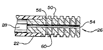
A :Flexible embodiment specifically designed to produce linear
lesions is shown schematically in Figures 15 and 16. The elongated
electrode 90 is preferably constructed from a porous or micraporous mesh
91 woven from small diameter metallic threads or merely configured with
an appearance of a fine weave. The porous mesh can also be constructed
JUN-18-1986 16:01
2119711
.1~.
P.22~42
from a series of small porous metal rungs Closely spaced to each other.
Preferably, the microporous mesh 91 covers an entire circumference near
the distal end 32 of the ablation catheter. End portions of the mesh 91 are
securely connected to the shaft theough mechanical clamps, connectors or
adhesive bonds 9~.
The elongated electrode 90 is electrically connected to the handle 24,
shown in Figure 1, through electrical connection means 64 preferably
comprising at least one conducting wire attached to the electrical interface
connection 40 at handle 24. Por ablation, appropriate electrical Current is
supplied to elongated electrode 90 through electrical connection means 64.
The electrical current can be direct current or alternating current, and
preferably is a radiofrequency signal. A, flexible, extended embodiment
electrode provides the capability to form deep, linear lesions along a
portion of a heart wall during ablation for the treatment of particular
arrhythmias. The fluid insulating/protecting character of the invention is
more important as the electrode length Increases due to the corresponding
increase in possible localized uneven heating along the length of the
electrode. Such uneven heating leads to the formation of hot spots which
result in biological tissue Coagulation. However, creation of this
continuous fluid protective layer reduces the possibility of areas of
coagulation by maintaining a more even temperature and, when using
conductive saline, creation of a conductive gap-filler material (the saline}
to provide more uniform electrical distribution of energy,
The inside surface 94 of the elongated electrode 90 is exposed to the
JUN-18-1996 16: L1
P.23i42
2179711
- 1e
central lumen 28 via a plurality of macroscopic holes 96. Holes 96 axe
preferably sized between about 0.1 millimeters (mm) to about 3 mm, and
preferably about D.2 mm to about 1.D mm. Fluid flows from the proximal
end 34 of the catheter dawn a fluid interface in the central lumen 28 to
macroscopic holes 96, The pressure of the fluid within the central lumen
28 forces water to disperse in the annular space 98 between the shaft of the
catheter and the fine weave forming the mesh 9I. The porosity of the
mesh 91 is selected such that the resistance to the flow of fluid through the
mesh 91 is significantly larger than the flow resistance at interconnecting
1D holes 96. This selection of porosity of the mesh 91 ensures that there is
an
essentially even flow of fluid over the outer surface IDO of the elongated
electrode 90.
Generally, the length LS of elongated electrode 90 is significantly
larger than the length LZ of the ring electrodes 30. The length of elongated
electrode 90 is selected to produce the size of the linear lesion appropriate
for the treatment o~ the patient. This length will preferably range from
about 5 mm to about 5 centimeters (cm). This length wilt often more
preferably range from about D.5 cm to about 1.5 cm.
A ring electrode 30 could not be constructed with a width
2D contemplated for the elongated electrode 9D because the ring electrode 30
would be too rigid. The elongated electrode 90 is flexible similar to or even
more than the catheter body 22. This flexibility allows the elongated
electrode 90 to have the appropriate width without limiting the capability
of passing the distal end 32 of the catheter conveniently through the
JUN-18-1996 16:01 - .. ~ .. P.24i42
2179711
_y9_
cardiovascular system info the heart.
The fluid introduced through ports 38, macroscopic holes 96 or
other orifices, of catheter 2U is preferably a biologically compatible fluid,
and znay be in a gaseous or liquid state. For example, the fluid may
comprise carbon dioxide, nitxog2n, helium, water, and/or saline. Fluid
enters through, for example, port 38 and is passed though central lumen 28
of catheter body 22. The fluid perfuses Eip structure 26 and/or ring
electrodes 30 through the path means in tip structure 2b and/or ring
electrodes 30, arid creates a fluid protective layer surrounding exterior
surfaces of tip structure 2( or exterior surfaces of electrodes 30, 90 thereby
minimizing contact of tip structure 26 or electrodes 30, 90 with biological
material, such as blood.
The rate of fluid flow through central lumen 2$ of catheter 20 may
vary and range From U.1 rnl/min, to 40 ml/min. Fluid flow through
catheter 2U may be adjusted by a fluid infusion pump, if the fluid is liquid,
or by pressure, if the fluid is a gas. The fluid flow is regulated by the
infusion pump for the liquid fluid, ox by a needle valve if a gas, so as to
maintain an optimal disbursing flow over the tip structure 26 and/or
electrodes 30, 90 and maintain a desired Hp temperature. Preferably, the
protective layer of fluid covers all or substantially all of the surface area
of
tip structure 26 and is betty een about O.U01 min arid 3. mm,, and more
preferably, about O.U1 mm. in thickness, although this may vary depending
an the application, and may vary in thickness during a given procedure.
Temperature sensing means 47 (for example as shown in Figures 3
JUN-18-1996 18:02 . P.25i42
2179711
and 4) may be incorporated into tip structure 26 ffor sensing and measuring
the temperature of tip structure 2b and for sensing and measuring the
temperature of the biological tissue in contact with tip structure 2b.
Temperature sensing means 47 may be incorporated in any of the tip
structure embodiments shown in Figures 2-Ip, I5-I6. The temperature
sensing means generally comprises at least one temperature sensor, such
as a thermocouple ox theravstor. In addition, temperature sensing means
47 array be utilized as a feedback system to adjust the flow rate off the
biologically compatible fluid to maintain the temperature of the tip
structure at a particular temperature within a designated range of
temperatures, such as 4D°C to g5°C. Also, temperature sensing
means 47
may be used as a feedback system to adjust the flow rate of the biologically
compatible fluid so as to maintain the temperature of the biological tissue
in contact with tip structure 26 at a particular temperature within a
'15 designated range of temperatures, such as 40°C to 95°C. The
temperature
of the tissue or tip structure 26 is controiIed by the temperature of the
fluid, the distribution of the fluid xeIative to internal and external
surfaces
to the tip structure, the energy applied to the catheter, and the fluid flow
rate.
Catheter 20 may include ablation means within tip structure 26.
Preferably, the ablation means may be a wire connected to an 1ZF energy
source, although other types of electrical energy, including microwave and
direct current, or ultrasound may be utilized. Alternatively, the ablation
means may include optical fibers fox delivery of Iaser energy. The ablation
,TUhi-18-1996 16:02 - P.26i~2
2179711
-zz -
means may be connected to an energy source through port 36, or an
additional port.
As shown in Figure 1, device 42 may be passed through central
luuten 28 of catheter 20. l7evice 42 may include, for example, a guidewire
for ease of entry of catheter 2D into the heart or vascular system; a
diagnostic device, such as an optical pressure sensor; a suction catkeeter for
biopsy of biological material near the distal tip; an endoscope For direct
viewing of the biological material in the vicinity o~ the distal tip of the
catheter; or other devices.
ID In one example of operation, catheter body 22 of catheter 20 is
preferably percutaneously inserted into the body. The catheter is
positioned so that it lies against cardiac tissue such that the flexible
poxnus
elongated electrode 90 makes contact along its length with the tissue area
that is to be ablated. tl,long the line of contact, energy will flow from the
conductive source to the electrode and into the cardiac tissue.
Simultaneous fluid flow is maintained around the electrode creating a
buffer between the tissue and the eleciaode. Tip structure ?6, as an
electrode, may also be utilized to measure electrical potentials of the tissue
and provide information regarding <ardfac signal measurement. Electrical
connection means 40 extends from tip struCfure 2fi, through port 36, and is
connected to monitoring equipment. Tip structure 26 may be utilized to
map, monitor, and measure the cardiac signals and electrical potentials of
the tissue, and locate arrhthymogenic sites.
A biologically compatible fluid is introduced through port 38. The
JUN-18-199b 3E:02 P. ~ 42
2179711
fluid passes through a central lumen of catheter body 22 and is directed to
tip structure 26. The fluid passes through Hp structure 26 and/or ring
electrodes 30 and/or elongated electrode 90 through path means 4t3, 52, 54,
61 or holes 96 in a manner determined by the embodiment of distal end 32
used. Tl~,e fluid perfuses tip structure 26 and forms a fluid protective layer
around exterior surface 50 of tip structure 26 and/or exterior surface 66 of
ring electrodes 30 and/or the exterior surface of the elongated electrode 90.
The fluid layer formed around catheter tip structure 26 and/or ring
electrodes 30 and/or elongated electrode 90 maintains biological materials,
such as blood, at a distance Erom catheter tip structure 2b, thereby
minimizing contact of catheter tip structure 26 witA the biological
material, as well as cooling tip structure 2G and jor elongated electrode 90.
Since there is a consistent, controlled buffer layer between the biological
material and catheter tip structure 26 and/or the elongated electrode 90,
the coagulation of biological materials is reduced and the impedance or
resistance to energy transfer of the tissue near the distal end 32 of the
catheter 20 is regulated and minimized during ablation.
Once the site has been located by the monitoring of the
electrophysiologicaI signals of the tissue, the ablative energy is activated.
As a result of the fluid protective layer, the transfer of electrical energy
to
the tissue is enhanced. increased destructfon of cardiac tissue also results
from tip structure cooling since larger and deeper lesions in the cardiac
tissue are achieved than have been previously possible. Zlse of the
eloxZgated electrode 90 allows the production of deep linear lesions.
SL'IV-18-1996 16.:03 -
P. 28%42
2179711
The ftow rate of the fluid over exterior surface 50 of Hp structure 26
ox exterior surface 66 of ring electrodes 30 or exterior surface of elongated
electrode 90 may be accomplished in a controlled manner so that a thin
fluid film is formed around exterior surface 50, 66, 100 of tip structure 26,
ring electrodes 30 and elongated electrode 90. The maintenance of a
controlled, stable, uniform fluid film along substantially the entire
exterior surface of Hp 26, ring electrodes 30 and elongated electrode 90 may
be accomplished by using the various embodiments of distal end 32
described above having a multiplicity of passages or path means 48, 52, 54,
61 or holes 96. Path means 4$, 52, S4, 61 and holes 96 permit an even,
consistent distribution of minute quantities of a biologically ~aanpatible
fluid over substantially the entire tip exterior surface 50 oz ring electrodes
exterior surface 66.
The fluid can be pumped through tip structure 26, or heat generated
by the electrical oz ablation process tare be Cased to expand the fluid and
create a movement of ftuid to the exterior surface 50, 66 of tip structure 26
or ring electrodes 30 ox elongated electrode 90. This movement of fluid
provides a buffer or protective insulating layer between the exterior
surface of tip structure 26 and/or ring electrode 30 and/or elongated
electrode 90 and the biologi:al matezial, such as blood, thereby reducing
the coagulation of biological materials on tip structure 26 and/or ring
electrode 30 andJor elongated electrode 90. In addition, the movement of
fluid over and around tip structure 26 may be aided by passages or
oha.~tnels 56, 58 on exterior surface, 50 of tip structure 26. Cooling of tip
SUN-18-1996.-16:03
P.29i42
2179711
-24-
structure 26 and/or ring electrode 3D and/or elongated electrode 90
increases the lesion size produced by the ablation means since the point of
maximum tissue temperature is likely moved away from tip structure 26,
which allows for an altered tissue heat profile, as further described below.
Another advantage of the fluid layer buffering the surface area of tip
structure 26 and/or ring electrodes 30 and/or elongated electrode 90 is that
the fluid layer also cools the tissue adjacent tip structure 26 and elongated
electrode 90 during ablation. In addition, the fluid aids in maintaining
the tissue adjacextt tip structure 26 and elongated electrode 90 in a coaler
and potentially more conductive state. which permits more electricity or
ablative energy to enter the tissue. As a result, larger lesions are produced
because a larger voltage can be applied, producing a larger electric field
without producing excessive temperatures and coagulum formation at the
tip/tissue interface. Lesions are produced with this invention in the form
of a line measuring about 1 cm to about 4 cm in length and about 3 mm to
about 5 mm in width while simultaneously maintaining the fluid
protective layer. This is accomplished without having to move the
catheter and without requiring several ablations. Also, the greater the
pressure of the fluid, the more biological products are kept from the field
of influence of, or area surrounding, tip structure 26 and/or elongated
electrode 90.
A control system may be included for controlling and regulating the
electrical potentials and temperatures in a mariner that allows for
determination of the ablation effects in the tissue. It is possible to control
SUhd-18-1996 16=03 .., -. ..
2179711
-25-
P.30i42
the distribution of tissue heating by controlling the temperature of tip
structure 26 and/or elongated electrode 90 and the radiofrequency voltage,
or other energy used, applied between tip structure 26 and/or elongated
electrode 90 and a reference electrode on the surface of the body. The
voltage may be set to achieve a desired electrical field strength, and the
temperature of tip structure 26 and/or elongated electrode 90 may be set to
provide a desired temperature distn'bution of the tissue. The temperature
distribution will then determine the size of the lesion, i_e., the denatured
pxatezn dimensions in the myocardium.
I0 The fluid flow rate can be regulated relative to biological
parameters, sucks as tissue temperature, by the temperature sensing means.
Por instance, if the temperature of the tissue increases, the fluid Flow rate
can be increased by the regulation of the fluid infusion pump or gas needle
valve. If the tissue temperature adjacent tip structure 26 andlor elongated
electrode 90 is not high eztough, the fluid flow rate can be decreased. This
permits pawer to be set independently of temperature. It is significant to
note that it is normally not necessary to remove the introduced fluid from
the body.
It is also possible to generate reversible affects of ablation by use of a
cooling fluid down the central lumen 28 of catheter 20 and tip structure 26,
or by use of a low temperature controlled or elevational heating. An area
in the heart tissue is quenched with a cold or icy fluid to produce a tissue
temperature of 0°C to 30°C, ox heated with electrical energy
with closed
loop temperature controls as described above to produce tissue
JUN-18-199b. .1.6:H3 P.31i42
2179711
-26-
teznperature5 ranging from 4D°C to 48°C_ Those cool and warm
temperatures slow the conduction o~ signals and ternpararily and
reversibly eliminate the conduction pathways. This technique may be
advantageously used to see the affect on the tissue before the tissue is
permanently of#ected. The heart tissue gradually heats or cools back to
normal. This technique is also advantageous since no catheter exchange
Would be required.
Various modifications and alterztivns of this invention will become
apparent to those skilled in the art without dep«rring from the scope and
spirit of this invention.