Note: Descriptions are shown in the official language in which they were submitted.
W O 95131947 PCT/US95/~D1655
;~i9,1Q89
INTERVERTEBRAL FUSION IMPLANT
I. BACKGROUND OF THE INVENTION
1. Field of the Invention
This invention pertains to intervertebral
fusion. More particularly, this invention pertains to
an implant to facilitate fusion between two vertebrae.
1D 2. - Description of the Prior Art
Back pain is extremely debilitating.
Individuals suffering from severe back pain are
frequently precluded from the full-enjoyment of-life
including gainful employment and leisure activities.
In
I5 addition to the substantial human costs associated with
back pain, society bears a substantial financial cost.
Lost employment time adversely impacts on productivity
as does medical insurance costs.
Frequently, the cause of back pain is
20 traceable to diseased disc material between opposing
vertebrae. When the disc material is diseased, the
opposing vertebrae-are not adequately. supported.
In order to address back pain resulting from
disc disease, prior art surgical techniques have been
25 developed to fuse the opposing vertebrae. Such
techniques include removing the diseased disc and
packing the disc space with bone. The intent of such
a
procedure is for the packed bone to grow together and
fuse with the bone of the opposing vertebra. If
30 successful, the two opposing vertebrae are now rigidly
linked thereby avoiding intervertebral instability.
In fusing opposing vertebrae, the prior art
has developed surgical techniques and apparatus to
' facilitate interbody fusion as well as to permit
35 stabilization of the vertebra while the fusion process
is occurring. To this end, surgical implants have been
developed.
An example of a surgical implant for
facilitating interbody fusion is shown in U.S. Patent
WO 95/31947 PCT/US95/01655
21~10~9
2
No. 5,015,247 to Michelson-dated May 14, 1991.
Michelson uses a circular cross-section cylindrical
implant which i-s of uniform diameter throughout its .
length and which includes an external thread. The
implant is hollow and has holes farmed through the ,
cylindrical wall of the implant. The implant is placed
within a prepared site between the vertebrae. The-
prepared site is a bore formed through the-disc material
as well as partially formed through the end plates of
the opposing vertebrae. Theimplant- is threaded into
the bore-and packed with bone chips or the like.
Another example of an interbody fusion device
is shown U.B. Patent No. 4;834,757 to Brantigan dated
May 30, 1989. The Brantigan device is a parallelepiped
plug which is forced into a complementarily shaped
cavity formed-between opposing-vertebrae.
Prior-art interbody fusion devices are not
trouble free. For example, prior art devices suffer
from uncontrolled subsidence of the device into the
vertebral body. By subsidence, it is meant that after
the implant is placed between the opposing vertebra, the
implant migrates into the vertebral body. Also, in many
prior_art implants, direct bone apposition only occurs
on two surfaces. In addition, unwanted invasion of disc
or cartilage material into-the implant may occur upon
insertion. Such prior art devices typically have
minimal surface area contact with the end plates of the
vertebra. In addition to the above, such priorart
devices have a geometry which-prevents close placement
when two implants are placed in a-side-by-side relation-
within a common disc space. A prior art device to -
increase the density of implant placement is-shown in
U.S. Patent No. 5,055,104 to Ray dated-DCtober 8, 1991.
In that patent, the device-is a helicalthread. Two
such devices are placed'side-by-side with the threads
intermeshing.
W0 95131947 , .' . ~ .. PCT/US951(II655
2 ~ ~ ~ aa9
3
It is an object of the present invention to
provide an implant for use in-interbody fusion. It is a
further obje-ct of the present invention to provide such
an implant which has reduced subsidence.
II. SUMMARY OF THE INVENTION
According to a preferred embodiment of the
present invention, an implant is provided for
facilitating intervertebral fusion between opposing
vertebrae. The implant includes first and second
bearing surfaces, each extending along a plane generally
parallel to the longitudinal axis-of the implant. The
first and second bearing surfaces are spaced apart in
generally parallel alignment. A first and second ridge
extend from the first and second bearing surfaces,
respectively. Openings are provided-through both the
first and second ridges with the openings in
communication with an interior of the implant. The
bearing surfaces and ridges are sized for-the first and
second bearing surfaces to oppose and abut cortical bone
while the ridges extend into cancellous bone.
III. BRIEF DBSCRTPTION DF THE DRAWINGS
Fig. 1 is a schematic view showing two
vertebral bodies separated by a disc material;
Fig. 1A is an end view of a prior art
interbody device opposing a vertebra;
Fig. 1B is an end view of a different prior
art intervertebral fusion device opposing a vertebra;
Fig. 1C is a side sectional view of an
interbody device received between two vertebrae;
~ Fig. 2 is a front, right side and top
perspective view of an implant according to the present
invention;
Fig-. 3 is an elevation view of a trailing end
of-the implant of Fig. 2;
Fig. 4 is a view taken along line 4-4 of Fig. 3;
WO 95131947 21910 8 9 PCTIUS95101655
i ~,
4
Fig. 5 is a perspective view of the sectioned
implant of Fig. 4;
Fig. 6 is a side-elevation view of the implant .
of Fig. 2;
Fig. 7 is a view taken along line--7-7 of-Fig.
6;
Fig. 8 is a top plan view of the implanb of
Fig. 2;
Figs 9 is an enlarged view of a portion--of the
anchor detail of Fig. 6;
Fig. 10 is an-end view of two vertebrae--formed
with a borethere between and stretched apart to receive
the implant of Fig. 2; -
Fig. 11 is the view of Fig. 10 showing the
implant of Fig. 2 inserted-within the bore of Fig_ 10;
Fig. 12 is a -view similar to that of Fig. 11
showing two implants disposed between: opposing
vertebrae;
Fig. 13 is a side sectional view of an implant
shown inserted between two-vertebrae;
Fig. 14 is a schematic representation of the
view of Fig. 13 showing radiused-surfaces of the implant
in great exaggeration-for purpose of llustration;
Fig. 15 is a perspective view of an implant
alternative embodiment; and
Fig. 16 is a view of a tool for placement of
the implant within a bore.-
IV. DESCRIPTION OF THE PREFERRED EMBODIMENT
Referring now to the several drawing figures
in which identical elements are numbered identically
throughout, a description of the preferred embodiment of -
the present invention-will now be provided.-
With-initial referenceto -Fig. 1, the implant
ofthe present invention is intended for use in
facilitating fusion between two vertebrae. -.Fig. 1
shows, in schematic format, an upper vertebra 10 and a
. 1.'~ l ~. ,:
W095131947 ~ z ~,~931 D 8 9 P~~S95/Ot655
lower vertebra 12. Each of the vertebrae 10,12 are
vertically aligned and present opposing end plates
10a,12a. The end plates IDa,l2a are separated by a disc
14. Thedisc 14 includes a fibrous inner material 16.
5 The periphery of the disc 14 is a fibrous material
conventionally referred to as the annulus 18. The
interior of each of the vertebrae-10,12 consists of
soft, cancellous bone 10b,12b. At the end plates
10a,12a, the vertebrae 10,12 include hard cortical bone
layers lOc,l2c. The-cortical bone layers commonly have
a thickness T of about 2 mm.
In interbody fusion placement, it is desirable
to provide a bore to receive the fusion implant with the
bore formed through the disc space 14 between the
opposing vertebrae 10,12 and withthe bore cutting
through the cortical bone 10c,12c of the end plates
10a,12a. As a result, the cancellous bone 10b,12b of
each of the vertebrae 10,12 is exposed and opposing the
implant. Exposure of the cancellous bone is desirable
since such cancellous bone lDb,l2b is substantially
laden with blood vessels. The cancellous bone is the
most rapidly growing and forms the bone growth linking
the vertebrae 10,12 upon completion of the spinal fusion
therapy.
While exposure of the cancellous bone is
desirable for the purpose of facilitating bone growth,
the opposition of the implant to the cancellous bone
increases the probability of subsidence since the
cancellous bone is-relatively soft and undesirable for
the purposes of weight bearing.
With the anatomy thus described, disadvantages
and problems associated with prior art implants can best
be understood with references to Figs. 1A, 1B and 1C.
Fig. 1A shows an implant 100' positioned within a bore
102' formed iri an upper vertebrae 10'. For purposes of
illustration, a lower vertebrae is not shown in Fig. 1A
but it will be appreciated that the lower vertebrae is
,., ~ . , ,-,..-.
. . . . .. , '.,':; ,
CA 02191089 1996-11-23
similarly bored as upper vertebrae 10'. The
intervertebral implant 100' shown in Fig. 1A is a
generally cylindrical threaded implant such as that
shown in U.S. Patent 5,015,247. The bore 102' is an arc
of a cylinder size to receive the implant 100'. So
formed, the cortical bone 10c' of end plate 10a' is
provided with angled surfaces 10d' opposing the implant
100'. The surfaces 10d' are the areas of greatest
support since this is where the implant 10' bears
against the cortical bone 10c'. Unfortunately, the
surfaces lOd are of small surface area when compared to
the total surface of the implant 100. Due to the angled
surfaces 10d', the implant 100' is susceptible to
slippage relative to the end plate 10a'. Upon
occurrence of such slippage, subsidence of the implant
100' into the soft cancellous bone 10b' occurs. This
subsidence causes the disc space to collapse and revert
back to its pre-surgical condition.
Fig. 1B also shows a cross sectional view with
an implant 100 " such as that shown in U.S. Patent
4,834,757. The implant 100 " is substantially
rectangular in cross section. The vertebra 10 " is
provided with a complementarily shaped bore 102 " . As a
result, the cortical bone lOc " of the end plate 10a "
opposes the implant 100" at generally vertical surfaces
lOd " . In this example, the cortical bone lOc "
provides no bearing surface opposing migration of the
implant 100 " into the cancellous bone lOb " .
To prevent migration of either cylindrical or
rectangular cross section implants into cancellous bone,
certain prior art devices size the implant to rest on
the vertical cortical bone surfaces of the vertebra.
For example, Fig. 1C shows an implant 100 " ' (which
could be either circular or rectangular in cross
section), positioned between an upper vertebra 10 " ' and
a lower vertebra 12 " '. Each of the vertebrae 10 " ',
12 " ' have outer vertical walls. of cortical bone
AMEt~EO
WO 95131947 21910 ~ 9 ; -; , ;,,~ ~, ~, PCT~S95/01655
7
10e" ' , 12e" ' . - A bore 102' " is formed between the
opposing vertebra 10" ',12 " '. The bore has a
longitudinal length such that the-ends 100a " ' and
100b " ' of implant 100 " ' bear directly against and
oppose the cortical bone 12e'-"-,10e " '_ In this method
of implantation, the implant 102' completely spans the
soft cancellous bone lOb " ',12b " '. In viewing Fig. 1C,
the reader will note that in-the absence of an implant
length spanning the vertebra, the-implant will subside
into soft cancellous bone.
Examples of prior art teachings showing
implants being sized and positioned to span soft
cancellous bone and bear directly against cortical bone
is shown in both U.S. Patent 4,834,757 and U.S. Patent
4,743,256. A disadvantage with the technique of having
an implant sized and positioned such that it spans the
soft cancellous bone and bears directly upon the outer
cortical walls of the vertebra is that the bore forming
-
operation for- placement on the implant must be precisely
controlled as to the length of the bore. The cortical
layer against which the implant bears is very thin
(approximately 2 millimeters). If the bore length is
too small, the implant will not bear on the cortical
bone. If the bore length is too great, the boring tool
will pierce through the end of the vertebra. If the
former occurs, subsidence is highly probable. The
latter can be extremely dangerous. A boring tool
piercing through a vertebra can puncture or sever
important anatomical features such as the spinal cord,
aorta or the Like. If such anatomical features are
damaged, severe consequences (including paralysis or
- death) can follow.
Having thus described the prior art implants
and disadvantages associated with such implants, a
description of an implant according to the present
invention will now be described. It will be noted that
the implant of the present invention provides full
R'O 95/31947 21910 8 9 ~ ~ , PCT~S95101655
8
bearing an--cortical bone while-avoiding the need for a
precisely controlled depth of cut.
With initial reference to Figs. 2-9, an-
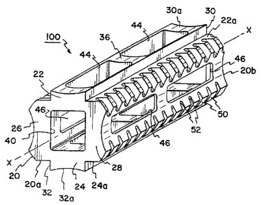
implant 100 according to the present invention is shown.
The implant 100 includes a body 20 which is
substantially square in cross-section. The body extends
along a longitudinal axis X-X. _.
The body 20 includes two generally flat upper
and lower bearing walls 22,24. Bearing walls 22,24 are
joined by side walls 26,28. Each of the bearing walls
22,24 present, outwardly facing-bearing surfaces 22a,24a.
Each of the bearing surfaces 22a,24a extend generally
parallel to each other and parallel to the longitudinal
axis X-X.
Projecting perpendicularly away from the
center of each of bearing walls 22,24 are raised ridges
30,32. Each of the ridges 30,32-extend parallel to axis
X-X and project outwardly from the body 20. The ridges-
30,32 are cent-rally positioned on the bearing surfaces
22a,24a such that the bearing walls 22,24 are exposed
along opposite sides of each of the raised ridges 30,32.
The ridges 30,32 terminate at concave faces-30a,32a.
The body 20 extends from a leading end 20a to
a trailing end 20b. Intermediate ends 20a,20b, side
walls 26,28 are joined by reinforcing ribs 36 (see-Figs.
4 and 5).
A bore 40 extends axially through body 20 and-
extends completely throughthe leadi.ngend 20a and
trailing end 2-Ob. Bore40 -is generally rectangular in
cross section as best shown in Figs. 3 and 7.
Formed completely through.ridges 30,32 and
bearing walls 22,24 are a plurality of openings 44. -
Openings 44 have an axis Z--Z which is perpendicular to
longitudinal axis X-X. -Each of the openings 44 is in -
direct communication with bore 40.
Side walls 26,28 -are concave and are prbvided
with openings 46 therethrough in communication with
R'O 95131947 } ' PCT1US95/OI655
~1g1~89
9
chamber40.Openings,46 extend along an axis Y-Y which
is mutually perpendicular to axes Z-Z and X-X (see Figs.
6 and 7 ) .
At the-edges of intersection-between walls
22,24, 26 and 28, a plurality of anchor segments 50 are
provided. Between each of the segments 50, a valley, or
recess 52 is formed to define-the anchor segments 50.
The anchor segments 50 are portions of a helix-pattern
surrounding the axis X-X. Also, as best shown in Fig.
3, the valleys-52 reside on the arc ofa circle having
radius Rl from axis X-X.
As shown best in Fig. 9, each of the anchor -
segments SO is generally square in cross section with an
end of the anchor 50 having an angled surface set at-an
angle A, relative to a line parallel to the axis X-X and
slanted downwardly towards the leading end 20a. In a
preferred embodiment, angle A1 is 10°.
In a preferred embodiment, the cross sectional
area of-the implant is not uniform throughout its
longitudinal-dimension. With best reference to Figs. 3
and 8, the outer faces 30a,32a of-the ridges 30,32 and
the side walls 26,28 are radiused-inwardly as indicated
at radii R2,R,. The benefits of the radii R,,R, will be
more fully described. - R3 equals the outside diameter of
anchors 50. Further, the leading end 20a is provided
with a taper.angle A2 (Fig. 6) which, in a preferred
embodiment, is 10°.
As will be more fully described, the implant
100 is placed within a bore formed between two vertebra.
The formation of the bore relative to the sizing of the
implant is important for reasons that will become
apparent.- Accordingly, for the purposes of illustrating
a preferred embodiment, the presently anticipated
dimensions of-the implants 100 will be given. It will
be appreciated that various sizes of implant 100 will be
available to accommodate different sized patients and
different regions in the spine.
'!~,.-..
CA 02191089 1996-11-23
1. Length of implant L (Fig. 4) : . 28
millimeters;
2. Size of bores 44 (L1 x W1, see Fig. 8)
10.6 mm by 5.49 mm (.416 inches by .216
5 inches) ;
3. Size of bores 46 (L2 x W2, see Fig. 6)
10.6 mm by 3.81 mm (.416 inches by .150
inches) ;
4. Radius R1 (Fig. 3) from axis X-X to
10 valleys 52: 7.5 millimeters;
' S. Size of cross section of bore 40 (W3 x
L3): 7 millimeters by 8 millimeters;
6. Width (W4, see Fig. 3) of ridges 30,32: 7
millimeters;
7. Height (H1, see Fig. 3) of ridges 30,32:
1 millimeter;
8 . Height (H2, Fig. 3 ) of convex area of side
walls 26,28: 8 millimeters;
9. Thickness (T1, Fig. 3) of bearing walls
26,28: 2 millimeters;
10. Pitch (P, Fig. 9) of anchors 50: 2.3 mm;
11. Thickness (T2, Fig. 9) of anchors 50: 1
mm;
12. Radius R2 (Fig. 8): 190 mm; and
13. Radius R3 (Fig. 3): 8.75 mm.
To place the implant 100 between the vertebra
10,12 attention is now directed to Fig. 10. The
vertebra 10,12 are distracted to stretch the annulus 18.
A bore 102 is formed with its cylindrical axis extending
parallel to and centrally positioned between the end
plates 10a,12a. The bore 102 is sized for its radius RB
to be equal to the radius (R1) of the implant 100 (as
shown in Fig. 3) to the v~0.11eys 52. The implant 100 and
bore 102 are sized such that the radius RB will extend
through the cortical layers lOc,l2c without extensive
penetration into the soft cancellous bone lOb,l2b.
For the reasons that will become apparent,
bore 102 must be precisely sized and accurately
At~EPIt?~ S'~EET
WO 95131947 PCT/US95101655
11
positioned with the axis of the bore:_-102 centrally
positioned between the end plates 10a,12a and parallel
to the end plates 10a,12a. A surgical method and lcit
for accomplishing such an accurate formation of a bore
between vertebra is the subject of commonly assigned and
co-pending U.S. Patent Application Ser. No. 08/015,863,
filed February 10, 1993.
With bore 102 thus formed, the implant 100 is
inserted into the bore 102 with the leading end 20a
-first-introduced into the bore 102_ The implant 100 is
rotated about its axis X-X to advance the implant 100
into the bore 102 to the position shown in Fig. 13.
Alternatively, implant 100 need not be-rotated but
simply can be impacted by driving-it axially along its
axis X-X. The implant 100 is positioned such that upon
full insertion into the bore 102, openings 44 are
directed toward the soft cancellous bone IOb,l2b. The
openings-46 are directed toward the space formerly
occupied-by removed disc material 16.
With the radius RH of the bore selected to
equal the radius R1 to the valleys 52, after insertion of
the implant, the bearing surfaces 22a,24a directly
oppose and-abut the cortical layer lOc,l2c of the end
plates l0a,l2a (see Fig. 11). Also, with the relative
sizing of the bore 102 thus described, the ridges 30,32
protrude beyond the cortical bone layer 10c,12c into the
soft cancellous bone lOb,l2b. With this structure and
positioning of the implant 100, a surgeon can place bone
chips within the bore 40. Accordingly, the bone 10b,12b
-is fused-together by a bone column formed through the
aligned bores 44,40. The load bearing of the surfaces
22a,24a against the cortical bone lOc,l2c prevents
subsidence of the implant 100 into the cancellous bone
lOb,l2b. The bearing surfaces 22a,24a are parallel to
theimplant 100 as opposed to current devices where a
rounded surface contacts the implant 100 at an angle
(e-a., U.S. Pat. No. 5,015,247) or rectangular devices
W0 95131947 ' PCT/US9510165.5
2? 91089
12
where there is no end platecontact except at the
extreme ends of the implant (e-Q., U.S. Pat. No.
4,834,757). Also, the present implant 100 has non- _
threaded ridges 30,32 that project through the end plate
10a 12a and directly contact the cancellous bone
lOb,l2b. The surface area of the bores 44 is made-as
large as possible while-permitting structural integrity
to the implant 100 to provide maximum porosity to
cancellous bone growing through the implant 100.
In Fig. 11, a single implant 100 is shown
inserted. Inmany applications (particularly in the
lumbar region of the spine), two implants disposed in
parallel alignment are preferred-- Such a positioning is
shown in Fig. 12. Also, in=Fig. 12, it will be noted
that the implants 100 are in close proximity. The
closeness of proximity is attained by the concave aide
walls 26,28.
Normally, with convex side walls such as that
shown in U.S. Patent-No. 5,015,247, implants cannot be
placed with their axes in close proximity. Also, with
threaded convex side walls, the implants of U.S. Patient
No. 5,015,247 cannot be allowed to touch. If the second
implant to be inserted touches the first previously
inserted implant, the second implant can cause the first
implant to unscrew as the second-implant is advanced.
This creates apotentially dangerous situation where the
previously inserted implant can be inadvertently
unthreaded into a major-vessel orthe spinal cord. As a
result in certain regions-of the spine;-only one implant
can be placed while two would otherwise_be desirable.
With the concave side-walls 26,28, the present
implants can be placed in closer proximity increasing
the likelihood that two implants can be-used at any disc
level .-
In-the embodiment-of Fig. 12, bone-dowels 200
are positioned between both implants 100 and opposing
the side walls 26,28 of the-implants 100 on both sides
WO 95!31947 t ,' PCTlUS9Sl01655
13
thereof. The dowels have convex accurate outer surfaces
202 shaped to conform with the concave surfaces of the
side walls -2b,28. Bone dowels 2QD-are placed on the
exterior side walls such that all bores 46 are in direct
opposition to a bone dowel 200. With this application,
disc material 16 is blocked by the bone dowels 200 from
entering into the interior of the implants 1D0 and
interfering with bone growth through the implants 100.
Further, thebone growth through the bores 44,40 fuses
IO with the bone growth through the side bares 46 and fuses
with thebone dowels 200. Accordingly, the linkage
between the vertebra 10,12 is enhanced since each of the
implants 100 is cross linked.
Fig. 14 illustrates the_value of the non-
uniform cross-section of implant 100. Namely, the dip R~
(shown exaggerated in Fig. 14 for purpose of
illustration)in both of the walls 30a,32a and the side
walls 26,28 prevents movement of the implant 100 along
its axis X-X after the bone growth is achieved.
In. the event a surgeon prefers not to use bone
dowels 200 in the manner indicated in Fig. 12, it is
desirable not to have the side wall openings 46 opposing
disc material in order to prevent_such disc material
from entering into the implant 100 and interfering with
bone growth through the implant 10~. Accordingly, Fig.
15 shows an alternative embodiment implant 100a where
the side walls 28a,26a are solid and do not include
openings 46. Accordingly, there is no direct
communication between the disc material and the interior
of the implant 100a.
Fig. 16 shows an insertion tool 300 for
inserting the implant 100. The insertion tool 300
includes four prongs 300a-300d. The prongs 30Da-300d
cover the openings 44,46 with the thicker prongs 300d,
300b having convex inner surfaces 301 sized to
complementary mate with the concave side walls 26,28.
Further, the thinner prongs 300a,30Dc have convex
.i ~irir~~it~ 1 ~~~ , ~ w t. ~V~r'v.~1
CA 02191089 1996-11-23
14
surfaces 302 sized to mate with the concave surfaces
30a,32a of the ridges 30,32. The insertion device 300
covers the holes 44,46 during insertion of the implant
100 to prevent disc material and other debris from
entering the interior 40 of the implant 100. Also, the
outer surfaces 304a-304d of each of the prongs is
generally the arc of a cylinder such that the device 100
within the insertion tool 300 presents a cylindrical
surface permitting the non-cylindrical implant 100 to be
implanted into a round bore 102. A handle 306 connects
the prongs and permits turning or axial driving of the
tool 300.
As indicated, it is desirable that bores 44 be
of maximum surface area as possible to increase the
surface porosity of the implant 100. Applicants,
through animal studies and human clinical experience,
have found that the larger the surface porosity the
greater the probability for successful bone ingrowth
into the implant 100.
The present invention utilizes the anchors 50
embedded within the end plates 10a,12a to hold the
implant 100 in position. Since the end plates 10a,12a
are formed of cortical bone 10a,12a, the embedded
anchors 50 within the end plates 10a,12a provide
substantial force against inadvertent movement of the
implant 100. Also, the anchors 50 permit either
threading the implant 100 by rotating it about its axis
X-X or by implanting while driving the implant 100 and
tool 300 with a hammer or the like along its axis X-X.
The square cross section anchor 50 is tapered (at A1) to
provide resistance to expulsion.
At~~NO~~ SHEET