Note: Descriptions are shown in the official language in which they were submitted.
21951 0 PCT/US96/07671
WO 96/41162
~ - 1 -
SYSTEMS AND METHODS FOR IDENTIFYING
AND CONTROLLING INTERFACES BETWEEN
BLOOD COMPONENTS
Field of the Invention:
The invention generally relates to blood
collection and processing systems and methods. In
a more particular sense, the invention relates to
systems and methods for locating interfaces between
different blood components.
Background of the Invention:
Most of the whole blood collected from do-
nors today is not itself stored and used for
transfusion. Instead, the whole blood is separated
into its clinically proven components (typically red
blood cells, platelets, and plasma), which are them-
selves individually stored and used to treat a
multiplicity of specific conditions and diseased
states. For example, the red blood cell component
is used to treat anemia; the concentrated platelet
component is used to control thrombocytopenic
bleeding; and the platelet-poor plasma component is
used as a volume expander or as a source of Clotting
Factor VIII for the treatment of hemophilia.
During centrifugal blood processing, an
interface develops between the red blood cell and
plasma components. Leukocytes occupy this interface,
which is also referred to as the buffy coat.
In collecting whole blood components for
transfusion, it is desirable to minimize the
presence of impurities or other materials that may
WO 96/41162 2195190 PCT/US96/07671
- 2 - 0
cause undesired side effects in the recipient. For
example, because of possible febrile reactions, it
is generally considered desirable to transfuse red
blood cells and plasma substantially free of
leukocytes, particularly for recipients who undergo
frequent transfusions.
It is therefore important during blood
processing to be able to accurately identify the
leukocyte-rich interface between red blood cell and
plasma components, so that processing can be
controlled to isolate the interface from the other
components. This need exists not only for automated
blood collection procedures; but also for manual
blood collection procedures.
Conventional systems and methods often
employ optical signal processing to identify and
control the interface. Such systems often have
limited tolerance to "noise", which leads to false
readings. Such noise can arise due to variations in
the performance of the optical elements, especially
when multiple optical elements are used in tandem,
since optical elements are known to have a high
degree of variability in gain, focus, and
directivity. Mechanical vibration is another source
of noise.
Blood components are "sticky" and can smear
along the sides of a separation chamber or bag. The
smearing is yet another category of noise, as it
leads to false readings and the incorrect
identification of the interface.
8ummarv of the Invention:
The invention provides systems and methods
that consistently provide accurate monitoring of a
volume of blood, despite the presence of noise of
all types.
WO 96/41162 219519'" PCT/US96/07671 -
~ - 3 -
The systems and methods locate at least
three spaced apart sensing units in association
with volume of blood comprising a first blood
component region, a second blood component region,
and an interface region between the first and second
' blood component regions. The systems and methods
locate at least one of the sensing units so that its
optical field lies above the interface region and at
least one of the sensing units so that its optical
field lies below the interface region. Each sensing
unit senses the attenuation of energy emitted into
blood in its optical field and generates a signal
relating to the attenuation.
The systems and methods convert the signals
to a signal vector of signal values having a shape
approximated by a function. The systems and methods
normalize the signal vector, and also create a
vector of convolution signal values by multiplying
the normalized signal vector by the function. The
systems and methods identify the sensing unit
associated with the highest convolution signal
value. It is this sensing unit that lies closest to
the interface.
In a preferred embodiment, the systems and
methods limit the signal values of the signal vector
according to prescribed criteria that eliminate the
effect of noise.
In a preferred embodiment, the systems and
methods generate an output relating to the identity
of the sensing element associated with the highest
convolution signal value. This output can be used,
for example, to limit travel of the interface region
within a container while the first or second blood
component regions are conveyed from the container.
In a preferred embodiment, the function is
CA 02195190 2006-05-08
-4-
a sigmoidal function, and the normalized signal vector has a shape symmetric
about -1 and 1.
According to an aspect of the invention, there is provided a system for
monitoring a volume of blood comprising a first blood component region, a
second blood component region, and an interface region between the first and
second blood component regions comprising:
at least three spaced apart sensing units that each senses the
attenuation of energy emitted into the blood and generates a signal relating
to
the attenuation, at least one of the sensing units being located with respect
to
the blood volume above the interface region and at least one of the sensing
units being located with respect to the blood volume below the interface
region; and
a processing element coupled to the sensing units that locates the
sensing unit closest to the interface region by converting the signals to a
signal vector of signal values having a shape approximated by a function,
normalizing the signal vector, creating a vector of convolution signal values
by
multiplying the normalized signal vector by the function, and identifying the
sensing unit associated with the highest convolution signal value.
According to another aspect of the invention, there is provided a
system for monitoring the contents of a container holding blood comprising a
first blood component region, a second blood component region, and an
interface region between the first and second blood component regions
comprising:
at least three spaced apart sensing units that each senses the
attenuation of energy emitted into the blood and generates a signal relating
to
the attenuation, at least one of the sensing units being located above the
interface region and at least one of the sensing units being located below the
interface region; and
a processing element coupled to the sensing units including means for
converting the signals to a signal vector of signal values having a shape
approximated by a function, means for normalizing the signal vector, means
for creating a vector of convolution signal values by multiplying the
normalized
signal vector by the function, and means for identifying the sensing unit
CA 02195190 2006-05-08
-4a-
associated with the highest convolution signal value to thereby identify the
sensing unit closest to the interface region.
According to another aspect of the invention, there is provided a blood
component collection system comprising:
a chamber holding blood comprising a first blood component region, a
second blood component region, and an interface region between the first and
second blood component regions;
an outlet path communicating with the chamber to convey at least one
of the first blood and second blood component regions from the chamber;
at least three spaced apart sensing units arranged along the chamber, each
of the sensing units sensing the attenuation of energy emitted into the blood
within the chamber and generating a signal relating to the attenuation, at
least
one of the sensing units being located above the interface region and at least
one of the sensing units being located below the interface region; and
a processing element coupled to the sensing units that locates the
sensing unit closest to the interface region, while the outlet path conveys
the
at least one first and second blood component region from the chamber, by
following the steps of converting the signals to a signal vector of signal
values
having a shape approximated by a function, normalizing the signal vector,
creating a vector of convolution signal values by multiplying the normalized
signal vector by the function, and identifying the sensing unit associated
with
the highest convolution signal value.
According to a further aspect of the invention, there is provided a
method for monitoring a volume of blood comprising a first blood component
region, a second blood component region, and an interface region between
the first and second blood component regions comprising the steps of:
locating at least three spaced apart sensing units with respect to the
blood volume, with at least one of the sensing units located above the
interface region and at least one of the sensing units located below the
interface region;
sensing with each sensing unit the attenuation of energy emitted into
the blood;
generating a signal relating to the attenuation sensed by each sensing
unit;
CA 02195190 2006-05-08
-4b-
converting the signals to a signal vector of signal values having a
shape approximated by a function;
normalizing the signal vector;
creating a vector of convolution signal values by multiplying the
normalized signal vector by the function; and
identifying the sensing unit associated with the highest convolution
signal value to thereby identify the sensing unit closest to the interface
region.
Other features and advantages of the invention will become apparent
upon review of the following description, drawings, and appended claims.
Brief Description of the Drawings:
Fig. 1 is a somewhat schematic view of a blood collection system that
includes an signal processor that identifies the location of the interface
between blood components according to the features of the invention;
Fig. 2 is an enlarged view of a portion of the system, showing one of
the multiple sensors that generates signals for processing by the signal
processor;
Fig. 3 is a schematic flow chart showing the operation of the signal
processor;
Fig. 4 is a somewhat schematic view of a blood processing system that
incorporates the signal processor shown in Fig. 1 for controlling the
collection
of blood components; and
Fig. 5 is a schematic flow chart showing the operation of a preferred
embodiment of the signal processor, which takes into account noise caused
by smearing of sticky blood components.
The invention may be embodied in several forms without departing
from its spirit or essential characteristics. The scope of the invention is
defined
in the appended claims, rather than in the specific description preceding
them.
All embodiments that fall within the meaning and range of equivalency
of the claims are therefore intended to be embraced by the claims.
Description of the Preferred Embodiments
WO 96/41162 2195190 PCT/US96/07671
= - 5 -
Fig. 1 shows a container 10 made of
flexible, transparent plastic material containing a
unit of whole blood. The whole blood has been
centrifugally separated into component parts within
the container 10 by conventional techniques.
As Fig. 1 shows, the heavier red blood cell
component 12 of whole blood collects where the
greatest g-field forces are generated, during
centrifugation, which Fig. 1 shows to be the bottom
of the container 10. The lighter plasma component
14 of whole blood collects where least g-field
forces are generated, which Fig. 1 shows to be the
top region of the container 10.
During centrifugal separation, an
intermediate layer 16 of leukocytes (commonly called
the "interface" or "buffy coat") forms between the
red blood cell component and the plasma component.
If the plasma component is platelet-poor plasma
(PPP), the interface 16 also includes a substantial
amount of platelets. If the plasma component is
platelet-rich plasma (PRP), substantially fewer of
platelets remain in the interface 16. Whether the
separation process provides PRP or PPP plasma
component depends upon the rotational speed and time
of processing. Slower rotational speeds over a given
time period (called a "soft" spin) produce PRP.
Higher rotational speeds over the same time period
(called a "hard" spin) yield fewer platelets in the
plasma, and produce PPP.
Fig. 1 shows the container 10 held in a
system 18 which optically identifies the location of
the interface 16. The system includes a holding
station 20 comprising a back plate 22 and a front
plate 24, which sandwich the container 10 and its
contents between them.
WO 96/41] 62 2195190 PCT/US96107671
- 6 -
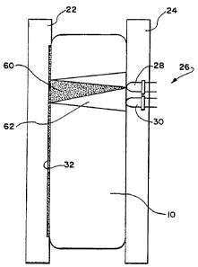
The front plate 24 includes an array of
sensors units 26. The number of sensor units 26 can
vary according to the size of the container and the
sensitivity required. Ten sensor units 26 are shown
for the purpose of illustration. The sensor units
26 are designated Sensor [1] to Sensor [N], where
Sensor [1] is the topmost sensor unit and Sensor N
is the bottommost sensor unit. The spacing between
sensor units 26 is selected such that Sensor [1]
will always be above the interface 16 and Sensor [N]
will always be below the interface 16.
In the illustrated embodiment, each sensor
unit 26 (see Fig. 2) comprises a light emitting
diode (LED) 28 and a photo transistor 30 vertically
stacked one on top of the other and both directed
toward the container 10. As Fig. 2 best shows, the
LED 28 emits energy through the transparent material
of the container 10 and the contents which lie in
its optical field 60. The surface of the back plate
22 is made of a material that reflects the energy
the LED 28 emits. The reflected energy passes back
through the container 10 and its contents and is
received by the photo transistor 30 within its
optical field 62.
In a preferred embodiment (see Fig. 2), a
label 32 is applied to the wall of the container 10
that sits against the back plate 22. The label 32
includes an interior surface that reflects the
energy emitted by the LED 28 back to the photo
transistor 30.
In an alternative embodiment, the LED 28
and photo transistor 30 of each sensor unit can be
arranged in facing, oppositely spaced relationship,
one on the front plate 24 and the other on the back
plate 22.
WO 96/41162 219519-0 PCT/US96/07671
i -' -
The photo transistor 30 generates an analog
signal, the magnitude of which is dependent upon the
level of attenuation that the energy emitted by the
LED 28 experiences upon passing through the contents
lying in its optical field 60. The emitted energy
is selected so that the red blood cell component 12
provides a high level of attenuation, while the
plasma component 14 provides a significantly lower
level of attenuation. The interface 16 lies at the
threshold between these two different attenuation
levels.
The system 18 includes a signal processor
34 (see Fig. 1), which analyses the attenuation of
the signals generated by the sensor units 26 for the
purpose of locating the interface 16. The signal
processor 34 includes a sensor controller 36 coupled
to each LED 28. The controller 36 turns the LEDs 28
on sequentially from top to bottom, or vice versa.
The signal processor 34 includes a data acquisition
element 38 coupled to the photo transistors 30. The
data acquisition element 38 periodically samples the
analog signals coming sequentially from each photo
transistor 30. The data acquisition element 38
converts each analog signal to digital form.
The signal processor 34 further includes a
processing element 40 that receives the digital
signals from the data acquisition element 38.
According to the invention, the processing element
40 converts the digital signals to a 1 x N column
vector, designated Signal [*]. The shape of the
Signal[*] vector gives a large relative value for
the sensor units 26 above the interface 16 and a
small relative value below the interface 16. The
shape of the Signal[*] vector is approximated by the
following sigmoidal function:
WO 96/41162 21951/ O PCT/US96/07671
- 8 - 0
S(Y) = K +
E (1)
+e-~tr Yo~
where
K is the maximum signal level through
the plasma component;
yo is the Sensor[yo] position where
the signal equals hK, which is the location of the
transition where the interface 16 resides;
a is a random variable that determines
the steepness of the transition of the function from
K to 0. The parameter is set by the operator. In
the preferred embodiment, c-_ 3; and
e is a parameter for noise, which
accounts for--the variability of optical components,
the non-homogeneous nature of blood components, and
variations in the reflective system including the
reflective nature of the back plate 22 or label 32
and any non uniform or curved surface in the back
plate 22 or front plate 24.
In estimating the location of the
transition (i.e., the interface), the processing
element 40 (see Fig. 3) first normalizes the
Signal[*) vector to be symmetrical between -1 and 1.
The normalization function SigNorm[i] is expressed
as follows:
'd i, i e {1, ..., N}
SrgNorm[i] = 2 Sigital[i]-min(Signal[ *]) (2)
max(Signal[ *])-min(Signal[=])
where:
Signal[i] is the digital signal of
Sensor[i];
2 1 " 5 1 ~ ~ PCT/US96/07671
WO 96/41162
9 -
min(Signal[*]) is the smallest digital
signal received from all the sensor units; and
max(Signal[*]) is the largest digital
signal received from all the sensor units.
Next, the processing element 40 creates an
N x N matrix, Sigmoid[*], as a series of row
vectors. Each row vector is constructed as the
value of the sigmoidal function (from Equation 1)
with K = 1, e= 0, yo = 0, and y ranging from -N+1 to
0 for the first row and from 0 to N for the last
row, expressed as follows:
S(-N+i) S(-N+2) ... S(O)
Srgmora[*] = S(-1) S(0) ... S(N-2) (3)
S(0) S(1) ... S(N-1)
The sigmoid[*] matrix needs to be
calculated only once, given the number of sensor
units 26 and assuming a value for a.
Next, the processing element 40 generates
a column vector, Conv[*], by performing a matrix
multiplication of the Sigmoid[*] matrix (from
Equation 3) and the SigNorm[*] column vector (from
Equation 2), expressed as follows:
Conv[ *] = Sigmoid[* ] x SigNorm[ *] (4)
where:
the operator "x" indicates matrix
multiplication and not the cross product operator.
Equation 4 is equivalent to performing a
= 25 convolution of the SigNorm[*] vector (Equation 2)
with the sigmoidal function of Equation 1.
WO 96141162 21" 5 190 PCT/US96/07671
- 10 - ~
The processing element 40 selects the index
of the maximum value of the Conv[*] vector (ImAx) as
the index closest to the level of the interface 16.
The position of the interface (P) can be
estimated to finer than integer resolution as
follows:
Conv[I~+1] - Com~[I~ 1]
P = N + 1 I~ (5)
2m
where:
m is the larger of the difference
between Conv[IMAX] - Conv[I MAX + 1] and Conv[I KAX] -
Conv[IKAX - 1].
The above methodology provides an accurate
estimate of the optical signal marking the
transition from the plasma component to the red
blood cell component under a wide range of operating
conditions. The methodology also provides a high
degree of -tolerance to the differences between
optical signal intensity due to variations in LED
intensities, variations in photo transistor
sensitivities, and changes in distance between the
back plate 22 and the sensor units 26 in the front
plate 24.
EXAMPLE
Ten sensor units arranged as shown in Fig.
1 acquire signals as set forth in the following
Table 1:
TABLE 1
DIGITAL SIGNALS BY SENSOR[i]
Sensor [i] Digital Signal
Sensor (1) 200
Sensor (2) 200
WO 96/41162 2195190 PCT11JS96107671
~ - 11 -
Sensor (3) 200
Sensor (4) 200
Sensor (5) 200
Sensor (6) 200
Sensor (7) 200
Sensor (8) 100
Sensor (9) 0
Sensor (10) 0
The normalized signal vector SigNorm[i]
computed according to Equation 2 (with a 3) is set
forth in the following Table 2:
TABLE 2
SigNorm[i] VECTOR
Sensor [i] SigNorm[i]
Sensor (1) 1
Sensor (2) 1
Sensor (3) 1
Sensor (4) 1
Sensor (5) 1
Sensor (6) 1
Sensor (7) 1
Sensor (8) 0
Sensor (9) -1
Sensor (10) -1
The 10 x 10 matrix Sigmoid[*] computed
according to Equation 3 (with a = 3) is set forth
the following Table 3:
TABLE 3
SIGMOID[*] MATRIX
WO 96/41162 21/519 O PCT/US96/07671
- 12 -
1.00 1.00 1.00 1.00 1.00 1.00 1.00 1.00 0.95 0.50
1.00 1.00 1.00 1.00 1.00 1.00 1.00 0.95 0.50 0.05
1.00 1.00 1.00 1.00 1.00 1.00 0.95 0.50 0.05 0.00
1.00 1.00 1.00 1.00 1.00 0.95 0.50 0.05 0.00 0.00
1.00 1.00 1.00 1.00 0.95 0.50 0.05 0.00 0.00 0.00
1.00 1.00 1.00 0.95 0.50 0.05 0.00 0.00 0.00 0.00
1.00 1.00 0.95 0.50 0.05 0.00 0.00 0.00 0.00 0.00
1.00 0.95 0.50 0.05 0.00 0.00 0.00 0.00 0.00 0.00
0.95 0.50 0.05 0.00 0.00 0.00 0.00 0.00 0.00 0.00
0.50 0.05 0.00 0.00 0.00 0.00 0.00 0.00 0.00 0.00
The column vector Conv[*) computed
according to Equation 4 is set forth in the
following Table 4:
TABLE 4
CONV[*] COLUMN VECTOR
Sensor [i] Conv[i]
Sensor (1) 5.5473
Sensor (2) 6.44997
Sensor (3) 6.90007
Sensor (4) 6.44738
Sensor (5) 5.49727
Sensor (6) 4.49986
Sensor (7) 3.5
Sensor (8) 2.50013
Sensor (9) 1.5026
Sensor (10) 0.55003
Based upon Table 4, Imm = 3.
Based upon Table 4:
Conv(IMAX - 1) = 6.44997
WO 96/41162 2195190 PCT/US96107671
~ - 13 -
Conv(ImX) = 6.90007
Conv(I.. + 1) = 6.44738
m = 0.4527
Based upon these values, P is calculated
according to Equation 5 as being 8.00.
Fig. 4 shows a blood collection apparatus
42 for the container 10, which uses the system 18 as
just described for transferring plasma component 14
and red blood cell component 12 from the container
10. The apparatus 42 includes the front and back
plates 22 and 24 to hold the container 10, as
already described. In Fig. 4, the container 10
includes a top port 44 with associated flexible
tubing 46 and a bottom port 48 with its own
associated flexible tubing 40. The apparatus 42 also
includes the array of sensor units 26 (again
numbering 10 for the purpose of illustration)
coupled in association with the signal processor 34,
as already described.
The apparatus 42 further includes an
actuator 52 for moving one of the plates 22/24 with
respect to the other plate. In Fig. 4, the
actuator 52 moves the front plate 24 toward the the
back plate 22. This movement squeezes the container
10 between the plates 22/24. The compression
expresses the plasma component 14 from the top port
44 into the associated tubing 46, for example, for
collection in a transfer bag (not shown). The red
blood cell component 12 is also expressed by the
same action from the bottom port 48 into the
associated tubing 50, for example, into another
transfer bag (not shown).
The apparatus 42 includes electrically
actuated solenoid clamps 54 and 56, which are
operatively associated, respectively, with the top
WO 96/41162 2195190 PCT/US96107671
- 14 - ~
and bottom tubing 46 and 50. The signal processor
34 monitors the position of the interface 16 in the
container 10 as the plasma and red blood cell
components 12 and 14 are expressed from the
container 10.
The apparatus 42 further includes a clamp
controller 58. The clamp controller 58 coordinates
operation of the clamps 54 and 56 in response to the
signal processor 34, to keep the interface 16
sandwiched between the plasma component 14 and the
red blood cell component 12 within the container 10,
while the plasma and red blood cell components 12/14
are expressed from the container. This technique
retains the interface 16 in the container 10 and,
with it, most of the leukocytes for subsequent
harvesting or disposal. This technique provides
from about 0.75 to about 1.00 log reduction in the
number of leukocytes in the plasma component and the
red blood cell component, when compared to the
leukocytes contained in the whole blood.
In the illustrated embodiment, the clamp
controller 58 opens both clamps 54/56 as the
actuator 52 moves the front plate 24 to begin the
expression of plasma and red blood cell components
12/14 from the container 10. The signal processor
34 continuously derives and outputs either I., or
P in the ..manner already described, thereby
identifying the position of the interface 16 in the
container 10. The clamp controller 58 compares this
output to a desired location for the interface 16.
The desired location is set by the operator,
typically near the middle sensor unit.
if the interface position derived by the
signal processor 40 is higher than the desired
location, the clamp controller 58 commands the top
WO 96/41162 2195190 PCTIUS96/07671
~ - 15 -
clamp 54 to close, blocking further exit of the
plasma component 14. The interface 16 will fall as
the red blood cell component 12 continues to be
expressed from the container 10 through the still
open tubing 46.
The clamp controller 58 continues to
compare the interface position derived by the
signal processor 40 with the desired location. When
the derived interface position is lower than the
desired position, the clamp controller 58 commands
the top clamp 54 to open, and plasma component 14
again exits the container 10 along with the red
blood cell component 12. The plasma component 14 is
less viscous than the red blood cell component 12,
and thus flows more quickly from the container 10,
so the interface 16 may again rise. Nevertheless,
working together, the signal processor 40 and clamp
controller 58 maintain the interface 16 at the
desired location within the container 10.
If it is desired to control the expression
of two fluids with similar viscosities, the clamps
54/56 can be controlled as follows: clamp 54 is
controlled as previously described, and clamp 56 is
controlled to be in the opposite state as clamp 54.
The methodology uses information from all
sensor units, not just the sensor unit where the
interface 16 is sought to be stabilized. Thus, a
smooth signal is generated should the interface 16
move from sensor unit to sensor unit above and below
the desired position.
As plasma and red blood cell components
12/14 are expressed from the container, the
interface 16 will move within a prescribed range
within the container 10. During this movement, red
blood cells and/or leukocytes in the interface 16
WO 96/41162 219" 194 PCT/US96/07671
- 16 -
can "stick" to the interior wall of the container.
This, in turn, can falsely attenuate the optical
signals when the interface 16 drops lower than the
,
region where the cells are stuck to the container
5 wall.
In a preferred embodiment (see Fig. 5), the
signal processor 40 takes into account the possible
presence of cells stuck to the interior wall of the
container. In this embodiment, the signal processor
10 40 truncates the values of the largest digital
signals by a prescribed amount before deriving the
normalized function SigNorm[i].
More particularly, in this embodiment, the
signal processor 40 first limits the vector
Signal[*], creating a limited signal vector
SigLim(*], as follows:
V i, i e{1, ..., N}
SigLim[r] = min[Signal[i], T x maao(Signal[ *])] (6)
where:
max(Signal[*]) is the largest digital
signal received from all the sensor units;
T is a truncation factor, where 0 < T
< 1, and is selected based upon the operating
parameters of the particular system based upon the
degree of false attenuation experienced. In a
system like that shown in Fig. 4, where the degree
of attenuation by sticky red blood cells and/or
leukocytes can be as much as about .85 (85%), T is
selected to be about 15% (0.15); and
the expression min[x,y] selects the
value of x or y that has the least numerical value.
In this embodiment, the signal processor 40
2195190
WO 96/41162 PCT/US96/07671
~ - 17 -
generates the vector SigNorm(i] based upon SigLim[*]
as follows:
V i, i < N
SigNorm[J] = 2 SigLim[i]-min(SfgLim[*]) -1 (7)
max(SigLim[ *])-min(SfgLim[ *])
where:
SigLim[i] is the limited digital
signal of Sensor(i] according to Equation 6;
min(SigLim[*]) is the smallest limited
digital signal according to Equation 6; and
max(SigLim[*]) is the largest limited
digital signal according to Equation 6.
In all other respects, the signal processor
40 manipulates the optical signal information to
derive I., or P as previously described.
Various features of the invention are set
forth in the following claims: