Note: Descriptions are shown in the official language in which they were submitted.
WO 96/15827 ' ' . ~ I 9 6 8 8 ~ PCT/US~5114807
RATE RE~PCI:S.VE CARDIAC PACEMAKER WITH
FILTERED IMPEDANCE SENSING
Technical Field
The invention relates to rate responsive cardiac pace",dhe,~, and more
particularly to cardiac pacell,dhe,~ which autu",dli-,a"; adjust their pacing
pdl dl I lelel ~, for example, the pacing rate in response to measured il, ,I,edance,
and most particularly in response to measured i,,,,uedance changes in the
5 heart.
Background Art
Implanted cardiac pace" Idhel :, are employed to assist patients suffering
10 from severe bradycardia or ~,hlul1uLIu,uk, i"~,u",,u~ .,ce. Originally, such
pace",dhe,~ restored a normal, at rest, heart rate by providing a fixed rate or
narrow range of externally p,uy,d"""dL,le rates. However, these pace",dhe,~
failed to meet patients' metabolic demands during exercise. Consequently, so-
called "rate adaptive" or "rate responsive" pace",ahe,~ were developed.
15 These pacel "aher:. sense some pa, dl I ,~.t~.r correlated to physiologic need and
adjust the pacing rate of the pace",dhel.
Numerous pdl dl I lelel ::- have been selected to attempt to correlate pacing
rate to the actual ul "Isiuloyic need of the patient. Blood pH, blood
20 temperature, QT interval, vibration, le~uildlioll rate, or accelerdliuns due to
physical activity have been employed with varying degrees of success. Among
these pd,d",eLer~ are the stroke volume of the heart and the minute volume
of le~ ildlion, both pdldlll~ . being inferred from i",,uedance measurements.
The stroke volume of the heart is def ned as the volume of biood expelled by
25 the ventricle in a single beat. It is equal to the difference between the enddiastolic volume and the end systolic volume. In normal human subjects with
healthy hearts, the stroke voiume of the heart has been found to remain
WO 96/15827 ~ 9 6 8 8 4 PCIIUS9S/14807
-2-
relatively constant over a wide range of exertion. Increases in cardiac output
required to meet ~Jh~,;Jluyil. needs are primarily provided by increased heart
rate. For certain patients with pace",akera whose heart rate is controlled by
the pact~",dh~" increased cardiac output during exertion is provided by the
5 heart dllt~ JIiug to increase its stroke volume. The stroke volume cannot
increase, however, by a factor more than about two to two and a half times.
Ill-;l~d~;llg the pacing rate is therefore still desired. It has been proposed to
utilize the body's tendency to attempt to increase stroke volume to adjust the
pacing rate of an implanted pace",dk~l, thereby providing an d~,UlU,Uli '
1û physiologic pacing rate.
For example, in Salo et al., U.S. 4,686,987 a stroke volume ~e:spùns~ve~
rate adjusting pa~,e:",dht:r is described. An AC signal is inserted through an
implanted lead. The changing volume of the heart alters the i",pedd"ce
15 between the lead electrode and another electrode or the can of the
pace",dh~l, and the changing i"".edd"ce modulates the detected AC signal.
By isolating the resulting amplitude envelope, an indication of the changing
i~l,ueddnce can be obtained. This fluctuation is deemed to be a function, at
least in part, of the action of the heart.
Chirife, U.S.Patent5,154,171,proposedthatmetabolicdemandsshould
be related to the ejection fraction, as a more accurate measure of true
physiologic need. The ejection fraction is the stroke volume divided by the end
diastolic volume. The stroke volume is taken to be the end diastolic volume
25 minus the end systolic volume. The observed i"".edance of the heart is
deemed to be a function of volume of the heart and therefore to be an
indication of the desired measurements when taken at an dyplu,uli~4t~ time.
The i" ,~edd"ce of the body, however, is not solely related to the beating
30 of the heart. Other motions and factors also change the il~l~Jeddln,
~;hdld~ Lhi::l. One example is change due to It::~,UildliUIl. It has been
proposed that the minute volume of ,t:a,ui,dlioll could be detected by an
-- -iP ~ 2 1 9 6 ~34
' '
d~,ululJlidLtN mpedance measurement. See, for example, U.S. Patent 4,901,725
entitled "Minute Volume Rate Responsive Pacemaker" to Nappholz et al
U.S. Patent 5,201,808 to Steinhaus et al., describes several attempts to
~ 5 detect the minute volume due to I I~Spi~ dLioll in an accurate manner. Steinhaus
et al. also proposes a relatively high frequency wave form as the appropriate
means for measuring the spatial impedance as a function of the patient's pleuralpressure. Steinhaus et al. notes that different frequencies for the testing pulse
are adapted to detecting different phenomenon. That is, one range of frequency
may be more appropriate for detecting changes due to heart beats, another
would be more appropriate for detecting minute volume.
WO 94/06512 l'Circuit for Measuring Impedance in the Heart" describes
the ., ~1 ' n of biphasic test pulses, that is, two pulses of opposite polarity,and using the difference of measurements taken during each of the two pulses
to derive a vlaue representative of the impedance of the heart.
Particularly relevant is the apparatus described in U.S. Patent 5,197,467
to Steinhaus, et al. In particular, Steinhaus, et al. describes charging a capacitor
(see particularly FIG. 2) and discharging the capacitor through the heart or a
portion of the body for a selected brief interval. The voltage remaining on the
capacitor after the period of discharge can be detected through a buffer,
converted to digital illru,llldLio,,, and used to estimate the impedance of thatportion of the patient's body between the cathode and anode electrodes.
However, a problem raised by the use of impedance as an indirect
measure of physiologic need is the i, IdcLt~ll l lil IdL~ current path. The impedance
of the body is generally measured between at least two points within the body,
perhaps an electrode in the heart and a second electrode or the can of an
3û implanted device. The path between these to points, however, is inherently
ind~ "i"dLu. Moreover, the measurement may be affected by motion of the
.2i q6884
-3a-
electrode tip, by conditions surrounding the tip or by electrical capacitances
adjacent electrodes (as described in Steinhaus et al. '808), or other factors Ingeneral, however, these factors are relatively slow to change, as compared to
changes in impedance due to the beating of the heart
AlllENDED SI~EEr
WO96/15827 " i '"'''' ~ 9 PCT/IJS9S/14807
Moreover it has observed that changes in i"l~,eda"ce due to heart beats are
usually on the order of 0.5 to 20 ohms whereas long-term changes
,t:,u,t:s~"li"9 a baseline i"",edd"ce have a magnitude of about 500 ohms and
tend to vary over a range of several hundred ohms. In addition, since the
5 i",~edd"- e is measured indirectly by measuring a voltage and deriving the
i",l,edd"ce the intrinsic electrical condition of the heart can distort the
measurement of i",~,edance. MyopuL~,,lidl~ pacing artifacts, pacing after
potentials and general electrical noise can all mask the desired measurement.
It is desirable therefore to eliminate or minimize the effect of background
10 i"lt,~ "ue or apparent baseline i~ue.ld"ce so that changes in i",uedd"ce
due to the relatively fast beating heart or to I~Jilaliun may be amplified and
more easily detected.
Disclosure of Invention
Disclosed herein is an illl~JldllLaL)le rate responsive pace",ah~:l
sensitive to i~l~,uedduce changes in the heart as an indicator of cardiac strokevolume wherein common interfering signals such as the illlldcdldidc
ele~L,uy,d"" myoelectric signals pacing artifacts and pacing after-potentials
20 are cli lliU..'U l from the measurement of i,lll.edanc~:. This enhances the
pa..e",dk~, ability to distinguish cardiac-related changes in i""~edd,l.e.
In a preferred el"L,odil"erll a cardiac paut",dh~l senses varying
il"~.edance of the heart by di~ l,a,yi"g an active capacitor through an
25 electrode implanted within the heart to a second electrode or to the case or
can of the pacc:",dhel. The active capacitor is di~:l,a,ycd for a selected shortperiod of time after which the voltage remaining on the capacitor is buffered
for further ~,, uuessing~ Prior to discharge of this active capacitor however the
cardiac pac~l",aker samples the electrical condition of the heart or the body of30 the patient between the two electrodes by charging a passive capacitor. The
voltage on this passive capacitor is also buffered and held in a sample and
hold circuit until the active capacitor has been .lia~;l,d,~ed. The voltage on the
-
WO 96/15827 i '. . ~ 6 ~ 8 4 PCT/US95/14807
passive capacitor is subtracted from the residual voltage on the active
capacitor and the resulting voltage is held in a sample and hold circuit. The
. voltage held in the sample and hold circuit is communicated to a
u,ulucessol for adjustment of the rate of the pace",dh~:,. To minimize
5 error in the measurement of voltage di~.,l,d,yed from the active capacitor, the
selected short period of time for discharge can be varied dynamically by the
cardiac pac~",ah~r.
It is the principal object, therefore, to provide a rate-responsive
10 pace~ ~dht:l which can more accurately detect i" ,,uedance changes in the heart.
A further object is to provide an i,,,,ueclanc~ sensitive pac~:",dhel which
can reject background and illl~lrt:l~"~,e signals such as the illLIdcaldidc
15 el~ uyldlll, myoelectric signals, pacing potential artifacts, and pacing after-
potentials, for example.
Another object is to provide a rate responsive pace",dh~l which can
amplify the effects of cardiac related i~lueddll~,e changes.
Another important object is to provide a rate responsive pacer which is
more selectively It::"Uoll_;./C to cardiac stoke volume changes, as indicated bychanges in cardiac i"",edd"ce.
A further object is to provide a rate responsive, i",,uedd"~,e sensing
pau~:" ,dh~r which varies a discharge time of an active capacitor to reduce error
in measurement of discharge voltages.
These and other objects and features will be apparent to the skilled
artisan from the following detailed desu,i~.liu,, taken with reference to the
~ccu",,ud"ying drawings.
W0 96rlS827 ; ~ ~ 7 ~ 9 6 8 8 4
Brief Des~ .Liol1 oF Drawings
FIG. 1 is a block diagram of a first preferred t:lllbo.li",~"L of a rate
adaptive p act:",dht:n
FIG. 2 is a block diagram of a prior art rate adaptive pac~",ak~r.
FIG. 3 is a graph of voltage as a measure of i"",edd"ce as detected by
the prior-art pact:,,,dh~l of FIG. 2.
FIG. 4 is a graph of voltage as a measure of i",ueddnce as detected by
a pace",dhe, according to FIG. 1.
FIG. 5 is a timing diagram.
FIG. 6 is a flow chart of an algorithm for ~ y enror in voltage
measurement on an active capacitor.
Best Mode of Carrying Out the Invention
A preferred ~",L,Od;"n:"l will now be described with reference to the
a.~o",,ud"ying fgures. Like numerals will be used to designate like parts
throughout.
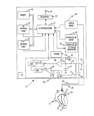
Referring now to FIG. 1 a pace",dher generally desiy,ldLt:d 10 is
illustrated in schematic fashion with conne- Iion to a human heart 12. For ease
of illustration the desu,i~,Lion is directed to a pac~:"ldht:~ having atrial sensing
and ventricular sensing and pacing. It should be ulld~l~luod however that
the invention can be employed for sensing in the atrium the ventricle or both
and that both atrial or ventricular pacing could be provided without departing
from the teachings thereof. In addition the features described herein could
aiso be combined with an illluldllLdble d~iibl -/cd,diuvertor.
W09611S8Z7 ij ' ~ 2 t 96~4 PCI/US95/14807
Vvith this u"de,aldnu;l,y, the illustrated pac~:",aher 10 co"".,iaes a
up~ucessor 14 which executes various control programs to regulate the
action of the ,.)act:",dht:r. The ~iu~u~Jluc~:ssul 14 is cunlle~ L~d to additional
memory 16 for the storage of programs and data as may be needed. As is
. 5 known in the art one or more internal clocks may be provided to permit timing
of various events. For example an A-V interval timer 18 may be provided.
Similarly a V-A interval timer 20 may also be provided as known in the art.
The ~iu~uulucessor is provided with a telemetry circuit 22 so that
communication can be had across an antenna 24 to an external p~uyldllllller
(not shown). Telemetry permits an attending physician to obtain data and
illfu~ ;Jll from the pact:",aht:l and to control the pd~.ellldh~, to set varioussele. I~ P pdlall~t:k:~a as known in the art.
The pa~ lldhel 10 is conl1e~ d to the heart 12 through a first lead 26
to an electrode 27 in the atrium 28 and through a second lead 30 to an
electrode 31 in the ventricle 32. An indifferent electrode is provided to
complete the electrical circuit. In the illustrated er"l,odi",e"l a can 60 or outer
casing of the pact:",dhe, serves as the indifferent electrode. Bipolar leads canalso be used as well as the unipolar leads illustrated here. Atrial sensing
through an atrial sense circuit 34 and ventricular sensing through a
ventricular sense circuit 36, provide i"ru""dlion to the microprocessor
conce",i"g the condition and responsiveness of the heart. In addition pacing
pulses are provided to the ventricle from a ventricular stimulus generator 38.
It is clearly within the scope of those skilled in the art to provide atrial pacing
should that be desired or to provide cardioversion/dc:ribrilldliùn ~ in
response to the detected condition of the heart. Stimulation of the heart is
passed through a coupling capacitor 40 in a conventional fashion. A switch
S5, con"eult:d to ground is peliu.lh a !y closed to discharge the capacitor 40
and balance stimulation pulses producing a net zero charge at the electrode.
To control the pulse rate of the ventricular stimulus generator 38, the
uulucessor acquires i"ru""dliu" on the condition of the heart through an
i" ,yedd"ce circuit 42. The i" ",edd"ce circuit 42 detects changes in i" ".eda"ce
. r~=7
~ r~ '9.6884 ,,
primarily due to the changing shape of the heart, which is related to the physical
shape of the heart as it beats and pumps blood. This information can be used
~ to derive a measure of the stroke volume or ejection fraction of the heart.
In addition to the measurement of impedance, a sensor 44 may also be
provided to obtain an indication of physiologic need and adjust the pacing rate.Such a sensor may be an accelerometer, as described by Dahl, U.S. Patent
4,140,132, a temperature sensor, as described by Alt, U.S. Patent 4,688,573,
or any other suitable sensor of a parameter which may be correlated to
physiologic need of the patient.
The impedance circuit 42 comprises a first capacitor 48 which I will call
a passive capacitor. This capacitor is connected to the lead 30 through a switchS1 and to ground through a second switch S2. The capacitor is also connected
to a buffer 48 in common with the two switches S1 and S2. On the other side of
the capacitor 46, the capacitor 46 is connected to ground. The buffer 48
communicates with a sample and hold circuit 50. The function of the separate
sample and hold circuit 50 can be performed by the passive capacitor 46 and the
buffer 48, if the sampling time (see FIG. 5) is short and the impedance of the
buffer 48 is high. Each of the two switches S1 and S2 and the sample and hold
circuit ~0 are controlled by the microprocessor 14. Such connections are well
krlown in the art and are not illustrated for the sake of clarity. A second capacitor
52, called herein an "active capacitor", is also connected to the lead 30 through
a switch S4. Preferably, the passive capacitor is of similar magnitude to the
active capacitor, and most preferably the passive capacitor has the same
capacitance as the active capacitor. This enables the passive capacitor to serveas an accurate model of the effect of background voltages on the active
capacitor, as will be more fully explained below.
AMENDED SHEE~
WO 96/158Z7 ,,, ~ ; 2 l 9 6 8 8 4 PCI/US95/14807
The side of the active capacitor 52 cu,,neuL~d to the lead is further
co"neult:d through a switch S3 to a voltage source, labeled VSRC in FIG. 1.
Finally, the capacitor is cG""e~ d in common with the two switches S4 and
S3 to a buffer 54. The other side of the capacitor 52 is co""e,,l~d to ground.
.~ 5 The output of the buffer 54 is combined with the output of the sample and hold
circuit 50, as will be more particularly described below, by subtracting the
voltage of the sample and hold circuit 50 from the output of the buffer 54. The
resulting voltage is held in a second sampie and hold circuit 56 until required
by the " ,iw u,u~ucessol . Typically, the analog value of the voltage held by the
sample and hold circuit 56 is converted to a digital value for further p, u~ess;l l9~
As explained above, the switches S3 and S4 and the sample and hold circuit
56 are controlled by the 1lliUlUplUC~:5501 14 in a manner similar to that of
switches S1 and S2 and sample and hold circuit 50.
The operation of the illlpeddnG~ circuit 42 can be u"de~:,luod with
respect to a timing diagram, FIG. 5. Preferably, the i,,,,ueclance circuit
d~ lllilles the illl,uedd~ue of the heart at a relatively high rate, on the order
of 100 times per second. A single ope,dtiondl cycle is described with respect
to FIG. 5. As each cycle begins, passive capacitor 46 is in a di~ dl yed state
while active capacitor 52 is charged to a p,~sele~ d voltage level, VSRC. which
may be about 0.5 V or less. Initially, during the cycle, S1 is closed for a
p,~seleuled period, for example, 15 usec. This is indicated in the timing
diagram of FIG. 5 by the line S1 going high. Simultaneousiy, switch S2 is
opened as indicated by the line S2 going low. This effectively connects the
passive capacitor 46 through the lead 30 to the electrode 31 within the heart
12. The passive capacitor 46 assumes the electrical value of the electrode 31
during the time that switch S1 is closed.
After switch S1 opens, the electrical condition of the passive capacitor
46 appears through the buffer 48 at the sample and hold circuit 50. The
sample and hold circuit 50 is therefore triggered by the ~nic~u~u~ ssol to
capture this voltage as indicated by the line S/H 50 going high. While the
WO 96/lS827 ~ 8 4 ~I
-10-
passive capacitor 46 is charged from the electrical condition of the heart, the
active capacitor 52 is charged from VSRC through S3 as indicated by the high
condition of line S3 in FIG. 5. When switch S1 opens, switch S3 also opens
as indicated by the low condition of line S3. Simultaneously, switch S4
5 closes, as shown by line S4 in FIG. 5, for a ,u,t:sele~ d period of time, for
example 15 u,sec. If the active capactitor 52 has the same ~,d~.a~,itd"~.e as the
passive capacitor 46, as described above, and if the resistance of the two
switches S4 and S1 are equal, then S1 is preferrably activated for the same
length of time as S4. The active capacitor 52 dia~ dlyes through switch S4
10 and lead 30 through the electrode 31 in the heart. Electrical current passes
from the electrode 31 within the heart to an anode on lead 30 or to the can 60
of the pac~:",dher which acts as an indifferent electrode.
When S4 opens, S3 does not i"""~ y close. Rather, the electrical
15 condition of the active capacitor 52 is passed through buffer 54. The electrical
value retained in the sample and hold circuit 50"t:~.,.is~"Li"g the electrical
condition of the heart, is subtracted from the output of buffer 54 and the
resulting value is captured by the sample and hold circuit 56, as l~yl~s~ d
by line S/H 56 going high. After the sampling by sample and hold circuit 56
20 is complete, initial conditions on the capacitors 46, 52 can be restored by
conl,e-,li"g the passive capacitor 46 to ground through S2 (indicated by line
S2 going high) and the active capacitor 52 to VSRC through switch S3 (indicated
by line S3 going high). In addition, pacing and i"" e.lance sensor pulses are
usually passed to the heart through an AC-coupling capacitor 40. Switch S5
25 is used to discharge this capacitor and to produce a balanced pulse which
results in zero net charge flow through the tissue. This is indicated by line S5going high, closing switch S5. Switch S5 opens when line S5 goes low.
S4 being closed (see FIG. 5) It~ tS a selected short period of time
30 during which the active capacitor 52 is dia~,lldlyed through the heart. The
voltage on the active capacitor 52 decays ex,col1e~ 'y according to the
following formula:
-
WO96115827 ~ 1 9 6 8 ~ 4 PCr/US95114807
VcA(t) = Voe
Where VCA is the voltage remaining on the active capacitor after a time t; V0
is the initial voltage on the capacitor; R is the lumped resistance of the circuit,
5 and Ca is the capduitd",,e of the active capacitor 52. There is an error
af-- ' with making the measurement of VCA as there is in making any
measurement. This error can be minimized, however, by making the
measurement after an elapsed time T equal to one time constant that is, at t
= T = RCa. The desired measured value is R d~'~.",i"ed as follows:
R = -V( Ca In (VcA(t) / V0) )
The fractional error in the measurement of R, that is, d(ln R), is a function
which has a minimum at t = T = RCa. The function is:
d(ln R) = -[In (VcA(t) / V0)] [VCA(t) / V0]
The value Ca, the ud~ a-,ild"-,e of the active capacitor, is constant, but the
20 value R, the i"",eddnce of the circuit including the heart, is changing. The
error ~~o~ d with the measurement of VCA (and thus also the error
a~so- ' with the i"",edd"ce) can be minimized by plUy,dl"",;"y the
,..o""~uter 14 to dynd",i~..,lly adjust the time during which S4 is open. A
suitable procedure, generally desiy"dL~d 80, is illustrated diay,d"""dlical'y in25 FIG. 6.
The procedure 80 is part of the general operation of the "~k.~ ucu"~uuter
14. When the procedure 80 begins 82, an average omt~ f,e" ~/c value of
the illl~ ddllce. ii is d~e:ll"i"ed 84. This could, for example be the rolling
30 average of the measured value of the i" ,pedance for a p, t:d~ . " ,i"ed number
of cycles. The fractional error d(ln R) is then computed 86. The fractional
error is compared 88 to an ~-c~ l,le value m. If the fractional error is less
1 9b884 ;;
: . .i, -12-
than the acceptable value m, the value t, that is the time switch S4 is open, isunchanged. If the fractional error is greater than the acceptable value m, a newvalue of t is calculated 90 such that t = R Ca. The microprocessor proceeds 92
with other processing, using the new value t to determine the impedance from
5 the measured value of VCA after a discharge time t.
Prior art devices, such as that described by Steinhaus, et al. in U.S.
Patent 5,197,467, did not provide the sampling circuitry for detecting the inherent
electrical condition of the heart as described herein. In FIG. 2, I have illustrated
10 a prior art device, such as described by Steinhaus, et al. All the componentsare labeled as in connection with FIG. 1. It can be seen that only the active
capacitor 52 with its ~C50~ h-1 switches S3 and S4, buffer 54 and sample and
hold circuit 56 have been provided. With the prior art circuit 52, the electrical
condition of the heart tends to mask or obscure the desired impedance
15 measurement resulting from the changing physical confguration of the heart.
This is illustrated in FIG. 3. FIG. 3 represents an impedance 70 corresponding
to measurements which would be obtained through the sample and hold circuit
56 in the prior art device of FIG. 2. Because the electrical polarization potentials,
pacing after potentials, and other components along the current path would also
20 be sensed, a large offset or baseline value 72 could be detected. In addition,
illLIdcaldiac electrogram artifacts, for example artifact 74, would also be
detected. The signal could also be distorted by myopul~l ILi~ls and other artifacts
not related to the impedance.
FIG 4, on the other hand, illustrates measurement of impedance using
an impedance circuit in accordance with my invention. Offset due to artifacts
and background electrical condition is eliminated, as are the effects arising from
myJ~.uL~nLials, cardiac pacing, and pacing after-potentials. The resulting signal
more nearly represents actual changes in impedance related to the physical
action of the heart.
AMENDED SHEET
WO 96/15827 ~ 9 6 8 8 4 PCINS9S/14807
-13-
Having identified i, l "~)edd"ce i"' I l Id~iUn r - - - ' ' with cardiac
cull~lduliui,s, this ill ~.llldliUIl can then be used to controi the pacing rate or
other pacing pdldlll~ such as A-V delay intervals. By co, 'rl " ,g the
pacing rate in such a manner as to keep the stroke volume relatively constant
from cycle to cycle, a physioluy;.,a"~/ d,u,ulu~J~idl~ pacing rate is selected.