Note: Descriptions are shown in the official language in which they were submitted.
CA 02199637 2005-08-17
-1-
CONICALLY-SHAPED ANTERIOR FUSION CAGE
AND METHOD OF IMPLANTATION
BACKGROUND
Field of the Invention
The present invention is directed to devices and methods for facilitating the
fusi gof bone structures and more particularly the fusing together of adjacent
vertebral bodies or bone structures.
Background of the Invention
Technical literature and patent documents disclose a number of devices
and methods for fusing bones together. One such device which has proven to be
successful is disclosed in U.S. Patent 4,961,740, entitled "V-THREAD FUSION
CAGE AND METHOD OF FUSING A BONE JOINT," which patent has been
assigned the present assignee. The referenced patent discloses a fusion cage
which is preferably cylindrical and has a thread formed as part of the
external
cylindrical surface. The fusion cage defines an internal cavity and apertures
through the wall of the cage which communicate the external cylindrical
surtace
with the internal cavity. The apertures are formed in the valleys of the
thread.
Normally two such cages are used to stabilize and fuse together adjacent
vertebral bodies or bone structures.
In practice, using a posterior approach, a patient's vertebral bone
structures are exposed and degenerate disk material located between the
vertebral bone structures is removed. A threaded tap is used to tap a
complementary thread in the upper and lower vertebral bone structures
preparatory to the insertion of the above fusion cage. Once such tapping has
been accomplished, using an introduction tool, the fusion cage is screwed into
the
space between the adjacent vertebral bone structures. The thread bites into
the
bone of the upper and lower vertebral bone structures, stabilizing the bone
structures, and preventing the fusion cage from working out of this position
due to
patient movement. Generally two such fusion cages are applied using this
technique. Once the two implants have been positioned, then bone growth
inducing substances, such as bone chips, are packed into the internal cavity
of the
fusion cages. These bone growth inducing substances come into immediate
contact with the bone from the vertebral bone structures which project into
the
CA 02199637 2006-06-22
-2-
internal cavity through the apertures. Such projection of bone is due to the
fact
that the apertures are formed in the valleys of the external thread of the
fusion
cage. Such immediate bone to bone contact between the vertebral bone
structures and the bone pack within the fusion cages results in more rapid
propagation of bone cells between the adjacent vertebral bone structures and
thus
a more rapid fusion of the adjacent vertebral bone structures.
Summary of the Invention
The present invention is directed to a fusion cage which has been designed
to be implanted using an anterior approach to the vertebral bone structures.
In accordance with one embodiment of the present invention there is
provided a cage device for promoting fusion with one or more bone structures
comprising: a conically-shaped cage body having an outer surface and a
plurality
of apertures extending through the outer surface, the cage body including a
proximal end having a first diameter and a distal end having a second
diameter,
wherein the first diameter of the proximal end is greater than the second
diameter
of the distal end; and threading provided on the outer surface of the cage
body for
anchoring the cage body to the one or more bone structures.
Preferably, the apertures increase in size from the distal end towards the
proximal end of the cage body.
In accordance with another embodiment of the present invention there is
provided a cage body for promoting fusion with one or more bone structure
comprising: a conically-shaped cage body having an outer surface surrounding
an
internal cavity of the cage body, the cage body including a proximal end
having a
first diameter and a distal end having a second diameter, wherein the first
diameter of the proximal end is greater than the second diameter of the distal
end;
at least one thread provided on the outer surface of the cage body adapted to
anchor the cage body to the one or more bone structures; and a plurality of
apertures extending through the outer surface of the cage body for providing
communication between the internal cavity and the outer surface, wherein the
cage body is continuously tapered between the distal end and the proximal end
of
the cage body.
Yet another embodiment provides for a cage body for promoting fusion with
one or more bone structures comprising: a conically-shaped cage body having an
CA 02199637 2006-06-22
-3-
outer surface and a plurality of apertures extending through the outer
surface, the
cage body including a proximal end having a first diameter and a distal end
having
a second diameter, wherein the first diameter of the proximal end is greater
than
the second diameter of the distal end; and continuous threads provided on the
outer surface of the cage body, wherein the outer surface having the
continuous
threads is tapered between the distal end and the proximal end of the cage
body.
The distal end, in preferred embodiments, is rounded with for example a
bull nose in order to facilitate the insertion of the cage body relative to
one or more
bone structures. The conically-shaped cage body is particularly advantageous
for
use with an anterior approach to vertebral bone structure fusion. This is due
to
the fact that the normal lordosis of the vertebral bone structures defines a
wedged-shape space for a vertebral disk between, for example, lumbar
vertebrae.
Accordingly, the conically-shaped body cage can be sized and selected in order
to
maintain or enlarge upon the normal lordosis.
In a preferred embodiment of the present invention, a fusion cage includes
a conically-shaped cage body having a proximal end and a distal end with the
distal end having a diameter which is smaller than the diameter of the
proximal
end. The conically-shaped cage body has a conically-shaped outer surface and
at least one flute formed in the conically-shaped outer surface. The flute
acts as a
relief much as the flute placed on self-tapping screws in order to facilitate
the
insertion of the fusion cage using a twisting motion between two vertebral
bone
structures.
In another preferred embodiment of the invention, a fusion cage includes a
conically-shaped cage body having a proximal end and a distal end, the distal
end
having a diameter which is smaller than the diameter of the proximal end. The
conically-shaped cage body has a conically-shaped outer surface and a thread
formed as part of the conically-shaped outer surface. The thread allows the
cage
body to be inserted using an anterior approach. Due to the fact that the cage
body is conically-shaped, the requirement for pretapping the vertebral bone
structures to receive the fusion cage is eliminated with the fusion cage being
self-
tapping. Also the cage gradually spreads apart the vertebral bone structures
as
the cage is inserted in order to regain or enlarge the natural lordosis of the
adjacent vertebral bone structures. As with other embodiments of the present
CA 02199637 2006-06-22
-3a-
invention, flutes can be provided through the thread in order to allow for
enhanced
thread tapping by the cage and for a smoother insertion of the fusion cage
between the vertebral bone structures. Preferably two or three flutes would be
formed spaced about the fusion cage in order than one flute would be engaging
with or adjacent to an upper vertebral bone structure with another flute being
engaging with or adjacent to a lower vertebral bone structure. Such a
relationship
maintains alignment of the fusion cage and prevent wandering as the fusion
cage
is introduced between the two vertebral bone structures. Without two or more
flutes, wandering might occur due to the fact that the thread is only
substantially
engaged with the vertebral bone structures and not with the disk material
between
the vertebral bone structures, which disk material does not provide support to
the
thread.
30
WO 96!08205 219 9 6 3 7 PcT/US95/11~
-4-
In a further aspect of the invention, any of the above embodiments
can be provided with a plurality of apertures through the fusion cage and
c
an internal cavity with the apertures communicating between the internal
cavity and the external surface of the fusion cage. Bone growth inducing
s substances, such as bone chips, can be packed into the internal cavity
either before the fusion cage is inserted or after the fusion cage has
reached a final insertion position. The bone chips come in contact with
the vertebral bone structures through the apertures in order to facilitate
fusion between the adjacent vertebral bone structures.
io In another aspect of the invention which can be included in any of
the above embodiments, the cage body can have a round or bull nose
distal end with one or more flutes formed in the round or bull nose distal
end in order to enhance the self-tapping nature of the fusion cage.
In yet another aspect of the invention, introduction tools allow the
i5 fusion cage to be accurately positioned between the vertebral bone
structures.
The method of the present invention affords access to adjacent
vertebral bone structures using an anterior approach and procedure. Such
anterior approach and procedure can be preferably performed
20 laparoscopically using an introduction set including a cannula. A
laparoscopic procedure is minimally invasive as the abdomen muscle tissue
can be spread using a set of cannula of increasing size and a small
opening thereby developed through which a fusion cage can be inserted.
Such a procedure is less traumatic to the tissue than an alternate anterior
25 approach and procedure, also known as ari anterior lumbar interbody
fusion, where an incision, perhaps up to five inches long is made, through
the abdomen muscle tissue. It is to be understood however that either
anterior approach and procedure can be used with the fusion cage and fall
within the scope of the invention.
3o After such access, using preferably a laparoscopic technique,
degenerate disk material can be removed and, using a cannula and
insertion tool, an appropriately shaped fusion cage can be screwed into
WO 96/08205 219 9 6 3 7 PCT/US95III28I
_5_
place between the vertebral bone structures in order to stabilize the
vertebral bone structures and allow for fusion. Either preparatory to
insertion of the fusion cage or after it has been inserted, bone chips or
other bone growth inducing substances can be inserted into the fusion
s cage to promote bone to bone contact and subsequent fusion.
It is to be understood that although the above-embodiments have
been described with respect to the fusion of adjacent vertebral bodies or
bone structures, that the present invention can be used to fuse together
a variety of bone structures, in addition to being fused to one bone
io structure and used as, for example, a base for an implant.
Other objects and advantages of the invention can be obtained
through a review of the specification and the figures.
Brief Description of the Figure
i5 Figure 1 is a partially sectional side view of an embodiment of the
fusion cage of the invention.
Figure 2 depicts a left end (distal end) view of the fusion cage of
Figure 1.
Figure 3 depicts a right end (proximal end) view of the fusion cage
20 of Figure 1 .
Figure 4 depicts a view through line 4-4 of the fusion cage of Figure
1.
Figure 5 depicts the fusion cage of Figure 1 in conjunction with an
introduction tool.
2s Figure 6 depicts an alternative embodiment of the introduction tool.
Figures 7, 8, and 9 depict the progressive stages in the method of
inserting the fusion cage between adjacent vertebral bone structures.
.. Figure 10 depicts a side view of an alternative embodiment of the
fusion cage of the invention.
so Figure 1 1 depicts the left end (distal end) view of the fusion cage
of Figure 10.
WO 96/08205 PCT/US95/112~
2199637
-6-
Figure 12 depicts the right end (proximal end) view of the fusion
cage of Figure 10.
Figure 13 depicts a side view of yet another embodiment of the
v
fusion cage of the present invention.
s Figure 14 depicts a left distal end (distal end) view of the fusion
cage of the invention of Figure 13.
Figure 15 depicts a right end (proximal end) view of the fusion cage
of the invention of Figure 13.
Figure 16 depicts a sectional view taken through line 16-16 of
to Figure 13.
Detailed Descriation of the Preferred Embodiment
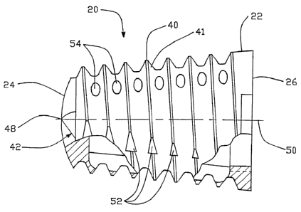
With respect to the figures in a particular Figure 1, a side view of
the preferred embodiment of the fusion cage 20 is depicted. Fusion cage
i5 20 includes a fusion cage body 22 which in this preferred embodiment is
provided in the shape of a cone. Fusion cage 20 includes a distal end 24
and a proximal end 26. The distal end 24 in a preferred embodiment is
rounded or bull nosed in order to facilitate the insertion of the fusion cage
20 relative to one or more bone structures. The proximal end 26 includes
ao an opening 28 which communicates with an internal cavity 30 defined by
the fusion cage 20. The opening 28 in a preferred embodiment is
threaded so that it can receive an end cap or plug 32 (Figure 5). End cap
32 is used to close off the proximal end 26 and retain bone growth
inducing substances packed therein as described herein-below. As can be
as seen in Figure 5, end cap 32 includes a threaded bore 34 which is
designed to receive an insertion tool. The threaded bore 34 has an initial
unthreaded, hex-shaped section 35 which can be used with a socket
wrench to tightly position end cap 32 in opening 28. The proximal end 26
further define first and second peripheral indentations 36, 38. These
3o peripheral indentations 36, 38 receive tangs from an insertion tool as
described hereinbelow for facilitating the insertion of the fusion cage 20.
CA 02199637 2005-08-17
7-
A thread 40 is defined as part of the outer cylindrical surface 41 of the body
22. It is to be understood that the thread can be replaced with a plurality of
discrete threads or a plurality of projections, ridges, protrusions, barbs, or
spurs
and be within the spirit and scope of the invention.
The rounded distal end 24, and at least some of the turns of thread 40
defined flutes or relief grooves 42, 44, and 46. (Figures 1, 2.) In a
preferred
embodiment, flutes 42, 44, and 46 meet at a central point 48 of the distal end
24
on the longitudinal axis 50 of the fusion cage 20. In other embodiments the
flutes
can be smaller and not extend all the way to the central point 48 on the
longitude
axis 50. Still in other embodiments, the flutes can be eliminated from the
distal
end 24 and such embodiments are still within the spirit and scope of the
invention.
The flutes extend from the distal end 24 toward the proximal end 26 as
shown in Figure 1 with respect to flute 42. These flutes are defined by the
sections 52 which are removed from the thread. In a preferred embodiment, the
flutes become narrower as they approach the proximal end 26 due to the fact
that
thread relief for purposes of self tapping becomes less important as the cage
reaches a final resting position. As shown in other embodiments, the flutes
can
be deeper and extend from the distal end completely to the proximal end. Still
further in other embodiments the flutes can be confined to the first several
turns of
the thread adjacent to the distal end and/or to just the distal end.
As can be seen in Figures 1, 4, a plurality of apertures 54 are provided
through wall 56 of the fusion cage 20. In a preferred embodiment, these
apertures 54 are formed by broaching grooves 58 in the internal surface 60 of
the
internal cavity 30. The effect of such broaching is to remove material from
the
valleys between the turns of the thread 40, thus defining the aperture 54. The
advantages of such an arrangement are taught by the above-referenced U.S.
Patent No. 4,961,740.which allows for immediate bone to bone contact between
the vertebral bodies or bone structures and the bone packed within the
internal
cavity 30 of the fusion cage 20.
The apertures 54 in a preferred embodiment increase in size from smaller
apertures closer to the distal end 24 to a larger aperture closer to the
proximal end
26. This increase in size allows for more bone to bone contact. Alternatively
in
the embodiment as shown in Figure 1, all the apertures are of the same size.
CA 02199637 2005-08-17
_$_
As can be seen in Figure 4, the apertures are clustered about a transverse
axis 51, both at the upper and lower ends of the axis. This is so that in
position,
the apertures come into contact with the upper and lower vertebral bone
structures (Figure 9) to encourage bone growth through the fusion cage from
the
vertebral bone structures. The lateral section of the fusion cage found along
the
outer transverse axis 53 do not have apertures in order to prevent growth of
disk
material which might interfere with the bone fusing process.
A preferred embodiment of the conically-shaped fusion cage 20 includes a
fusion cage which is 23 millimeters in length having a distal end 24 with a
diameter of 14 millimeters and a proximal end 26 with a diameter of 18
millimeters. The cage body is a right circular cone. The thread has a pitch of
30°
and there are ten turns per inch with a thread depth of 0.53 inches. Further
the
cage is made of a titanium material. Preferably this and other disclosed
fusion
cages are machined. However, the processes such as molding can be used to
accomplished formation of the fusion cages.
The cage is inserted between vertebral bodies using an insertion tool 62
(Figure 5). Insertion tool 62 includes an inner handle 64 and an outer handle
66.
the outer handle includes a bore 68 for receiving the inner handle 64. Handles
64,
66 include knobs 70, 72 respectively. The distal end of inner handle 64
defines a
threaded shaft 74, having a reverse thread to facilitate easy removal, and the
distal end of handle 66 define a cylindrical disk 76 which has first and
second
tangs 78, 80, projecting from the peripheral edge of the cylindrical disk 76.
These
tangs 78, 80 are designed to mate with the peripheral indentation 36, 38 of
the
fusion cage 20. For purposes of inserting the fusion cage between the
vertebral
bodies, the end cap 32 is inserted into the fusion cage 20 as shown in Figure
5.
Then the threaded shaft 74 of the inner handle is introduced into the threaded
bore 34 of the end cap 32. After this is accomplished, the outer handle 66 is
slid
over the inner'handle 64 and the tangs 78, 80 are positioned into engagement
with the indentations 36, 38. In this arrangement, the fusion cage 20 can be
anteriorly inserted into the space befinreen the vertebral body structure
using the
insertion tool 62.
An alternative embodiment of the insertion tool is shown in Figure 6. In this
figure, insertion tool 82 includes a handle 84 with a knob 86. At the end of
the
CA 02199637 2005-08-17
_g_
insertion tool 82 distal from the knob 86 is a cylindrical disk 88 which has
first and
second tangs 90, 92, which have the same function as the above tangs 78, 80.
Extending from the center of the cylindrical disk 88 along the centerline of
the
insertion tool 82 is a shaft 94 which has a ball detent 96. For use with
insertion
tool 82, the threaded bore 34 of the end cap 32 would be replaced with a bore
having a lip which could engage with the ball detent 96 of the insertion tool
82.
The method for inserting the fusion cage 20 of Figure 1 using an anterior
approach and procedure to the vertebral bodies is as follows. It is to be
understood that although the focus of this discussion is on a laparoscopic
procedure, that the anterior approach and procedure can also include a more
invasive procedure where a long incision is made in the abdomen wall.
With an anterior approach, using an introduction set such s described by
way of example only, in U.S. Patent 4,863,430, entitled "INTRODUCTION SET
WITH FLEXIBLE TROCAR WITH CURVED CANNULA," but however with larger
diameter instruments, an amount of disk material is removed between the two
vertebral bodies or bone structures which are to be fused together. This
procedure is accomplished through a cannula position adjacent to the vertebral
bone structures. With the same or a larger diameter cannula, the
30
WO 96/08205 ~ 219 9 6 3 7 pCT~S95/11~
-ZO-
fusion cage 20 can be introduced adjacent to the vertebral bone
structures. In a first procedure, the fusion cage is packed with bone
growth substances and the end cap 32 is affixed to the fusion cage 20.
Insertion tool 62 is then secured to the fusion cage 20 and the fusion cage
s is guided through the cannula to a location adjacent to the upper and
lower vertebral body such as presented schematically in Figures 7, 8, 9,
by upper body 98 and lower body 100. In the initial position as shown in
Figure 7, the fusion cage 20 is adjacent to the anterior sections 102, 104
of the vertebral bodies 98, 100. As the introduction tool is turned, the
io thread 40 of the fusion cage 20 bites into the vertebral bodies 98, 100.
Further turning of the introduction tool causes the fusion cage to move
through the position shown in Figure 8 to the final resting position shown
in Figure 9, where the distal end 24 is moved adjacent to the posterior
sections 106, 108 of the vertebral bone structures 98, 100. As this
15 occurs, the fusion cage 20 increases the lordosis or spacing between the
vertebral bodies, basically distracting the vertebral bodies and causing the
vertebral bodies to pivot about the posterior sections 106, 108, with such
posterior sections acting like a hinge. It is noted that most of the
distraction occurs adjacent to the anterior sections, but that distractions
2o also occur at the posterior sections where the hinged effect is exhibited.
Preferably, the lordosis is increased over the normal lordosis in order to
stabilize the vertebral bone structures prior to fusion occurring.
Stabilization occurs due to the fact that increased lordosis places
additional stress on the anterior longitudinal ligaments which are part of
2s the anatomy holding the vertebral bodies in place.
Once the fusion cage 20 is appropriately positioned, the handle 64
of the insertion tool 62 is unscrewed from the cap 32 and the insertion
tool 62 is pulled away from the fusion cage.
An alternative embodiment of a fusion cage 200 is shown in Figures
so 10, 1 1, and 12. Fusion cage 200 includes a distal end 202 and an a
proximal end 204. Fusion cage 200 includes an internal cavity 206. End
caps not shown can be used to close the ports 208, 210 of distal and
W O 96!08205 219 9 6 3 7 PCT/US95111281
-11-
proximal ends 202, 204. A plurality of threads 212 is defined on the
external conical surface 214 of the fusion cage 200. Defined by the
thread 212 are first and second flutes 216, 218, which in this embodiment
extend from the distal end 202 to the proximal end 204. These flutes
s provide thread relief allowing the fusion cage 200 to be self-tapping.
The fusion cage 200 includes a plurality of elongated apertures 220
which are formed through the side walls of a fusion cage 200. The
elongated apertures 202 are formed in such a way that the internal conical
surface 214 is spaced away from the internal surface 224 of the internal
io cavity 206 by the thickness of the sidewall 222.
A further embodiment of the invention is shown in Figures 13, 14,
15 and 16. fn Figure 16 the fusion cage 300 has distal and proximal ends
302 and 304 respectively. The fusion cage 300 defines an internal cavity
306, and ports 308 and 310 defined through the distal and proximal ends
i5 302 and 304 respectfully. A thread 312 is defined as part of the external
conical surface 314 of the fusion cage 200. First, second and third flutes
316, 318, and 320, are defined in the thread 312 from the distal end 302
to the proximal end 304. These flutes give the fusion cage 300 an
enhanced self-tapping advantage. These flutes are equally spaced about
2o the fusion cage 300 in a manner similar to the flutes of the fusion cage
embodiment 20 in Figure 1.
A plurality of aperture 322 is provided through the external conical
surface 314 of the fusion cage 300 and through the side wall 324 opening
into the internal cavity 306. Accordingly, at the location of the aperture
25 322 the external surface 314 is held away from the internal surface 326
by the thickness of the side wall 324.
Industrial Aaplicabilitv
The present invention affords the advantages of a fusion cage
so which can be introduced through an anterior approach in order to maintain
or increase lordosis between adjacent vertebral bodies. The fusion cage
has the advantage of being conically-shaped and self-tapping through the
WO 96/08205 PCT/US95/112~
219963 ,7
-12-
use of external flutes. The flutes additionally assist in keeping the fusion
cage aligned and centered as the cage is being inserted between the
vertebral bone structures.
Other advantages, aspects, and objects of the invention can be
s obtained through a review of the claims and the appended figures.
It is to be understood that additional embodiments of the invention
can be constructed and fall within the spirit and scope of the claims.