Note: Descriptions are shown in the official language in which they were submitted.
WO96/14027 2 2 0 ~ 3 1 g PCTIUS95/14327
-
DESCRIPTION
INTRALUMINAL STENTING GRAFT
5 Backaround Art
The present invention is directed to an intraluminal stenting graft.
More specifically, the invention is directed to an intraluminal stenting
graft for implantation in a blood vessel including a collapsible tube
member formed from a plurality of cylinders. The invention is further
10 directed to a method for making such a stenting graft.
Intraluminal stenting grafts are known in the art. An example of
an intraluminal stenting graft/stent is disclosed in U.S. Patent No.
5,156,620, which is incorporated herein by reference. Intraluminal
stenting grafts are implanted in a blood vessel to repair, for example,
15 aortic aneurysms. They are also used to support sections of a blood
vessel that are diseased or have become narrowed by arteriosclerosis.
Disclosure of Invention
The present invention is directed to an intraiuminal stenting graft
20 for implantation in a blood vessel and a method for making same. The
intraluminal stenting graft includes a collapsible tube member having a
first end and a second end. An outer layer and an inner layer extend
between the ends. The outer layer is more flexible than the inner layer.
The outer layer is joined to the inner layer to form a plurality of cylinders
25 longitudinally extending between the first end and the second end.
The method of the present invention includes the steps of:
(a) placing a first layer of material on a substantially flat
surface;
(b) placing a second layer of material on a shaped surface;
WO96/14027 2 2 0 1 3 1 9 Pcr/uss5ll4327
(c) maintaining the second layer on said shaped surface by use
of reverse pressure;
(d) moving the second layer to the first layer;
(e) joining the second layer to the first layer to form a plurality
of longitudinally extending cylinders; and
(f) shaping the first and second layers to form a tube member.
The primary object of the present invention is to provide an
intraluminal stenting graft that is efficient.
An important object of the present invention is to provide an
intraluminal stenting graft that is relatively easy to use.
Other objects and advantages of the invention will become
apparent upon a review of the accompanying drawings and the following
detailed description of the invention.
Brief Description of the Drawings
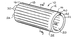
Fig. 1 is a perspective view of a first embodiment of an
intraluminal stenting graft according to the present invention;
Fig. 2 is a cross-sectional view of the plurality of cylinders of the
present invention taken along line 2-2 of Fig. 1;
Fig. 3 is a cross-sectional view taken along line 3-3 of Fig. 2
showing the one-way valve of the present invention positioned in the
opening in the end wall of the tube member;
Fig. 4 is a cross-sectional view taken along line 4-4 of Fig. 2
showing one of the cylinders according to the present invention;
Fig. 5 is a cross-sectional view of the intraluminal stenting graft of
the present invention positioned in a blood vessel at the site of
implantation in a collapsed condition;
Fig. 6 is a cross-sectional view similar to the view of Fig. 5
showing the intraluminal stenting graft implanted in a blood vessel;
2 ~ O ~ 3 ~ g
WO 96/14027 PCT/US95/14327
3
Fig. 7 is a second embodiment of an intraluminal stenting graft
- according to the present invention;
Fig. 8 is a side elevational view of the first layer of material on a
platen being treated according to the method of the present invention;
Fig. 9 is a side elevational view showing the second layer of
material on a shaped surface being maintained on the surface by reverse
pressure according to the method of the present invention;
Fig. 10 is a view similar to the view of Fig. 9 showing the joining
of the second layer to the first layer; and
Fig. 11 is a side elevational view showing the second layer joined
to the first layer.
Best Mode For Carrying Out Invention
Referring now to the drawings, the present invention will now be
described in detail. Referring to Figs. 1 and 2, the intraluminal stenting
graft of the present invention is indicated by the reference number 10.
The stenting graft 10 includes a collapsible tube member 12 having a
first end 14 and a second end 16. An outer layer of material 18 and an
inner layer of material 20 extend between said first end 14 and said
second end 16. A first end wall 22 extends between the outer layer 18
and the inner layer 20 at the first end 14. A second end wall 24 extends
between the outer layer 18 and the inner layer 20 at the second end 16.
As shown in Figs. 1, 2 and 4, the outer layer 18 is joined to the
inner layer 20 to form a plurality of cylinders 30 that extend
longitudinally between the first end 14 and the second end 16. As
shown in Fig. 1, the tube member 12 can include a radially extending
chamber 32 that is in communication with the plurality of cylinders 30.
In the present embodiment, the chamber 32 is positioned adjacent the
first end 14. However, it should be understood that the chamber 32 can
be positioned in a variety of locations along the length of the chamber.
,
Wo96~l4027 ~ 2 n 1 3 1 9 PCT/USg5/14327
Referring to Fig. 1, the tube member 12 can include an opening 40
in the first end wall 22. The opening 40 can receive a fluid, such as air.
As described below, the fluid causes the collapsed tube member 12 to
expand for implantation in a blood vessel. As shown in Fig. 3, a one-
5 way vaive 42, such as a check valve, can be positioned in the opening
40. The valve 42 allows for the introduction of the fluid into the tube
member 12. The valve prevents the escape of the fluid from the tube
member 12 after introduction into the tube member. The fluid can be
introduced into the tube member 12 through the valve 42 by a fluid
10 conduit 44.
Referring to Fig. 2, the outer layer 18 and the inner layer 20 are
composed of a polymer material that is biocompatible. An example of
such a material is polytetrafluoroethylene. The outer layer 18 is
constructed of a more flexible or lighter weight material than the inner
15 layer 20. This allows the outer layer 18 to be more compliant when the
tube member 12 is expanded. The inner layer 20 can be treated or
coated with a material such as expanded polytetrafluoroethylene (ePTFE)
to create a surface more conducive to blood flow.
As shown in Fig. 2, each of the cylinders 30 includes a centerline
20 C that extends longitudinally through the cylinder when the tube member
12 is in an expanded condition. The centerline C is a point from which
two radii Rl and R2 extend. The radii Rl and R2 define an angle ,B. The
angle ,B can be an obtuse angle being more than 90 and less than 180.
When the plurality of cylinders 30 are positioned adjacent one another to
25 form the tube member 12, as shown in Fig. 2, the radius R1 of one of the
cylinders bisects the radius R2 of the adjacent cylinder. This arrangement
causes the plurality of cylinders 30 to cooperate to maintain the tube
member 12 in a stable, expanded condition for implantation in a blood
vessel. It has been found that the greater compliance of the outer layer
30 18 and the greater amount of material of the outer layer 18 as compared
_
2 2 0 1 3 1 9
WO 96/14027 PCT/US95/14327
to the inner layer 20 causes the angle ,~ to be less than 180. When the
tube member 12 is expanded, the plurality of cylinders 30 interfere with
one another to force the tube member into a round configuration as
shown in Fig. 1. This provides an open pathway 46 for the flow of blood
in a blood vessel.
Referring now to Figs. 5 and 6, the intraluminal stenting graft 10
of the present invention is implanted in a blood vessel 50 by manipulating
the collapsed tube member 12 through the vessel to an implantation site
52. The tube member can be manipulated by the conduit 44, which is
in communication with the valve 42, or by some other suitable
apparatus. As shown in Fig. 6, when the stenting graft 10 is in the
proper position, fluid from the conduit 44 is introduced through the
opening 40 and into the chamber 32 and cylinders 30. The chamber 32
allows for an efficient distribution of fluid into the cylinders 30. As
described above, the plurality of cylinders 30 and the outer and inner
layers 18 and 20, respectively, cooperative to maintain the tube member
12 in a round and open configuration. After filling, the conduit 44 is
removed. The stenting graft 10 allows blood flow through the pathway
46 at the site of implantation 52.
A second embodiment of the intraluminal stenting graft 10 of the
present invention is shown in Fig. 7. The stenting graft 10 includes a
trunk portion 60 and branch portions 62 and 64. This embodiment can
be used, for example, at the bifurcation of the aorta and iliac arteries.
The trunk portion 60 can be positioned in the aorta and the branch
portions 62 and 64 can be positioned in the iliac arteries. Many other
configurations can be constructed depending on the application.
Referring now to Figs. 8 through 1 1, the method for manufacturing
an intraluminal stenting graft according to the present invention will be
described in detail. Referring to Fig. 8, a first layer of material 70, which
corresponds to the inner layer 20, is placed on a flat surface such as a
WO96/14027 2 2 û ~ 3 1 ~ Pcr/uss5/14327
platen 72. A bonding agent such as adhesive 76 is applied to the first
layer 70 by applicators 78.
As shown in Fig. 9, a second layer of material 80, which
corresponds to the outer layer 18, is placed on a shaped surface 82. The
5 shaped surface 82 includes longitudinally extending indentations 84
having, for example, partially cylindrical shapes. The indentations include
a coating 86 of synthetic resin polymers and products, such as Teflon~,
to prevent the second layer 80 from adhering to the shaped surface 82.
The second layer 80 is maintained on the shaped surface 82 by the use
10 of reverse pressure or vacuum created by a reversible pump P.
As shown in Fig. 10, the second layer 80 is moved to the first
layer 70. The layers 70 and 80 are fixedly joined together by the
adhesive 74. The layers can also be joined by a heat sealing process
(not shown).
As shown in Fig. 11, the joining of the first layer 70 to the second
layer 80 forms a plurality of longitudinally extending cylinders 30, as
described above. A chamber 30, end walls 22 and 24 and opening 40
can also be formed in the method. The longitudinally extending ends of
the joined layers can be brought together and joined by adhesive or
20 otherwise to form the cylindrical tube member 12 shown in Figs. 1 and
5.
The first layer 70 and second layer 80, as used in the method, can
be constructed of a polymer material, as described above for the outer
layer 18 and inner layer 20. The second layer 80 is more flexible and is
25 lighter weight than the first layer 70. The cylinders 30 that are formed
as a result of the method have the same characteristics as described
above concerning the centerline C, radii R1 and R2 and the angle ,~ being
less than 180.
-
22013 1 9
Wo 96/14027 Pcr/USs5/14327
The present invention can be modified and changed in a variety ofways with the scope of the invention being defined by the appended
claims.