Note: Descriptions are shown in the official language in which they were submitted.
CA 02207659 2002-06-05
60950-245
DRUG RELEASE STENT COATING AND pROCESS
HACRGRODND OF THE INVENTION
I. Cross-Reference to Related Patent
Cross-reference is made to United States Patent
No. 5,837,313 to Ding, entitled "DRUG RELEASE STENT COATTNG
PROCESS", issued on November 17, 1998.
II. Field of the Invention
The present invention relates generally to_providing
biostable elastomeric coatings on~the surfaces of implants
which incorporate biologically active species having
20 controlled release characteristics in the coating
particularly to providing a non-thrombogenic surface during
and after timed release of the biologically active species.
The invention is particularly described in terms of coatings
on therapeutic expandable stent prostheses for implantation
25 in body lumens, e.g., vascular implantation.
II. Related Art
In surgical or other related invasive procedures, the
insertion and expansion of stent devices in blood vessels,
i
CA 02207659 2002-06-05
60950-245
urinary tracts or other locations difficult: to otherwise
access for the purpose of preventing reste:nosis, providing
vessel or lumen wall sug~port or reinforcement and for other
therapeutic or restorative functions has become a common foam
of long-term treatment. Typically, such prostheses are
applied to a location of interest utilizing a vascular
catheter, or similar transluminal device, t:o carry the stem=
to the location of interest where it is thereafter released
to expand or be expanded in situ. These devices are genera:lly
designed as permanent implants which may bEacome incorporated
in the vascular or othez- tissue which they contact at
implantation.
One type of self-expanding stent has a flexible tubular
body formed of several ~.ndividual flexible thread elements
each of which extends in a helix configuration with the
centerline of the body serving as a common axis. The elements
are wound in the same direction but are displaced axially
relative to each other and meet, under crossing,a like number
of elements also so axially displaced, but having the opposite
direction of winding. 'his configuration provides a resilient
braided tubular structure which assumes stable dimensions upon
relaxation. Axial tension produces elongation and
corresponding diameter contraction that allows the stent to be
mounted on a catheter device and conveyed i;,hrough the vascular
system as a narrow elongated device. Once tension is relaxed
in situ, the device at least substantially reverts to its
original shape. Prostheses of the class including a braided
flexible tubular body ax-e illustrated and described in U.S.
2
CA 02207659 2002-06-05
60950-245
Patents 4'655 771 and 4 954 126 to Wallste:n and 5 061 275 to
Wallsten et al.
Implanted stents have been used to carry medicinal
agents, such as thrombolytic agents. U.S. Patent 5 163 952 to
Froix discloses a thermal memoried expanding plastic scent
device formulated to carry a medicinal agent in the material
of the stent itself. Pinchuk, in U.S. Patent 5 092 877,
discloses a stent of a polymeric material which may have a
coating associated with the delivery of drugs. Other patents
which are directed to devices of the class utilizing bio-
degradable or bio-sorbable polymers include Tang et al, U.S.
Patent 4 916 193, and MacGregor, U.S. Patent 4 994 071.
A patent to Sahatjian namely United States
Patent No. 5 304 121 issued April 19, 1994, discloses a
coating applied to a stmt consisting of a hydrogel polymer
and a preselected drug such as cell growth inhibitors or
heparin. A further method of making a coated intravascular
stent carrying a therapeutic material is described in Berg et
al., U.S. Patent No. 5 464 650, issued on November 7, 1995 and
corresponding to European Patent Application No. 0 623 354 A1
published 09 November 1994. In that disclosure, a polymer
coating material is dissolved in a solvent and the
therapeutic material dispersed in the solvent; the solvent
evaporated after application.
An article by Michael N. Helmus (a ca-inventor of the
present invention) entitled "Medical Device Design--A Systems
Approach: Central Venous Catheters", 22nd. International
Society for the Advancement of Material anal Process
Engineering Technical Conference (1990) relates to
3
CA 02207659 1997-06-12
polymer/drug/membrane systems for releasing heparin.. Those
polymer/ drug/membrane systems require two distinct types of
layers to function.
It has been recognized that contacting blood with the
surface of a foreign body in vivo has a tendency to induce
thrombogenic responses and that as the surface area of a
foreign device in contact with host blood increases, the
tendency for coagulation and clot forming at these surfaces
also increases. This has led to the use of immobilized
systemic anti-coagulant or thrombolytic agents such as heparin
on blood contacting surfaces such as oxygen uptake devices to
reduce this phenomenon. Such an approach is described by .
Winters, et~al., in U.S. Patents 5 182 317; 5 262 451 and 5
338 770 in which the amine functional groups of the active
material are covalently bonded using polyethylene oxide (PEO)
on a siloxane surface.
Another approach is described in U.S. Patent 4 613 665 to
Larm in which heparin is chemically covalently bound to
plastic surface materials containing primary amino groups to
impart a non-thrombogenic surface to the material. Other
approaches for bonding heparin are described in Barbucci, et
al., "Coating of commercially available materials with a new
heparinizabhe material", JOLrrial_ Of BinmaAir~a1 Materialc
Research, Vol 25, 1259-1274 (1991); Hubbell, J.A.,
"Pharmacologic Modification of Materials", cardiovascular
Patholoav, Vol 2, No 3(Suppl.), 121S-127S (1993); Gravlee,
G.P., "Heparin-Coated Cardiopulmonary Bypass Circuits", Journal
4
CA 02207659 2002-06-05
60950-245
of Cardiothoracic and V.~~scular Anesthesia, Vol 8, No 2, pp
213-222 (1994).
Although, polymeric stems are effective,
they may have mechanical properties that are inferior to
those of metal stems of like thickness and weave. Metallic
vascular stents braided of even relatively fine metal can
provide a large amount of strength to resist inwardly direcaed
circumferential pressure. A polymer material of comparable
strength requires a much thicker-walled structure or heavier,
denser filament weave, which in turn, reduces the cross-
sectional area available for flow through the stent and/or
reduces the relative amount of open space in the weave. A7.so,
it is usually more difficult to load and deliver polymeric:
stents using catheter delivery systems.
While certain types of stents such as braided metal
scents may be preferred fc~r some.applications, the coating and
coating modification process of the present invention is not
so limited and can be used on a wide variety of prosthetic
devices. Thus, in the case of stents, the. present invention
also applies, for example, to the class of stents that are not
self-expanding including those which can b~e expanded, for
instance, with a balloon; and is applicable polymeric stenta
of all kinds: Other medical devices that can benefit from the
present invention include blood exchanging' devices, vascular
access ports, central versus catheters, cardiovascular
catheters, extracorpeal circuits, vascular grafts, pumps,
heart valves, and cardiovascular sutures, to name a few.
Regardless of detailed embodiments, applicability of the
- 5
CA 02207659 1997-06-12 ,
invention=should not be considered limited with respect to
' implant design, implant location or materials of construction.
Further, the present invention may be.used with other types of
implantable prostheses.
Accordingly, it is a primary object of the present
invention to provide a coating and process for coating a stent
to be used as a deployed stent prostheses, the coating being
capable of effective controlled long-term delivery of '
biologically active materials.
.. 10 Another object of the invention is to provide a coating
and process for coating a stent prostheses using a biostable
hydrophobic elastomer in which biologically active species are
incorporated within a coating.
Still another object of the present invention is to
provide a multi-layer coating and process for the delivery of
biologically active species in~which the percentage of active
material can vary from layer to layer.
Yet another object of the present invention is to provide
a multi-layer coating and process for the delivery of
biologically active species from a coating with a non-
thrombogenic surface.
A further object of the invention is to provide a multi-
layer coatiisg for the delivery of biologically active species
such as heparin having a fluorosilicone top layer.
A still further object of the invention is to provide a
multi-layer coating for the delivery of biologically active
species such as heparin having a surface containing
immobilized polyethylene glycol (PEG).
6
CA 02207659 1997-06-12
w ' Other objects and advantages of the present invention
will become apparent to those skilled in the art upon
familiarization with the specification and appended claims.
SUMMARY OF THE INVENTION
The present invention provides a relatively thin layered
coating of biostable elastomeric material containing an amount
of biologically active material dispersed therein in
combination with a non-thrombogenic surface that is useful for '
- coating the surfaces of prostheses such as deployable stems.
-- 10 The preferred stent to be coated is a self-expanding,
open-ended tubular stent prostheses. Although other
materials, including polymer materials, can be used, in the
preferred embodiment, the tubular body is formed of a self-
expanding open braid of fine single or polyfilament metal wire
which flexes without collapsing, readily axially deforms to an
elongate shape for transluminal insertion via a vascular
catheter and resiliently expands toward predetermined stable
dimensions upon removal in situ.
In the process, the initial coating is preferably applied
as a mixture, solution or suspension of polymeric material and
finely divided biologically active species dispersed in an
organic vehicle or a solution or partial solution of such
species in x~-solvent or vehicle for the polymer and/or
biologically active species. For the purpose of this
application, the term "finally divided" means any type or size
of included material from dissolved molecules through
suspensions, colloids and particulate mixtures. The active
material is dispersed in a carrier material Which may be the
7
CA 02207659 1997-06-12 -
- polymer, a solvent, or both. The coating is preferably
applied as a plurality of relatively thin layers sequentially
applied in relatively rapid sequence and is preferably applied
with the stent in a radially expanded state.
In many applications the layered coating is referred to
or characterized as including an undercoat and topcoat. The
coating thickness ratio of the topcoat to undercoat may vary
with the desired effect and/or the elution system. Typically '
these are of different formulations with most or all of the
active.material being contained in the undercoat and a non-
thrombogenic surface is found in the topcoat.
The coating may be applied by dipping or spraying using
evaporative solvent materials of relatively high vapor
pressure to produce the desired viscosity and quickly
establish coating layer thicknesses. The preferred process is
predicated on. reciprocally spray coating a rotating radially
expanded stent employing an air brush device. The coating
process enables the material to adherently conform to and
cover the entire surface of the filaments of the open ,
- 20 structure of the stent but in a manner such that the open
lattice nature of the structure of the braid or other pattern
- is preserved in the coated device.
The coating is exposed to room temperature ventilation
for a predetermined time (possibly one hour or more) for
solvent vehicle evaporation. In the case of certain
undercoat materials, thereafter the polymer material is cured
at room temperature or elevated temperatures. Curing is
defined as the process of converting the elastomeric or
8
CA 02207659 2002-06-05
60950-245
polymericrnaterial into the finished or useful state by the
application of heat and,/or chemical agents which induce
physico-chemical changes. Where, for example, .polyurethane
thermoplastic elastomers are used as an undercoat material,
solvent evaporation can occur at room temperature rendering
the undercoat useful for controlled drug release without
further curing.
The applicable ventilation time and t~smperature for cure
are determined by the particular polymer involved and
l0 particular drugs used. For example, silicone or polysiloxane
materials (such as polydimethylsiloxane) have been used
successfully. Urethane pre-polymers can also be utilized.
Unlike the polyurethane thermoplastic elas~tomers, some of
these materials are applied as pre-polyiner;s in the coating
composition and must thereafter be heat cured. The preferred
silicone species have relatively low cure. temperatures and
are known as a room temperature vulcanizable (RTV) materials.
Some polydimethylsiloxane materials can be cured, for example,
by exposure to air at about 90°C for a period of time such as
16 hours. A curing step may be implemented both after
application of the undercoat or a certain number of lower
layers and the top layers or a single curing step used after
coating is completed.
The coated stents may thereafter be slabjected to a
postcure process which includes an inert gas plasma treatment,
and sterilization which may include gamma :radiation, ETO
treatment, electron beam or steam treatment.
9
CA 02207659 1997-06-12
In the plasma treatment, unconstrained coated stents are
placed in a reactor chamber and the system is purged with
nitrogen and a vacuum applied to 20-50 mTorr. Thereafter,
inert gas (argon, helium or mixture of them) is admitted to
the reaction chamber for the plasma treatment. One method
uses argon (Ar) gas, operating at a power range from 200 to
400 watts, a flow rate of 150-650 standard ml per minute,
which is equivalent to about 100 - 450 mTorr, and an exposure
.: time from 30 seconds to about 5 minutes. The stents can be
_ 10 removed immediately after the plasma treatment or remain in
the argon atmosphere for an additional period of time,
typically five minutes.
In accordance with the invention, the top coat or surface
coating may be applied in any of several ways to further
control thrombolitic effects and optionally, control the
release profile especially the initial very high release rate
associated with the elution of heparin.
In one embodiment, an outer layer of fluorosilicone (FSi)
. is applied to the undercoat as a topcoat. The outer layer
can also contain heparin. In another embodiment, polyethylene
glycol (PEG) is immobilized on the surface of the coating. In
- this process, the underlayer is subjected to inert gas plasma
treatment ahd immediately thereafter is treated by ammonia
(NHS) plasma to aminate the surface. Amination, as used in this
application, means creating mostly imino~groups and other
vitro containing species on the surface. This is followed by
immediate immersion into electrophillically activated
CA 02207659 2002-06-05
60950-245
polyethyldne glycol(PEG) solution with a reductive agent,
i.e., sodium cyanoborohydride.
The coated and cured stents having the modified outer
layer or surface are subjected to a final gamma radiation
sterilization nominall~~ at 2.5-3.5 Mrad. Argon (Ar) plasma
treated stents enjoy full resiliency after' radiation whether
exposed in a constrained or non-constrained status, while
constrained stents subjected to gamma sterilization without Ar
plasma pretreatment lose resiliency and do not recover at a
sufficient or appropriate rate.
The elastomeric materials that form t:he stent coating
underlayers should possess certain properties.
The layers may be composed of suitable hydrophobic biostable
elastomeric materials which do not degrade. Surface layer
material should minimize tissue rejection and tissue
inflammation and permit: encapsulation by tissue adjacent the
stent implantation site. Exposed material is designed to
reduce clotting tendenc:i:es in blood contacted and . the surface
is preferably modified accordingly. Thus,. underlayers of the
above materials are preferably provided with a fluorosilicone
outer coating layer wh~.ch may or may not contain imbedded
bioactive material, such as heparin. Alternatively, the outer
coating may~.consist essentially of polyethylene glycol (P,E~),
polysaccharides, phospholipids, or combinations of the
foregoing.
Polymers generally suitable for the undercoats or
underlayers~include silicones (e.g., poly:~iloxanes and
substituted polysiloxanes), polyurethanes, thermoplastic
11
CA 02207659 2002-06-05
60950-245
elastomers in general, ethylene vinyl acetate copolymers,
polyolefin elastomers, polyamide elastomers, and EPDM rubbers.
The above-referenced materials are considered hydrophobic with
respect to the contemplated environment of the invention.
Surface layer materials include fluorosilic;ones and
polyethylene glycol (PEG), polysaccharides, phospholipids, and
combinations of the foregoing.
While heparin is preferred as the incorporated active
material, agents possibly suitable for incorporation include
1o antithrobotics, anticoagulants, antibiotics,antiplatelet
agents, thorombolytics, antiproliferatives, steroidal and non-
steroidal antinflammatories, agents that inhibit hyperplasia
and in particular restenosis, smooth muscle cell inhibitors,
growth factors, growth factor inhibitors, cell adhesion
inhibitors, cell adhesion promoters and drugs that may enhance
the formation of healthy neointimal tissue, including
endothelial cell regeneration. The positive action may come
from inhibiting particular cells (e. g., smooth muscle cells)
or tissue formation (e. g., fibromuscular tissue) while
encouraging different cell migration (e.g., endothelium) and
tissue formation (neoint:imal tissue) .
Suitable materials for fabricating the braided stent
include stainless steel, tantalum, titanium alloys including
nitinol (a nickel titanium, thermomemoried alloy material),
and certain cobalt alloys including cobalt-chromium-nickel
alloys such as Elgiloy~ and Phynox~. Further details
concerning the fabrication and details of other aspects of the
stents themselves may be gleaned from the above referenced
12
CA 02207659 2002-06-05
60950-245
U.S. Patents 4 655 771 and 4 954 126 to Wallsten and 5 061 275
to Wallsten et al.
Various combinations of polymer coating materials can be
coordinated with biologically active species of interest to
produce desired effects when coated on stents to be implanted
in accordance with the invention. Loadings of therapeutic
materials may vary. The mechanism of incorporation of the
biologically active species into the surface coating., and
egress mechanism depend both on the nature: of the surface
coating polymer and the material to be incorporated. The
mechanism of release also depends on the mode of
incorporation. The mat.erial-may elute via interparticle paths
or be administered via transport or diffusion through the
encapsulating material itself.
For the purposes of this specification, "elution" is _
defined as any process of release that involves extraction or
release by direct contact of the material with-bodily fluids
through the interpartic:le paths connected with the exterior of
the coating. "Transport" or "diffusion" are defined to include
a mechanism of release in which the material released
traverses through another material.
The desired relea:oe rate profile can be tailored by
varying the'coating thickness, the radial distribution (layer
to layer) of bioactive materials, the mixing method, the
amount of bioactive material, the combination of different
matrix polymer materials at different layers,.and the
crosslink density of the polymeric material. The crosslink
density is related to the amount of crosslinking which takes
13
CA 02207659 2002-06-05
60950-245
place and also the relative tightness of the matrix created
by the particular crosslinking agent used. This, during the
curing process, determines the amount of crosslinking and
also the crosslink density of the polymer material. For
bioactive materials released from the crosslinked matrix,
such as heparin, a denser crosslink structure will result in
a longer release time and reduced burst effect.
It will also be appreciated that an unmedicated
silicone thin top layer provides some advantage and
additional control over drug elusion; however, in the case
of heparin, for example, it has been found that a topcoat or
surface coating modified to further control the initial
heparin release profile or to make the surface more non-
thrombogenic presents a distinct advantage.
In accordance with the present invention, there is
provided an implantable medical device having an outer
surface covered at least in part by a conformal undercoat of
a hydrophobic elastomeric material incorporating an amount
of biologically active material therein for timed delivery
therefrom; and a topcoat comprising a biostable, non-
thrombogenic material, disposed over the undercoat, said
non-thrombogenic material being substantially free of an
elutable material.
In accordance with the present invention, there is
further provided a medical device having at least a portion
which is implantable into the body of a patient, wherein at
least a part of the portion is covered with a coating for
release of at least one biologically active material,
wherein said coating comprises an undercoat comprising a
hydrophobic elastomerir_ material incorporating an amount of
biologically active material therein for timed release
14
CA 02207659 2002-06-05
60950-245
therefrom, and wherein said coating further comprises a
topcoat which at least partially covers the undercoat, said
topcoat comprising a b.iostable, non-thrombogenic material
which provides long-team non-thrombogenic:ity to the device
portion during and after release of the biologically active
material, and wherein .said topcoat is substantially free of
an elutable material.
In accordance with the present invention, there: is
further provided a ste:nt for implantation in a vascular
lumen comprising a tubular body having open ends and a
sidewall and a coating on at least a part: of a surface of
said sidewall, said coating comprising an undercoat
comprising a hydrophobic elastomeric material incorporating
an amount of a biologically active material therein for
timed delivery therefrom, and wherein said coating further
comprises a topcoat comprising an amount of a biostable,
non-thrombogenic material, which is capable of providing
long term non-thrombogenicity to the surface during and
after release of the biologically active material, wherein
said topcoat at least partially covers the undercoat, and.
wherein said topcoat i;~ substantially free of an elutable
material.
In accordancE= with the present invention, there is
further provided a method of making a ste~nt having a surface
covered at least in pal=t with a coating f:or timed delivery
of a biologically active material wherein the coating
comprises an undercoat and a topcoat, said method comprises
the steps of: (a) applying an undercoat formulation
comprising a hydrophobic elastomeric material and an amount
of a biologically active material to forrri the undercoat; and
(b) applying a topcoat formulation comprising a biostable,
non-thrombogenic mater~_al which provides long term non-
14a
CA 02207659 2002-06-05
60950-245
thrombogenicity to forrn the topcoat that at least partially
covers the undercoat, wherein the topcoat formulation is
substantially free of an elutable material.
BRIEF DESCRIPTION OF THE DRAWINGS
In the drawings, wherein like numerals designate
like parts throughout i:he same:
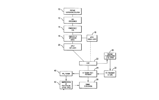
FIGURE 1 is a schematic flow diagram illustrating
the steps of the process of the invention;
FIGURE 2 represents a release profile for a multi-
layer system showing the percentage of heparin released over
a two-week period;
FIGURE 3 represents a release profile for a multi-
layer system showing the relative release rate of heparin
over a two-week period;
FIGURE 4 illustrates a profile of release kinetics
for different drug loadings at similar coating thicknesses
illustrating the release of heparin over a two-week period
14b
CA 02207659 1997-06-12
without associated means to provide a long term non-
thrombogenic surface thereafter;
FIGURE 5 illustrates drug elution kinetics at a given
loading of heparin over a two-week period at different coating
thicknesses without associated means to provide a long term
non-thrombogenic surface thereafter;
FIGURE 6 illustrates the release kinetics for a given
undercoat and topcoat material varied according to thickness
in which the percentage heparin in the undercoat and topcoats
are kept constant;
FIGURE 7 is a plot of heparin release kinetics in
phosphate buffer system at PH 7.4 with and without
fluorosilicone (FSi) topcoat; and
FIGURE 8 is another plot of heparin release kinetics in
phosphate buffer system in which a topcoat containing
fluorosilicone (FSi) only is compared with an FSi topcoat
containing 16.7% imbedded heparin.
DETAILED DESCRIPTION
According to the present invention, the stent coatings
incorporating biologically active materials for timed delivery
in situ in a body lumen of interest are preferably sprayed in
many thin layers from prepared coating solutions or
suspensions The steps of the process are illustrated
generally in Figure 1. The coating solutions or suspensions
are prepared at 10 as will be described later. The desired
amount of crosslinking agent (if any) is added to the
suspension/solution as at 12 and material is then agitated or
stirred to produce a homogenous coating composition at 14
CA 02207659 1997-06-12
- which is thereafter transferred to an application container or
device which may be a container for spray painting at 16.
- Typical exemplary preparations of coating solutions that were
used for heparin and dexamethasone appear next.
Ge_n_e_ra_1 preoarati nn of Hey~a i n 11n~3Prcnat i nq Cpmpos; t i nn
Silicone was obtained as a polymer precursor in solvent
(xylene) mixture. For example, a 35% solid silicone weight
content in xylene was procured from Applied Silicone, '
Part #40,000. First, the silicone-xylene mixture was weighed.
The solid silicone content was determined according to the
vendor's analysis. Precalculated amounts of finely divided
heparin (2-6 microns) were added into the silicone, then
tetrahydrofuron (THF) HPCL grade (Aldrich or Vii) was added.
For a 37.5% heparin coating, for example: Wsilicone = 5 g; solid
percent = 35%; W ,,ep = 5 x 0.35x .375/(0.625) - 1.05 g. The
amount of THF needed (44 ml) in the coating solution was
calculated by using the equation Wsilicone solid/VTHF = 0.04 for a
37.5% heparin coating solution). Finally, the manufacturer
crosslinker solution was added by~using Pasteur P-pipet. The
amount of crosslinker added was formed to effect the release
rate profile. Typically, five drops of crosslinker solution
were added for each five grams of silicone-xylene mixture.
The solutio~-was stirred by using the stirring rod until.the
suspension was homogenous and milk-like. The coating solution
was then transferred into a paint jar in condition for
application by air brush.
General Preparation of Dexametl,acnnA Undercoating Composition
16
CA 02207659 1997-06-12
Silicone (35% solution as above) was weighed into a
beaker on a Metler balance. The weight of dexamethasone free
alcohol or acetate form was calculated by silicone weight
multiplied by 0.35 and the desired percentage of dexamethasone
(1 to 40%) and the required amount was then weighed. Example:
Wsilicone = 5 g; for a 10 % dexamethasone coating, Wdex = 5 x
0. 35 x
0.1/0.9 = 0.194 g and THF needed in the coating solution
_ calculated. Wsilicone solid/V'fHF = 0.06 for a 10% dexamethasone
'
coating solution. Example: Ws;ll~o"e = 5 g; VTHF = 5 x 0.35/0.06
-- 29 ml. The dexamethasone was weighed in a beaker on an
analytical balance and half the total amount of THF was
added. The solution was stirred well to ensure full
dissolution of the dexamethasone. The stirred DEX-THF
solution was then transferred to the silicone container. The
beaker was washed with the remaining THF and this was
transferred to the silicone container. The crosslinker was
added by using a Pasteur pipet. Typically, five drops of
crosslinker were used for five grams of silicone.
.- The application of the coating material to the stent was
quite similar for all of the materials and the same for the
heparin and dexamethasone suspensions prepared as in the above
Examples. The suspension to be applied was transferred to an
application'device, at 16 in FIGURE 1. Typically a paint jar
attached to an air brush, such as a Badger Model 150, supplied
with a source of pressurized air through a regulator (Norgren,
0-160 psi) was used. Once the brush hose was attached to the
source of compressed air downstream of the regulator, the air
was applied. The pressure was adjusted to approximately 15-25
17
CA 02207659 1997-06-12
psi and the nozzle condition checked by depressing the
trigger.
Any appropriate method can be used to secure the stent
for spraying and rotating fixtures were utilized successfully
in the laboratory. Both ends of the relaxed stent were
fastened to the fixture by two resilient retainers, commonly
alligator clips, with the distance between the clips adjusted
so that the stent remained in a relaxed, unstretched
condition. The rotor was then energized and the spin speed
adjusted to the desired coating speed, nominally about 40 rpm.
With the stent rotating in a substantially horizontal
plane, the spray nozzle was adjusted so that the distance from
the nozzle to the stent was about 2-4 inches and the
composition was sprayed substantially horizontally with the
brush being directed along the stent from the distal end of
the stent to-the proximalwendwand then from the proximal end
to the distal end in a sweeping motion at a speed such that
one spray cycle occurred in about three stent rotations.
Typically a pause of less than one minute, normally about one-
half minute, elapsed between layers. Of course, the number of
coating layers did and will vary with the particular
application. For example, typical tie-layers as at 18 in
FIGURE l, f~ a coating level of 3-4 mg of heparin per cm'of
projected area, 20 cycles of coating application are required
and about 30 ml of solution will be consumed for a 3.5 mm
diameter by 14.5 cm long stent.
The rotation speed of the motor, of course, can be
adjusted as can the viscosity of the composition and the flow
18
CA 02207659 1997-06-12
rate of tie spray nozzle as desired to modify the layered
structure. Generally, with the above mixes, the best results
have been obtained at rotational speeds in the range of 30-50
rpm and with a spray nozzle flow rate in the range of 4-l0 ml
of coating composition per minute, depending on the stent
size. It is contemplated that a more sophisticated, computer-
controlled coating apparatus will successfully automate the
process demonstrated as feasible in the laboratory.
Several applied layers make up what is called the
undercoat as at 18. In one process, additional upper
undercoat layers, which may be of the same or different
composition with respect to bioactive material, the matrix
polymeric materials and crosslinking agent, for example, may
be applied as the top layer as at 20. The application of the
top layer follows the same coating procedure as the undercoat
with the number and thickness of layers being optional. Of
course, the thickness of any layer can be adjusted by
adjusting the speed of rotation of the stent and the spraying
conditions. Generally, the total coating thickness is
controlled by the number of spraying cycles or thin coats
.: which make up the total coat.
As shown at 22 in Figure 1, the coated stent is
thereafter subjected to a curing step in which the pre-polymer
and crosslinking agents cooperate to produce a cured polymer
matrix containing the biologically active species. The curing
process involves evaporation of the solvent xylene, THF, etc. _
and the curing and crosslinking of the polymer. Certain
silicone materials can be cured at relatively low
19
CA 02207659 1997-06-12
temperatures, (i.e. RT-50°C) in what is known as a room
temperature vulcanization (RTV) process. More typically,
however, the curing process involves higher temperature curing
materials and the coated stents are put into an oven at
approximately 90°C or higher for approximately 16 hours. The
temperature may be raised to as high as 150°C for
dexemethasane containing coated stents. Of course, the time
- and temperature may vary with particular silicones,
crosslinkers and biologically active species.
- 10 Stents coated and cured in the manner described need to
be sterilized prior to packaging for future implantation. For
sterilization, gamma radiation is a preferred method
particularly for heparin containing coatings; however, it has
been found that stents coated and cured according to the
process of the invention subjected to gamma sterilization may
be too slow to recover their original posture when delivered
to a vascular or other lumen site using a catheter unless a
pretreatment step as at 24 is first applied to the coated,
cured stent.
The pretreatment step involves an argon plasma treatment
- of the coated, cured stents in the unconstrained
configuration. In accordance with this procedure, the stents
are placed in a chamber of a plasma surface treatment system
such as a Plasma Science 350 (Himont/Plasma Science, Foster
City, CA). The system is equipped with a reactor chamber and
RF solid-state generator operating at 13.56 mHz and from 0-500
watts power output and being equipped with a microprocessor
controlled system and a complete vacuum pump package. The
CA 02207659 1997-06-12
reaction chamber contains an unimpeded work volume of 16.75
inches (42.55 cm) by 13.5 inches (34.3 cm) by 17.5 inches
(44.45 cm) in depth.
In the plasma process, unconstrained coated stents are
placed in a reactor chamber and the system is purged with
nitrogen and a vacuum applied to 20-50 mTorr. Thereafter,
inert gas (argon, helium or mixture of them) is admitted to
the reaction chamber for the plasma treatment. A highly
- preferred method of operation consists of using argon gas,
. 10 operating at a power range from 200 to 400 watts, a flow rate
of 150-650 standard ml per minute, which is equivalent to 100
- 450 mTorr, and an exposure time from 30 seconds to about 5
minutes. The stents can be removed immediately after the
plasma treatment or remain in the argon atmosphere for an
additional period of time, typically five minutes.
After this, as shown at 26, the stents may be exposed to
gamma sterilization at 2.5-3.5 Mrad. The radiation may be
carried out with the stent in either the radially non-
constrained status - or in the radially constrained status.
Preferably, however, the surface is modified prior to
plasma treatment or just prior to sterilization by one of
several additional processing methods of which some are
described ir~-relation to the following examples.
Example 1. Fluorosilicone surface treatment of eluting heparin
coating
The undercoat of a stent was coated as multiple applied
layers as described above thereafter and cured as described at
22. The heparin content of the undercoat was 37.5% and the
21
CA 02207659 1997-06-12
- coating thickness was about 30-40~. Fluorosilicone (FSi)
spray solution was prepared at 30 from a fluorosilicone
suspension (Applied Silicone X40032) by weighing an amount of
fluorosilicone suspension and adding tetrahydrofuran (THF)
according to the relation equation of VTHf= 1.2 x the weight of
fluorosilicone suspension. The solution was stirred very well
and spray-coated on the stent at 32 using the technique of the
application of the undercoat process at 18 and the coated
- stents were cured at 90°C for 16 hours. The coated stents are
-- l0 argon plasma treated prior to gamma sterilization according to
the procedures described above in accordance with steps 22-26.
Figure 7 is a plot of heparin release kinetics in
phosphate buffer system with fluorosilicone topcoat and
without any topcoat. The thickness of the topcoat. is about
10-15~. While it does not appear on the graph of FIGURE 7, it
should be noted that the release rate for the coating without
FSi is initially about 25 times higher than that with FSi,
- i.e., during the first 2 hours. This is, of course, clearly
.. off the scale of the graph. It is noteworthy, however, that
the coating with the FSi top layer or diffusion barrier does
- show a depressed initial release rate combined with an
enhanced ehi'sion rate after the first day and through the
first week up until about the tenth day. In addition, the
fluorosilicone (FSi) topcoat, by virtue of the high electro-
negativity of fluorination maintains non-thrombogenic surface
qualities during and after the elusion of the biologically
active heparin species. In addition, because of the negative
22
CA 02207659 1997-06-12
charges on the heparin itself, the electro-negativity of the
f luorosilicone topcoat may be, at least in part, responsible
for the modified heparin release kinetic profile.
FIGURE 8 compares a plot of fluorosilicone (FSi) top
coating containing 16.7% imbedded heparin with one containing
fluorosilicone (FSi) only. An undercoating is identical to
that utilized in FIGURE 7 containing about 37.5% heparin to a
thickness of about 30-40 microns. These elution kinetics are
quite comparable with the heparin-free FSi top layer greatly
l0 reducing the initial burst of heparin release and otherwise
the heparin in the FSi top layer imparts a slightly greater
release over the period of the test.
Example 2. Immobilization of polyethylene glycol (PEG) on
drug eluting undercoat
An undercoat was coated on a stent and cured at 22 as in
Example 1. The stent was then treated by argon gas plasma as
at 24 and ammonium gas plasma at 40. The equipment and the
process of.argon gas plasma treatment was as has been
described above. The ammonium plasma treatment was
implemented immediately after the argon gas plasma treatment,
_- to aminate the surface of the coating. The ammonium flow rate
was in the range of 100-700 cubic centimeter per minute (ccM)
in preferabf'y in the range of 500-600 ccM. The power output
of radio frequency plasma was in the range of 50-500 watts,
preferably in -200 watts. The process time was in the range
of 30sec-lOmin, preferably -5min.
Immediately after amination, the stents were immersed
into electrophilically activated polyethylene glycol (PEG)
23
CA 02207659 1997-06-12
_y solution at 42. PEG is known to be an inhibitor of protein
absorption. Examples of electrophilically activated PEG are
PEG nitrophenyl carbonates, PEG trichlorophenyh carbonates,
PEG tresylate, PEG glycidyl ether, PEG isocyanate, etc.,
optionally with one end terminated with methoxyl group.
Molecular weight of PEG ranged from about 1000-6000, and is
preferable about 3000. It has been observed that simple
ammonium amination will not generate large quantities of
primary and secondary amines on the elastomeric polymer
surface (for example silicone). Instead, imine (>C=N-H), and
other more oxidative nitro containing groups will dominate the
surface. It is generally necessary to add reductive agent'
such as NaBH3CN into the reaction media so that the functional
group on PEG can react with imine and possibly other nitro-
containing species on the surface, and therefore immobilize
PEG onto the surface. The typical concentration of NaBH3CN is
about 2mg/ml. Since PEG and its derivatives dissolve in water
and many polar and aromatic solvents, the solvent used in the
coating must be a solvent for PEG but not for the drug in the
undercoat to prevent the possible loss of the drug through
. leaching. In the case of eluting-heparin coating, a mixed
solvent of formamide and methyl ethyl ketone (MEK) or a mixed
solvent of formamide and acetone are preferred solvents
(preferably at ratios of 30 formamide: 70 MEK or acetone by
volume), since they will not dissolve heparin. The
concentration of PEG, the reaction time, the reaction
temperature and the pH value depend on the kind of PEG
employed. In the case of eluting heparin coating, 5% PEG
24
CA 02207659 2002-06-05
60950-245
tresylate~in (30-70j Formamide/MEK was used successfully. The
reaction time was 3 hours at room temperature. PEG was then
covalently bound to the surface. Gamma r<~diation was then
used for sterilization of this embodiment as previously
described.
With respect to tha_ anticoagulant material heparin, the
percentage in the undercoat is nominally from about 30-50% and
that of the topcoat from about 0-30~ active material. The
coating thickness ratio of the topcoat to the undercoat varies
l0 from about 1:10 to 1:2 and is preferably in the range of from
about 1:6 to 1:3.
Suppressing the burst effect also enables a reduction in
the drug loading or in other words, allows a reduction in the
coating thickness, since the physician will give a bolus
injection of antiplatelet/anticoagulation drugs to the patient
during the stenting process. As a result, the drug imbedded
in the stent can be fully.used without waste. Tailoring the
first day release, but raximixing second day and third day
release at the, thinnest possible coating configuration will
reduce the acute or subacute thrombosis.
Figure 4 depicts the general effect of drug loading for
coatings of similar thickness. The initia.I elution rate
increases with the drug loading as shown in Figure 5. The
release rate also increases with the thickness of the coating
at the same loading but tends to be inversely proportional to
the thickness of the topcoat as shown by the same drug loading
and similar undercoat thickness in Figure 6.
CA 02207659 1997-06-12
What'is apparent from the data gathered to date, however,
is that the process of the present invention enables the drug
elution kinetics to be controlled in a manner desired to meet
the needs of the particular stent application. In a similar
manner, stent coatings can be prepared using a combination of
two or more drugs and the drug release sequence and rate
controlled. For example, antiproliferation drugs may be
combined in the undercoat and antiplatelet drugs in the
. topcoat. In this manner, the antiplatelet drugs, for example,
heparin, will elute first followed by antiproliferation drugs
to better enable safe encapsulation of the implanted stent.
The heparin concentration measurement were made utilizing
a standard curve prepared by complexing azure A dye with
dilute solutions of heparin. Sixteen standards were used to
compile the standard curve in a well-known manner.
For the elution test, the stents were immersed in a
phosphate buffer solution at pH 7.4 in an incubator at
approximately 37°C. Periodic samplings of the solution were
processed to determine the amount of heparin eluted. After
each sampling, each stent was placed in heparin-free buffer
.. solution.
As stated above, while the allowable loading of the
elastomeric material with heparin may vary, in the case of
silicone materials heparin may exceed 60% of the total weight
of the layer. However, the loading generally most
advantageously used is in the range from.about 10% to 45% of
the total weight of the layer. In the case of dexamethasone,
the loading may be as high as 50% or more of the total weight
26
CA 02207659 1997-06-12
of the layer but is preferably in the range of about 0.4% to
45%.
It will be appreciated that the mechanism of
incorporation of the biologically active species into a thin
surface coating structure applicable to a metal stent is an
important aspect of the present invention. The need for
relatively thick-walled polymer elution stents or any membrane
overlayers associated with many prior drug elution devices is '
obviated, as is the need for utilizing biodegradable or
reabsorbable vehicles for carrying the biologically active
species. The technique clearly enables long-term delivery and
minimizes interference with the independent mechanical or
therapeutic benefits of the stent itself.
Coating materials are designed with a particular coating
technique, coating/drug combination and drug infusion
mechanism in mind. Consideration of the particular form and
mechanism of release of the biologically active species in the
coating allow the technique to produce superior results. In
this manner, delivery of the biologically active species from
the coating structure can be tailored to accommodate a variety
of applications.
Whereas the above examples depict coatings having two
different dz~'ug loadings or percentages of biologically active
material to be released, this is by no means limiting with
respect to the invention and it is contemplated that any
number of layers and combinations of loadings can be employed
to achieve a desired release profile. For example, gradual
grading and change in the loading of the layers can be
27
CA 02207659 1997-06-12
utilized in which, for example, higher loadings are used in
the inner layers. Also layers can be used which have no drug
loadings at all. For example, a pulsatile heparin release
system may be achieved by a coating in which alternate layers
containing heparin are sandwiched between unloaded layers of
silicone or other materials for a portion of the coating. In
other words, the invention allows untold numbers of
combinations which result in a great deal of flexibility with '
respect to controlling the release of biologically active
materials with regard to an implanted stent. Each applied
layer is typically from approximately 0.5 microns to 15
microns in thickness. The total number of sprayed layers, of
course, can vary widely, from less than 10 to more than 50
layers; commonly, 20 to 40 layers are included. The total
thickness of the coating can also vary widely, but. can
generally be~from about 10 to 200 microns.
Whereas the polymer of the coating may be any compatible
biostable elastomeric material capable of being adhered to the
stent material as a thin layer, hydrophobic materials are
preferred because it has been found that the release of the
biologically active species can generally be more predictably
controlled with such materials. Preferred materials include
silicone rubber elastomers and biostable polyurethanes
specifically.
This invention has been described herein in considerable
detail in order to comply with the Patent Statutes and to
provide those skilled in the art with the information needed
to apply the novel principles and to construct and use
28
CA 02207659 1997-06-12
embodiments of the example as required. However, it is to be
understood that the invention can be carried out by
specifically different devices and that various modifications
can be accomplished without departing from the scope of the
invention itself.
29