Note: Descriptions are shown in the official language in which they were submitted.
CA 02221330 1997-11-17
WO 96/39914 PCT/US96/08077
SYSTEM AND METHOD FOR
ELECTROSURGICAL CUTTING AND ABLATION
BACKGROUND OF THE INVENTION
1. Field of the Invention
The present invention relates generally to the field
of electrosurgery and, more particularly, to surgical devices
and methods which employ high frequency voltage to cut and
ablate tissue.
The field of electrosurgery includes a number of
loosely related surgical techniques which have in common the
application of electrical energy to modify the structure or
integrity of patient tissue. Electrosurgical procedures
usually operate through the application of very high frequency
currents to cut or ablate tissue structures, where the
operation can be monopolar or bipolar. Monopolar techniques
rely on external grounding of the patient, where the surgical
device defines only a single electrode pole. Bipolar devices
comprise both electrodes for the application of current
between their surfaces.
Electrosurgical procedures and techniques are
= 35 particularly advantageous since they generally reduce patient
bleeding and trauma associated with cutting operations.
Additionally, electrosurgical ablation procedures, where
tissue surfaces and volume may be reshaped, cannot be
CA 02221330 1997-11-17
WO 96/39914 PC'd'/US96/08077
2
duplicated through other treatment modalities.
Current electrosurgical devices and procedures,
however, suffer from a number of disadvantages. For example,
monopolar devices generally direct electric current along a
defined path from the exposed or active electrode through the
patient's body to the return electrode, which is externally ,
attached to a suitable location on the patient. This creates
the potential danger that the electric current will flow
through undefined paths in the patient's body, thereby
increasing the risk of unwanted electrical stimulation to
portions of the patient's body. In addition, since the
defined path through the patient's body has a relatively high
impedance (because of the large distance or resistivity of the
patient's body), large voltage differences must typically be
applied between the return and active electrodes in order to
generate a current suitable for ablation or cutting of the
target tissue. This current, however, may inadvertently flow
along body paths having less impedance than the defined
electrical path, which will substantially increase the current
flowing through these paths, possibly causing damage to or
destroying surrounding tissue.
Bipolar electrosurgical devices have an inherent
advantage over monopolar devices because the return current
path does not flow through the patient. In bipolar
electrosurgical devices, both the active and return electrode
are typically exposed so that they may both contact tissue,
thereby providing a return current path from the active to the
return electrode through the tissue. One drawback with this
configuration, however, is that the return electrode may cause
tissue desiccation or destruction at its contact point with
the patient's tissue. In addition, the active and return
electrodes are typically positioned close together to ensure
that the return current flows directly from the active to the return
electrode. The close proximity of these electrodes
generates the danger that the current will short across the =
electrodes, possibly impairing the electrical control system
and/or damaging or destroying surrounding tissue.
The use of electrosurgical procedures (both
CA 02221330 1999-09-24
3
monopolar and bipolar) in electrically conductive environments
can be further problematic. For example, many arthroscopic
procedures require flushing of the region to be treated with
isotonic saline (also referred to as normal saline), both to
maintain an isotonic environment and to keep the field of
viewing clear. The presence of saline, which is a highly
conductive electrolyte, can also cause shorting of the
electrosurgical electrode in both monopolar and bipolar modes.
Such shorting causes unnecessary heating in the treatment
environment and can further cause non-specific tissue
destruction.
In response to the various problems associated with
electrosurgical procedures in electrically conductive
environments, new methods and devices have been developed by
the applicant. These methods and devices provide selective
power delivery to the target tissue while minimizing power
delivery to the surrounding electrically conductive irrigant.
These methods are particularly useful in isotonic saline
filled body cavities, such as arthroscopic, urologic or
gynecologic cavities. The irrigant flooded body cavity
provides good visibility, facilitates the removal of bubbles
or other debris, minimizes the possibility of air embolism and
protects certain tissue from dehydration. Such methods and
devices are more fully described in PCT/US94/05168 and United
States Patents 5,697,909; 5,366,443; 5,697,882; and,
5,697,281.
Many surgical procedures, such as oral, laparoscopic
and open surgical procedures, are not performed with the
target tissue submerged under an irrigant. In laparoscopic
procedures, such as the resection of the gall bladder from the
liver, for example, the abdominal cavity is pressurized with
carbon dioxide (pneumoperitoneum) to provide working space for
the instruments and to improve the surgeon's visibility of the
surgical site. Other procedures, such as the ablation of
muscle or gingiva tissue in the mouth or the ablation and
necrosis of diseased tissue, are also typically performed in a
"dry" environment or field (i.e., not submerged under an
CA 02221330 1999-07-21
r ` =
4
electrically conducting irrigant).
For these and other reasons, improved systems and
methods are desired for the electrosurgical ablation and
cutting of tissue. These systems and methods should be
capable of providing a direct return current path from the
active electrode, through the target site, to the return
electrode to minimize the dangers of electrical current
flowing through undefined paths in the patient's body. The
system should also be configured to minimize contact between
the return electrode and surrounding tissue and to avoid
current shorting between the active and return electrodes.
Preferably, the system will be configured to apply high
frequency voltage for the cutting and ablation of tissue in
relatively dry environments, such as those encountered in
oral, laparoscopic and open surgical procedures.
2. Description of the Backcround Art
Devices incorporating radio frequency electrodes for
use in electrosurgical and electrocautery techniques are
described in Rand et al. (1985) J. Arthro. Surg. 1:242-246 and
U.S. Patent Nos. 5,281,216; 4,943,290; 4,936,301; 4,593,691;
4,228,800; and 4,202,337. U.S. Patent Nos. 4,943,290 and
4,036,301 describe methods for injecting non-conducting liquid
over the tip of a monopolar electrosurgical electrode to
electrically isolate the electrode, while energized, from a
surrounding electrically conducting irrigant. U.S. Patent
Nos. 5,195,959 and 4,674,499 describe monopolar and bipolar
electrosurgical devices, respectively, that include a conduit
for irrigating the surgical site.
35
CA 02221330 1999-07-21
4a
SUMMARY OF THE INVENTION
This invention provides the use of a return electrode and an electrode
terminal
electrically coupled to a high frequency voltage source for applying
electrical energy to a
target site on a structure within or on a patient's body, wherein an
electrosurgical probe is
adjacent to the body structure so that an electrode terminal is in at least
partial contact or
,.. close proximity with the target site, an electrically conducting fluid is
directed to the target
1 o site and the electrode terminal to generate a current flow path between
the electrode
terminal and the return electrode, and high frequency voltage is applied
between the
electrode terminal and the return electrode such that an electrical current
flows from the
electrode terminal, through the region of the target site, and to the return
electrode through
the current flow path.
This invention also provides the use of a return electrode and an electrode
terminal
electrically coupled to a high frequency voltage source for applying
electrical energy to a
target site on a structure within or on a patient's body, wherein an
electrosurgical probe is
adjacent to the body structure so that an electrode terminal is in at least
partial contact or
close proximity with the target site, an electrically conducting fluid is
directed to the target
site and the electrode terminal to surround the electrode terminal with
electrically
conducting fluid and to locate electrically conducting fluid between the
electrode terminal
and the target site, and high frequency voltage is applied between the
electrode terminal
and the return electrode such that an electrical current flows from the
electrode terminal
through the region of the target site to the return electrode.
This invention also provides an electrosurgical system for use with a high
frequency
power supply and an electrically conducting fluid supply, the system
comprising:
an electrosurgical probe comprising a shaft having a proximal end and a distal
end,
an electrode terminal disposed near the distal end, and a connector near the
proximal end of
the shaft for electrically coupling the electrode terminal to the
electrosurgical power supply;
a return electrode adapted to be electrically coupled to the electrosurgical
power
supply; and
a fluid delivery element having an inlet adapted to be fluidly coupled to the
CA 02221330 1999-07-21
4b
electrically conducting fluid supply for directing fluid to the target site to
generate a current
flow path between the return electrode and the electrode terminal.
This invention also provides an electrosurgical system for applying electrical
energy
to a target site on a structure within or on a patient's body, the system
comprising:
a high frequency power supply;
an electrosurgical probe comprising a shaft having a proximal end and a distal
end,
an electrode terminal disposed near the distal end, and a connector near the
proximal end of the shaft electrically coupling the electrode terminal to the
electrosurgical power supply;
a return electrode electrically coupled to the electrosurgical power supply;
and
an electrically conducting fluid supply for directing electrically conducting
fluid to the
target site such that the electrically conducting fluid generates a current
flow path between
the return electrode and the electrode terminal.
This invention also provides an electrosurgical system for applying electrical
energy
to a target site on a structure within or on a patient's body, the system
comprising:
a high frequency power supply;
an electrosurgical probe comprising a shaft having a proximal end and a distal
end,
an electrode terminal disposed near the distal end, and a connector near the
proximal end of the shaft electrically coupling the electrode terminal to the
electrosurgical power supply;
a return electrode electrically coupled to the electrosurgical power supply;
an electrically conducting fluid supply;
a fluid delivery element defining a fluid path electrically coupled to the
electrode terminal
for directing electrically conducting fluid to the target site and the
electrode terminal to
substantially surround the electrode terminal with electrically conducting
fluid and to locate
electrically conducting fluid between the electrode terminal and the target
site.
The present invention provides an apparatus and method for selectively
applying
electrical energy to structures within a patient's body. The apparatus and
method allow the
surgical team to perform electrosurgical interventions, such as ablation and
cutting of body
structures, without requiring the tissue to be submerged in an electrically
conducting
irrigant, such as isotonic saline.
CA 02221330 1997-11-17
WO 96/39914 PCT/US96/08077
The apparatus and method of the present invention are
particularly useful for treating and shaping gingiva, for
tissue dissection, e.g. separation of gall bladder from the
liver, and ablation and necrosis of diseased tissue, such as
5 tumors.
The method of the present invention comprises
positioning an electrosurgical probe adjacent the target
tissue so that at least one active electrode is brought into
at least partial contact or close proximity with the target
site. Electrically conducting liquid, such as isotonic
saline, is directed through a fluid path past a return
electrode and to the target site to generate a current flow
path between the target site and the return electrode. High
frequency voltage is then applied between the active and
return electrode through the current flow path created by the
electrically conducting liquid in either a bipolar or
monopolar manner. The probe may then be translated,
reciprocated or otherwise manipulated to cut the tissue or
effect the desired depth of ablation.
The above described method is particularly effective
in a dry environment (i.e., the tissue is not submerged in
fluid), such as open, laparoscopic or oral surgery, because
the electrically conducting liquid provides a suitable current
flow path from the target site to the return electrode. The
active electrode is preferably disposed at the distal end of
the probe and the return electrode is spaced from the active
electrode and enclosed within an insulating sheath. This
minimizes exposure of the return electrode to surrounding
tissue and minimizes possible shorting of the current between
the active and return electrodes. In oral procedures, the
probe may be introduced directly into the cavity of the open
mouth so that the active electrode is positioned against
= gingival or mucosal tissue. In laparoscopic procedures, the
probe will typically be passed through a conventional trocar
cannula while viewing of the operative site is provided
through the use of a laparoscope disposed in a separate
cannula.
The apparatus according to the present invention
CA 02221330 1997-11-17
WO 96/39914 PCTIUS96/08077
6
comprises an electrosurgical probe having a shaft with a
proximal end, a distal end, and at least one active electrode
at or near the distal end. A connector is provided at or near
the proximal end of the shaft for electrically coupling the
active electrode to a high frequency voltage source. A return
electrode coupled to the voltage source is spaced a sufficient
distance from the active electrode to substantially avoid or
minimize current shorting therebetween and to shield the
return electrode from tissue. The return electrode may be
provided integral with the shaft of the probe or it may be
separate from the shaft (e.g., on a liquid supply instrument).
In both cases, the return electrode defines an inner passage
for flow of electrically conducting liquid therethrough. The
liquid is directed through the return electrode and over the
active electrode to thereby provide a return current flow path
between the tissue target site and the return electrode.
In a preferred aspect of the invention, the active
electrode comprises an electrode array having a plurality of
electrically isolated electrode terminals disposed over a
contact surface, which may be a planar or non-planar surface
and which may be located at the distal tip or over a lateral
surface of the shaft, or over both the tip and lateral
surface(s). The electrode array will include at least two and
preferably more electrode terminals, and may further comprise
a temperature sensor. In a preferred aspect, each electrode
terminal will be connected to the proximal connector by an
electrically isolated conductor disposed within the shaft.
The conductors permit independent electrical coupling of the
electrode terminals to a high frequency power supply and
control system with optional temperature monitor for operation
of the probe. The control system preferably incorporate
active and/or passive current limiting structures, which are
designed to limit current flow when the associated electrode =
terminal is in contact with a low resistance return path back
to the return electrode.
The use of such electrode arrays in electrosurgical
procedures is particularly advantageous as it has been found
to limit the depth of tissue necrosis without substantially
CA 02221330 1997-11-17
WO 96/39914 PCT/US96/08077
7
reducing power delivery and ablation rates. The voltage
applied to each electrode terminal causes electrical energy to
be imparted to any body structure which is contacted by, or
comes into close proximity with, the electrode terminal, where
a current flow through all low electrical impedance paths is
preferably but not necessarily limited. It will be
appreciated that such low impedance paths generally occur when
an electrode terminal does not contact or come into close
proximity with the body structure, but rather is in contact
with a low impedance environment, such as the saline, or other
electrolyte being introduced past the return electrode. The
presence of an electrolyte provides a relatively low impedance
path back to the common or return electrode.
The apparatus and method of the present invention
provide a number of advantages, particularly in respect to the
ablation or cutting of tissue. The ability to control current
flow through individual electrode terminals minimizes power
dissipation into the surrounding medium. Limited power
dissipation, in turn, permits the use of electrolytic
irrigants, such as isotonic saline, to create a current flow
path between the active electrode terminals and the return
electrode. The isotonic saline may also be used to
simultaneously irrigate the surgical site, which provides a
number of well know physiological advantages. In addition,
the ability to operate in a bipolar or quasi-bipolar mode
reduces the risk of unwanted electrical stimulation from
return current flowing through the patient's body, which can
cause muscle spasms and can limit the depth of tissue necrosis
during ablative resection.
A further understanding of the nature and advantages
of the invention will become apparent by reference to the
remaining portions of the specification and drawings.
BRIEF DESCRIPTION OF THE DRAWINGS
Fig. 1 is a perspective view of the electrosurgical
system including an electrosurgical probe, an electrically
conducting liquid supply and an electrosurgical power supply
constructed in accordance with the principles of the present
CA 02221330 1997-11-17
WO 96/39914 PCTIUS96/08077
8
invention;
Fig. 2A is an enlarged, cross-sectional view of the
distal tip of the electrosurgical probe of Fig. 1 illustrating
an electrode arrangement suitable for rapid cutting and
ablation of tissue structures;
Fig. 2B is an enlarged end view of the distal tip of
the electrosurgical probe of Fig. 1;
Fig. 2C is a cross-sectional view of the proximal
end of the electrosurgical probe, illustrating an arrangement
for coupling the probe to the electrically conducting liquid
supply of Fig. 1;
Fig. 3 is a detailed cross-sectional view of an
alternative embodiment of the electrosurgical probe of Fig. 1;
Fig. 4 is an end view of the distal end of the
electrosurgical probe of Fig. 3;
Fig. 5 is an end view of an another embodiment of
the electrosurgical probe of Fig. 1;
Fig. 6 is a partial cross-sectional side view of a
further embodiment of the electrosurgical probe with the
electrode array disposed transversely to the axis of the
probe;
Fig. 7 is a partial front cross-sectional view of an
electrosurgical probe and an electrically conductive liquid
supply shaft illustrating use of the probe and the shaft in
ablating target tissue;
Fig. 8 is an enlarged, cross-sectional view of the
distal tip of yet another embodiment of the electrosurgical
probe of Fig. 1;
Fig. 9 is a detailed end view of the probe of Fig.
8;
Fig. 10 is a side view of an electrosurgical probe
having a shaft with an angled distal portion;
Fig. 11 is a side view of an electrosurgical probe having a shaft with a
perpendicular distal portion;
Fig. 12 is a schematic view of an electrosurgical ~
probe having two screwdriver-shaped electrodes extending from
the distal end;
Fig. 13 is an end view of the probe of Fig. 12; and
CA 02221330 1997-11-17
WO 96/39914 PCT/US96/08077
9
Fig. 14 illustrates use of the probe of Fig. 12 for
the rapid cutting of tissue.
DESCRIPTION OF THE PREFERRED EMBODIMENTS
The present invention provides an apparatus and
method for selectively applying electrical energy to a target
location within a patient's body, such as solid tissue or the
like, particularly including gingival tissues and mucosal
tissues located in the mouth. In addition, tissues which may
be treated by the system and method of the present invention
include tumors, abnormal tissues, and the like. For
convenience, the remaining disclosure will be directed
specifically to the cutting, shaping or ablation of gingival
or mucosal tissue in oral surgical procedures, but it will be
appreciated that the system and method can be applied equally
well to procedures involving other tissues of the body, as
well as to other procedures including open surgery,
laparoscopic surgery, thoracoscopic surgery, and other
endoscopic surgical procedures.
The present invention uses an electrode array
including a plurality of independently current-limited and/or
power-controlled electrode terminals distributed over a distal
contact surface of a probe to apply electrical energy
selectively to the target tissue while limiting the unwanted
application of electrical energy to the surrounding tissue and
environment resulting from power dissipation into surrounding
electrically conductive liquids, such as blood, normal saline,
and the like.
The electrosurgical probe will comprise a shaft
having a proximal end and a distal end which supports an
electrode array near its distal end. The shaft may assume a
wide variety of configurations, with the primary purpose being
to mechanically support the electrode array and permit the
treating physician to manipulate the array from a proximal end
of the shaft. Usually, the shaft will be a narrow-diameter
rod or tube, more usually having dimensions which permit it to
be introduced into a body cavity, such as the mouth or the
abdominal cavity, through an associated trocar or cannula in a
CA 02221330 1997-11-17
WO 96/39914 PCT/US96/08077
minimally invasive procedure, such as arthroscopic,
laparoscopic, thoracoscopic, and other endoscopic procedures.
Thus, the shaft will typically have a length of at least 5cm
for oral procedures and at least 10 cm, more typically being
5 20 cm, or longer for endoscopic procedures. The shaft will
typically have a diameter of at least 1 mm and frequently in
the range from 1 to 10 mm. The shaft may be rigid or flexible, with flexible
shafts optionally being combined with
a generally rigid external tube for mechanical support.
10 Flexible shafts may be combined with pull wires, shape memory
actuators, and other known mechanisms for effecting selective
deflection of the distal end of the shaft to facilitate
positioning of the electrode array. The shaft will usually
include a plurality of wires or other conductive elements
running axially therethrough to permit connection of the
electrode array to a connector at the proximal end of the
shaft. Specific shaft designs will be described in detail in
connection with the figures hereinafter.
The circumscribed area of the electrode array is in
the range from 0.25 mm2 to 75 mm2, preferably from 0.5 mm2 to
40 mm2, and will usually include at least two isolated
electrode terminals, more usually at least four electrode
terminals, preferably at least six electrode terminals, and
often 50 or more electrode terminals, disposed over the distal
contact surfaces on the shaft. By bringing the electrode
array(s) on the contact surface(s) against or in close
proximity with the target tissue and applying high frequency
voltage between the array(s) and an additional common or
return electrode in direct or indirect contact with the
patient's body, the target tissue is selectively ablated or
cut, permitting selective removal of portions of the target
tissue while desirably minimizing the depth of necrosis to
surrounding tissue. In particular, this invention provides a =
method and apparatus for effectively ablating and cutting
tissue which may be located in close proximity to other
critical organs, vessels or structures (e.g., teeth, bone) by
simultaneously (1) causing electrically conducting liquid to
flow between the common and active electrodes, (2) applying
CA 02221330 1997-11-17
WO 96/39914 PCT/US96/08077
11
electrical energy to the target tissue surrounding and
immediately adjacent to the tip of the probe, (3) bringing the
active electrode(s) in contact or close proximity with the
target tissue using the probe itself, and (4) optionally
moving the electrode array axially and/or transversely over
the tissue.
= Each individual electrode terminal in the electrode
array is electrically insulated from all other electrode
terminals in the array within said probe and is connected to a
power source which is isolated from each of the other
electrodes in the array or to circuitry which limits or
interrupts current flow to the electrode when low resistivity
material (e.g., blood or electrically conductive saline
irrigant) causes a lower impedance path between the common
electrode and the individual electrode terminal. The isolated
power sources for each individual electrode may be separate
power supply circuits having internal impedance
characteristics which limit power to the associated electrode
terminal when a low impedance return path is encountered, may
be a single power source which is connected to each of the
electrodes through independently actuatable switches or may be
provided by independent current limiting elements, such as
inductors, capacitors, resistors and/or combinations thereof.
The tip region of the probe is thus composed of many
independent electrode terminals designed to deliver electrical
energy in the vicinity of the tip. The selective application
of electrical energy to of the target tissue is achieved by
connecting each individual electrode terminal and the common
electrode to a power source having independently controlled or
current limited channels. The common electrode may be a
tubular member of conductive material proximal to the
electrode array at the tip which also serves as a conduit for
= the supply of the electrically conducting liquid between the
active and common electrodes. The application of high
frequency voltage between the common electrode and the
electrode array results in the generation of high electric
field intensities at the distal tips of the electrodes with
conduction of high frequency current from each individual
CA 02221330 1997-11-17
WO 96/39914 PCTIUS96/08077
12
electrode terminal to the said common electrode. The current
flow from each individual electrode terminal to the common
electrode is controlled by either active or passive means, or
a combination thereof, to deliver electrical energy to the
target tissue while minimizing energy delivery to surrounding
(non-target) tissue and any conductive fluids which may be
present (e.g., blood, electrolytic irrigants such as saline,
and the like).
In a preferred aspect, this invention takes
advantage of the differences in electrical resistivity between
the target tissue (e.g., gingiva, muscle, fascia, tumor or
other connective tissue) and the surrounding conductive liquid
(e.g., isotonic saline irrigant). By way of example, for any
selected level of applied voltage, if the electrical
conduction path between the common electrode and one of the
individual electrode terminals within the electrode array is
isotonic saline irrigant liquid (having a relatively low
electrical impedance), the current control means connected to
the individual electrode will limit current flow so that the
heating of intervening conductive liquid is minimized. On the
other hand, if a portion of or all of the electrical
conduction path between the common electrode and one of the
individual electrode terminals within the electrode array is
gingival tissue (having a relatively higher electrical
impedance), the current control circuitry or switch connected
to the individual electrode will allow current flow sufficient
for the deposition of electrical energy and associated
ablation or electrical breakdown of the target tissue in the
immediate vicinity of the electrode surface.
The application of a high frequency voltage between
the common or return electrode and the electrode array for
appropriate time intervals effects ablation, cutting or
reshaping of the target tissue. The tissue volume over which
energy is dissipated (i.e., a high voltage gradient exists)
may be precisely controlled, for example, by the use of a
multiplicity of small electrodes whose effective diameters
range from about 2 mm to 0.01 mm, preferably from about 1 mm
to 0.05 mm, and more preferably from about 0.5 mm to 0.1 mm.
CA 02221330 1997-11-17
WO 96139914 PCT/US96108077
13
Electrode areas for both circular and non-circular terminals
will have a contact area (per electrode) below 5 mm2,
preferably being in the range from 0.0001 mm2 to i mm2, and
more preferably from 0.005 mm2 to .5 mm2. The use of small
diameter electrode terminals increases the electric field
intensity and reduces the extent or depth of tissue necrosis
as a consequence of the divergence of current flux lines which
emanate from the exposed surface of each electrode terminal.
Energy deposition in tissue sufficient for irreversible damage
(i.e., necrosis) has been found to be limited to a distance of
about one-half to one electrode diameter. This is a
particular advantage over prior electrosurgical probes
employing single and/or larger electrodes where the depth of
tissue necrosis may not be sufficiently limited.
In previous electrosurgical devices, increased power
application and ablation rates have been achieved by
increasing the electrode area. Surprisingly, with the present
invention, it has been found that the total electrode area can
be increased (to increase power delivery and ablation rate)
without increasing the depth of necrosis by providing multiple
small electrode terminals. Preferably, the terminals will be
spaced-apart by a distance in the range from about one-half
diameter to one diameter for optimum power delivery, as
discussed below. The depth of necrosis may be further
controlled by switching the applied voltage off and on to
produce pulses of current, the pulses being of sufficient
duration and associated energy density to effect ablation
and/or cutting while being turned off for periods sufficiently
long to allow for thermal relaxation between energy pulses.
In this manner, the energy pulse duration and magnitude and
the time interval between energy pulses are selected to
achieve efficient rates of tissue ablation or cutting while
allowing the temperature of the treated zone of tissue to
"relax" or return to normal physiologic temperatures (usually
to within 10 C of normal body temperature [37 C], preferably
to within 5 C) before the onset of the next energy (current)
pulse.
The rate of energy delivery to the target tissue is
CA 02221330 1997-11-17
WO 96/39914 PCT/US96/08077
14
controlled by the applied voltage level and duty cycle of the
voltage pulse. The use of high frequency current minimizes
induced stimulation of muscle tissue or nerve tissue in the
vicinity of the body structure being treated. In addition,
high frequencies minimize the risk of interfering with the
natural pacing of the heart in circumstances where the probe
of the present invention is used near the heart.
The power applied to the common electrode and the
electrode array will be at high or radio frequency, typically
between about 20 kHz and 20 MHz, usually being between about
30 kHz and 2 MHz, and preferably being between about 50 kHz
and 400 kHz. The RMS (root mean square) voltage applied will
usually be in the range from about 5 volts to 1000 volts,
preferably being in the range from about 50 volts to 800
volts, and more preferably being in the range from about 10
volts to 500 volts. Usually, the current level will be
selectively limited or controlled and the voltage applied will
be independently adjustable, frequently in response to the
resistance of tissues and/or fluids in the pathway between an
individual electrode and the common electrode. Also, the
applied current level may be in response to a temperature
control means which maintains the target tissue temperature
with desired limits at the interface between the electrode
arrays and the target tissue. The desired surface temperature
along a propagating surface just beyond the region of ablation
will usually be in the range from about 40 C to 100 C, and
more usually from about 50 C to 60 C. The tissue being
ablated immediately adjacent the electrode array may reach
even higher temperatures.
The preferred power source of the present invention
delivers a high frequency current selectable to generate
average power levels ranging from tens of milliwatts to tens
of watts per electrode, depending on the target tissue being
ablated, the rate of ablation desired or the maximum allowed
temperature selected for the probe tip. The power source
allows the user to select the current level according to the
specific requirements of a particular oral surgery, open
surgery or other endoscopic surgery procedure.
CA 02221330 1999-09-24
The power source will be current limited or
otherwise controlled so that undesired heating of electrically
conductive fluids or other low electrical resistance media
does not occur. In a presently preferred embodiment of the
5 present invention, current limiting inductors are placed in
series with each independent electrode terminal, where the
inductance of the inductor is in the range of 20uH rto 5000uH,
depending on the electrical properties of the target tissue,
the desired ablation rate and the operating frequency.
10 Alternatively, capacitor-inductor (LC) circuit structures may
be employed, as described previously in co-pending PCT
application No. PCT/US94/05168.
Additionally, current
limiting resistors may be selected having a large positive
15 temperature coefficient of resistance so that, as the current
level begins to rise for any individual electrode in contact
with a low resistance medium (e.g., saline irrigant), the
resistance of the current limiting resistor increases
significantly, thereby minimizing the power delivery from said
electrode into the low resistance medium (e.g., saline
irrigant).
As an alternative to such passive circuit
structures, regulated current flow to each electrode terminal
may be provided by a multi-channel power supply. A
substantially constant current level for each individual
electrode terminal within a range which will limit power
delivery through a low resistance path, e.g., isotonic saline
irrigant, would be selected by the user to achieve the desired
rate of cutting or ablation. Such a multi-channel power
supply thus provides a substantially constant current source
with selectable current level in series with each electrode
terminal, wherein all electrodes will operate at or below the
same, user selectable maximum current level. Current flow to
all electrode terminals could be periodically sensed and
stopped if the temperature measured at the surface of the
electrode array exceeds user selected limits. Particular
control system designs for implementing this strategy are well
within the skill of the art.
CA 02221330 1997-11-17
WO 96/39914 PCTIUS96/08077
16
Yet another alternative involves the use of one or
several power supplies which allow one or several electrodes
to be simultaneously energized and which include active
control means for limiting current levels below a preselected
maximum level. In this arrangement, only one or several
electrodes would be simultaneously energized for a brief
period. Switching means would allow the next one or several
electrodes to be energized for a brief period. By
sequentially energizing one or several electrodes, the
interaction between adjacent electrodes can be minimized (for
the case of energizing several electrode positioned at the
maximum possible spacing within the overall envelope of the
electrode array) or eliminated (for the case of energizing
only a single electrode at any one time). As before, a
resistance measurement means may be employed for each
electrode prior to the application of power wherein a
(measured) low resistance (below some preselected level) will
prevent that electrode from being energized during given
cycle. By way of example, the sequential powering and control
scheme of the present invention would function in a manner
similar to an automobile distributor. In this example, an
electrical contact rotates past terminals connected to each
spark plug. In this example, each spark plug corresponds to
the exposed surface of each of the electrodes. In addition,
the present invention includes the means to measure the
resistance of the medium in contact with each electrode and
cause voltage to be applied only if the resistance exceeds a
preselected level.
The electrode array is formed over a contact surface
on the shaft of the electrosurgical probe. The common
(return) electrode surface will be recessed relative to the
distal end of the probe and may be recessed within the conduit
provided for the introduction of electrically conducting
liquid to the site of the target tissue and array of active
electrodes. In the exemplary embodiment, the shaft will be
cylindrical over most of its length, with the contact surface
being formed at the distal end of the shaft. In the case of
laparoscopic or endoscopic applications, the contact surface
CA 02221330 1997-11-17
WO 96/39914 PCT/US96/08077
17
may be recessed since it helps protect and shield the
electrode terminals on the surface while they are being
introduced, particularly while being introduced through the
working channel of a trocar channel or a viewing scope.
The area of the contact surface can vary widely, and
the contact surface can assume a variety of geometries, with
particular areas in geometries being selected for specific
applications. Electrode array contact surfaces can have areas
in the range from 0.25 mm2 to 50 mm2, usually being from 1 mm2
to 20 mm2. The geometries can be planar, concave, convex,
hemispherical, conical, or virtually any other regular or
irregular shape. Most commonly, the electrode arrays will be
formed at the distal tip of the electrosurgical probe shaft,
frequently being planar, disk-shaped, or hemispherical
surfaces for use in reshaping procedures or being linear
arrays for use in cutting. Alternatively or additionally, the
electrode arrays may be formed on lateral surfaces of the
electrosurgical probe shaft (e.g., in the manner of a
spatula), facilitating access to certain body structures in
electrosurgical procedures.
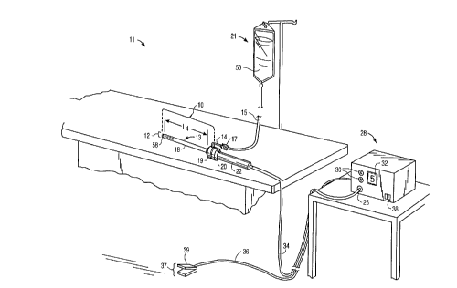
Referring to the drawings in detail, wherein like
numerals indicate like elements, an electrosurgical system 11
is shown constructed according to the principles of the
present invention. Electrosurgical system 11 generally
comprises an electrosurgical probe 10 connected to a power
supply 28 for providing high frequency voltage to a target
tissue 52 and a liquid source 21 for supplying electrically
conducting fluid 50 to probe 10.
In an exemplary embodiment as shown in Fig. 1,
electrosurgical probe 10 includes an elongated shaft 13 which
may be flexible or rigid, with flexible shafts optionally
including support cannulas or other structures (not shown).
Probe 10 includes a connector 19 at its proximal end and an
array 12 of electrode terminals 58 disposed on the distal tip
of shaft 13. A connecting cable 34 has a handle 22 with a
connector 20 which can be removably connected to connector 19
of probe 10. The proximal portion of cable 34 has a connector
26 to couple probe 10 to power supply 28. The electrode
CA 02221330 1997-11-17
WO 96/39914 PCT/US96/08077
18
terminals 58 are electrically isolated from each other and
each of the terminals 58 is connected to an active or passive
control network within power supply 28 by means of a plurality
of individually insulated conductors 42 (see Fig. 2C). Power
supply 28 has a selection means 30 to change the applied
voltage level. Power supply 28 also includes means for
energizing the electrodes 58 of probe 10 through the
depression of a pedal 39 in a foot pedal 37 positioned close
to the user. The foot pedal 37 may also include a second
pedal (not shown) for remotely adjusting the energy level
applied to electrodes 58. The specific design of a power
supply which may be used with the electrosurgical probe of the
present invention is described in parent application PCT US
94/051168, the full disclosure of which has previously been
incorporated herein by reference.
Referring to Figs. 2A and 2B, the electrically
isolated electrode terminals 58 are spaced-apart over an
electrode array surface 82. The electrode array surface 82
and individual electrode terminals 58 will usually have
dimensions within the ranges set forth above. In the
preferred embodiment, the electrode array surface 82 has a
circular cross-sectional shape with a diameter D (Fig. 2B) in
the range from 1 mm to 10 mm. Electrode array surface 82 may
also have an oval shape, having a length L in the range of 1
mm to 20 mm and a width W in the range from 0.5 mm to 7 mm, as
shown in Fig. 5. The individual electrode terminals 58 will
protrude over the electrode array surface 82 by a distance (H)
from 0 mm to 2 mm, preferably from 0 mm to 1 mm (see Fig. 3).
As described above, electrode terminals which are flush with
the surface, or protrude by a minimum distance, will provide
less aggressive ablation and are particularly suitable for
smoothing of treated tissue surfaces and providing hemostasis
to inhibit or prevent bleeding of treated surfaces.
The electrode terminals 58 are preferably composed
of a refractory, electrically conductive metal or alloy, such
as platinum, platinum alloys, titanium, titanium alloys and
the like. Platinum is the preferred choice for electrode
terminal material since it is biocompatible, has a low erosion
CA 02221330 1997-11-17
WO 96/39914 PCT/US96/08077
19
rate, and can be readily fabricated and attached to conductors
42 within the shaft 13 of electrosurgical probe 10. As shown
in Fig. 2B, the electrode terminals 58 are anchored in a
support matrix 48 of suitable insulating material (e.g.,
ceramic or glass material, such as alumina, zirconia and the
like) which could be formed at the time of manufacture in a
flat, hemispherical or other shape according to the
requirements of a particular procedure. The preferred support
matrix material is alumina, available from Kyocera Industrial
Ceramics Corporation, Elkgrove, Illinois, because of its high
thermal conductivity, good electrically insulative properties,
high flexural modulus, resistance to carbon tracking,
biocompatibility, and high melting point.
As shown in Fig. 2A, the support matrix 48 is
adhesively joined to a tubular support member 78 that extends
most or all of the distance between matrix 48 and the proximal
end of probe 10. Tubular member 78 preferably comprises an
electrically insulating material, such as an epoxy or
silicone-based material. In a preferred construction
technique, electrode terminals 58 extend through pre-formed
openings in the support matrix 48 so that they protrude above
electrode array surface 82 by the desired distance H (Fig. 3).
The electrodes are then bonded to the distal surface 82 of
support matrix 48, typically by an inorganic sealing material
80. Sealing material 80 is selected to provide effective
electrical insulation, and good adhesion to both the alumina
matrix 48 and the platinum or titanium electrode terminals.
Sealing material 80 additionally should have a compatible
thermal expansion coefficient and a melting point well below
that of platinum or titanium and alumina or zirconia,
typically being a glass or glass ceramic.
In the embodiment shown in Figs. 2A and 2B, probe 10
includes a return electrode 56 for completing the current path
between electrode terminals 58 and power supply 28. Return
electrode 56 is preferably an annular member positioned around
the exterior of shaft 13 of probe 10. Return electrode 56 may
fully or partially circumscribe tubular support member 78 to
form an annular gap 54 therebetween for flow of electrically
CA 02221330 1997-11-17
WO 96/39914 PCTIUS96/08077
conducting liquid 50 therethrough, as discussed below. Gap 54
preferably has a width in the range of 0.25 mm to 4 mm.
Return electrode 56 extends from the proximal end of probe 10,
where it is suitably connected to power supply 28 via
5 connectors 19, 20, to a point slightly proximal of electrode
array surface 82, typically about 1mm to 10 mm.
Return electrode 56 is disposed within an
electrically insulative jacket 18, which is typically formed
as one or more electrically insulative sheaths or coatings,
10 such as polytetrafluoroethylene, polyamide, and the like. The
provision of the electrically insulative jacket 18 over return
electrode 56 prevents direct electrical contact between return
electrode 56 and any adjacent body structure. Such direct
electrical contact between a body structure (e.g., tendon) and
15 an exposed common electrode member 56 could result in unwanted
heating and necrosis of the structure at the point of contact
causing necrosis.
Return electrode 56 is preferably formed from an
electrically conductive material, usually metal, which is
20 selected from the group consisting of stainless steel,
platinum or its alloys, titanium or its alloys, molybdenum or
its alloys, and nickel or its alloys. The return electrode 56
may be composed of the same metal or alloy which forms the
electrode terminals 58 to minimize any potential for corrosion
or the generation of electrochemical potentials due to the
presence of dissimilar metals contained within an electrically
conductive fluid 50, such as isotonic saline (discussed in
greater detail below).
As shown in Fig. 2A, return electrode 56 is not
directly connected to electrode terminals 58. To complete
this current path so that terminals 58 are electrically
connected to return electrode 56 via target tissue 52,
electrically conducting liquid 50 (e.g., isotonic saline) is
caused to flow along liquid paths 83. Liquid paths 83 are
formed by annular gap 54 between outer return electrode 56 and
tubular support member 78 and an inner lumen 57 within an
inner tubular member 59. The electrically conducting liquid
50 flowing through fluid paths 83 provides a pathway for
CA 02221330 1997-11-17
WO 96/39914 PCT/US96/08077
21
electrical current flow between target tissue 52 and return
electrode 56, as illustrated by the current flux lines 60 in
Fig. 2A. When a voltage difference is applied between
electrode array 12 and return electrode 56, high electric
field intensities will be generated at the distal tips of
terminals 58 with current flow from array 12 through the
target tissue to the return electrode, the high electric field
intensities causing ablation of tissue 52 in zone 88.
Figs. 2C, 3 and 4 illustrate an alternative
embodiment of electrosurgical probe 10 which has a return
electrode 55 positioned within tubular member 78. Return
electrode 55 is preferably a tubular member defining an inner
lumen 57 for allowing electrically conducting liquid 50 (e.g.,
isotonic saline) to flow therethrough in electrical contact
with return electrode 55. In this embodiment, a voltage
difference is applied between electrode terminals 58 and
return electrode 55 resulting in electrical current flow
through the electrically conducting liquid 50 as shown by
current flux lines 60 (Fig. 3). As a result of the applied
voltage difference and concomitant high electric field
intensities at the tips of electrode terminals 58, tissue 52
becomes ablated or transected in zone 88.
Fig. 2C illustrates the proximal or connector end 70
of probe 10 in the embodiment of Figs. 3 and 4. Connector 19
comprises a plurality of individual connector pins 74
positioned within a housing 72 at the proximal end 70 of probe
10. Electrode terminals 58 and the attached insulating
conductors 42 extend proximally to connector pins 74 in
connector housing 72. Return electrode 55 extends into
housing 72, where it bends radially outward to exit probe 10.
As shown in Figs. 1 and 2C, a liquid supply tube 15 removably
couples liquid source 21, (e.g., a bag of fluid elevated above
the surgical site or having a pumping device), with return
electrode 55. Preferably, an insulating jacket 14 covers the
exposed portions of electrode 55. One of the connector pins
76 is electrically connected to return electrode 55 to couple
electrode 55 to power supply 28 via cable 34. A manual
control valve 17 may also be provided between the proximal end
CA 02221330 1997-11-17
WO 96/39914 PCT/US96/08077
22
of electrode 55 and supply tube 15 to allow the surgical team
to regulate the flow of electrically conducting liquid 50.
Fig. 6 illustrates another embodiment of probe 10
where the distal portion of shaft 13 is bent so that electrode
terminals extend transversely to the shaft. Preferably, the
distal portion of shaft 13 is perpendicular to the rest of the
shaft so that electrode array surface 82 is generally parallel
to the shaft axis, as shown in Fig. 6. In this embodiment,
return electrode 55 is mounted to the outer surface of shaft
13 and is covered with an electrically insulating jacket 18.
The electrically conducting fluid 50 flows along flow path 83
through return electrode 55 and exits the distal end of
electrode 55 at a point proximal of electrode surface 82. The
fluid is directed exterior of shaft to electrode surface 82 to
create a return current path from electrode terminals 58,
through target tissue 52, to return electrode 55, as shown by
current flux lines 60.
Fig. 7 illustrates another embodiment of the
invention where electrosurgical system 11 further includes a
liquid supply instrument 64 for supplying electrically
conducting fluid 50 between electrode terminals 58 and return
electrode 55. Liquid supply instrument 64 comprises an inner
tubular member or return electrode 55 surrounded by an
electrically insulating jacket 18. Return electrode 55
defines an inner passage 83 for flow of fluid 50. As shown in
Fig. 7, the distal portion of instrument 64 is preferably bent
so that liquid 50 is discharged at an angle with respect to
instrument 64. This allows the surgical team to position
liquid supply instrument 64 adjacent electrode surface 82 with
the proximal portion of supply instrument 64 oriented at a
similar angle to probe 10.
Figs. 8 and 9 illustrate another embodiment of probe
10 where the return electrode is an outer tubular member 56
that circumscribes support member 78 and conductors 42.
Insulating jacket 18 surrounds tubular member 56 and is spaced
from member 56 by a plurality of longitudinal ribs 96 to
define an annular gap 54 therebetween (Fig. 9). Annular gap
preferably has a width in the range of .25 mm to 4 mm. Ribs
CA 02221330 1997-11-17
WO 96/39914 PCTlUS96/08077
23
96 can be formed on either the jacket 18 or member 56. The
distal end of return electrode 56 is a distance L1 from
electrode surface 82. Distance L1 is preferably about .5 to
mm and more preferably about 1 to 10 mm.
5 As shown in Fig. 8, electrically conducting liquid
50 flows through annular gap 54 (in electrical communication
with the return electrode) and is discharged through the
distal end of gap 54. The liquid 50 is then directed around
support member 78 to electrode terminals 58 to provide the
10 current pathway between the electrode terminals and return
electrode 56. Since return electrode 56 is proximally
recessed with respect to electrode surface 82, contact between
the return electrode 56 and surrounding tissue is minimized.
In addition, the distance Li between the active electrode
terminals 58 and the return electrode 56 reduces the risk of
current shorting therebetween.
The present invention is not limited to an electrode
array disposed on a relatively planar surface at the distal
tip of probe 10, as described above. Referring to Figs. 12-
14, an alternative probe 10 includes a pair of electrodes 58a,
58b mounted to the distal end of shaft 13. Electrodes 58a,
58b are electrically connected to power supply as described
above and preferably have tips 100a, 100b with a screwdriver
shape. The screwdriver shape provides a greater amount of
"edges" to electrodes 58a, 58b, to increase the electric field
intensity and current density at the edges and thereby improve
the cutting ability as well as the ability to limit bleeding
from the incised tissue (i.e., hemostasis).
As shown in Fig. 12, current flows between electrode
tips 100a and 100b as indicated by current flux lines 60 to
heat the target tissue 52. The surgical team then moves probe
10 transversely across tissue 52 to effect an incision 102 in
tissue 52, as shown in Fig. 14.
Other modifications and variations can be made to
disclose embodiments without departing from the subject
invention as defined in the following claims. For example,
shaft 13 of probe 10 may have a variety of configurations
other than the generally linear shape shown in Figs. 1-8. For
CA 02221330 1997-11-17
WO 96/39914 PCTIUS96/08077
24
example, shaft 13 may have a distal portion that is angled, in
the range of 100 to 30 (Fig. 10) or 90 (Figs. 11 and 6), to
improve access to the operative site of the tissue 52 being
ablated or cut (see Fig. 10). A shaft having a 90 bend angle
may be particular useful for accessing gingiva located in the
back portion of the patient's mouth and a shaft having a 10
to 30 bend angle may be useful for accessing gingiva near or
in the front of the patient's mouth.