Note: Descriptions are shown in the official language in which they were submitted.
CA 02222328 2010-02-05
LIPID-NUCLEIC ACID PARTICLES PREPARED VIA A
HYDROPHOBIC LIPID-NUCLEIC ACID COMPLEX INTERMEDIATE
AND USE FOR GENE TRANSFER
FIELD OF THE INVENTION
This invention relates to lipid-nucleic- acid particles which are useful for
the introduction of nucleic acids into cells, and methods of making and using
them. The
invention provides a circulation-stable, characterizable delivery vehicle for
the
introduction of plasmids or antisense compounds into cells. These vehicles are
safe,
stable, and practical for clinical use.
BACKGROUND OF THE INVENTION
Gene transfer into genetically impaired host cells in order to correct the
genetic defects has vast potential for succesfully treating a variety of thus
far hitherto
untreatable medical conditions. There are currently six major non-viral
methods by
l0 which genes are introduced into host cells: (i) direct microinjection, (ii)
calcium
phosphate precipitation, (ii) DEAE-dextran complexes, (iv) electroporation,
(v) cationic
lipid complexes and (vi) reconstituted viruses and virosomes (see Chang, et
al., Focus
10:88 (1988)).
Most reported examples of gene transfer have been performed in vitro. In
vivo gene transfer is complicated by serum interactions, immune clearance,
enzymatic
degradation of the genes, toxicity and biodistribution. In in vivo
administration, selection
is not possible, and a reasonably high frequency of transformation is
necessary to achieve
sufficient expression to compensate for a defective endogenous gene.
The in vivo gene transfer methods tinder study in the clinic consist almost
entirely of viral vectors. Although viral vectors have the inherent ability to
transport
nucleic acids across cell membranes and some can integrate exogenous DNA into
the
CA 02222328 1997-11-25
WO 96/40964 PCT/US96/09949
2
chromosomes, they can carry only limited amounts of DNA. In addition, their
use poses
significant risks. One such risk is that the viral vector may revert to a
pathogenic
genotype either through mutation or genetic exchange with a wild type virus.
In view of these limitations and risks, alternative non-viral-based gene
transfer methods have been developed. These methods use often plasmid vectors,
which
are small circular sequences of DNA, as vectors for DNA delivery. However,
most
plasmids do not possess the attributes required for intracellular delivery and
therefore
sophisticated delivery systems are required.
Cationic lipid complexes are presently the most effective generally used
means of introducing non-viral nucleic acids into cells. A number of different
formulations incorporating cationic lipids are commercially available. These
include:(i)
LIPOFECTIN (which uses 1,2-dioleyloxy-3- (N,N,N-trimethylamino)propane
chloride, or
DOTMA, see Eppstein, et al., U.S. Patent No. 4,897,355); LiPOFECTAMINE (which
uses DOSPA, see Hawley-Nelson, et al., Focus 15(3):73 (1993)); and LIPOFECTACE
(which uses N,N-distearyl-N,N-dimethyl-ammonium bromide, or DDAB, see Rose,
U.S.
Patent No. 5,279,833). Others have reported alternative cationic lipids that
work in
essentially the same manner but with different efficiencies, for example
1,2-dioleoyloxy-3-(N,N,N-trimethylamino) propane chloride, or DOTAP (see
Stomatatos,
et al., Biochemistry 27:'3917-3925 (1988)); glycerol based lipids (see
Leventis, et al.,
Biochem. Biophys. Acta 1023:124 (1990); lipopolyamines (see, Behr, et a!.,
U.S. Patent
No. 5,171,678) and cholesterol based lipids (see Epand, et al., WO 93/05162,
and U.S.
Patent No. 5,283,185). It has been reported that DOTMA and related compounds
are
significantly more active in gene transfer assays than their saturated
analogues (see,
Felgner, et al., WO91/16024). However, both DOTMA and DOSPA based
formulations, despite their efficiency in effecting gene transfer, are
prohibitively
expensive. DDAB on the other hand is inexpensive and readily available from
chemical
suppliers but is less effective than DOTMA in most cell lines. Another
disadvantage of
the current lipid systems is that they are not appropriate for intravenous
injection.
Lipid-based vectors used in gene transfer have generally been formulated
in one of two ways. In one method, the nucleic acid is introduced into
preformed
liposomes made of mixture of cationic lipids and neutral lipids. The complexes
thus
formed have undefined and complicated structures and the lipofection
efficiency is
severely reduced by the presence of serum. A second method involves the
formation ;.r
DNA complexes with mono- or poly-cationic lipids without the presence of a
neutral
CA 02222328 1997-11-25
WO 96/40964 PCT/US96/09949
3
lipid. These complexes are often prepared in the presence of ethanol and are
not stable
in water. Additionally, these complexes are adversely affected by serum (see,
Behr,
Acc. Chem. Res. 26:274-78 (1993)).
An examination of the relationship between the chemical structure of the
carrier vehicle and its efficiency of gene transfer has indicated that the
characteristics
which provide for effective gene transfer would make a carrier unstable in
circulation
(see, Ballas, et al., Biochim. Biophys. Acta 939:8-18 (1988)). Additionally,
degradation
either outside or inside the target cell remains a problem (see, Duzghines,
Subcellular
Biochemistry 11:195-286 (1985)). Others who have attempted to encapsulate DNA
in
lipid-based formulations have not overcome these problems (see, Szoka et al.,
Ann. Rev.
Biophys. Bioeng. 9:467 (1980); Deamer, U.S. Patent No. 4,515,736, and
Legendre,
Pharm. Res. 9:1235-1242 (1992)).
Ideally, a delivery vehicle for a nucleic acid or plasmid will have the
following characteristics: a) ease of preparation, b) capable of carrying a
large amount of
DNA per particle to enable gene transfer of all sizes of genes and. reduce the
volume of
injection, c) homogenous, d) reproducible, e) is serum stable with minimal
serum
interactions and shields DNA from extracellular degradation, and f) is capable
of
transfecting target cells in such a way that the DNA is not digested
intracellularly.
The present invention provides such compositions and methods for their
preparation and use.
SUMMARY OF THE INVENTION
The present invention comprises novel, lipid-nucleic acid particles. The
invention also comprises methods of making and using these particles.
In some embodiments, the particles are made by formation of hydrophobic
intermediate complexes in either detergent-based or organic solvent-based
systems,
followed by removal of the detergent or organic solvent. Preferred embodiments
are
charge-neutralized.
In one embodiment, a plasmid is combined with cationic lipids in a
detergent solution to provide a coated plasmid-lipid complex. The complex is
then
contacted with non-cationic lipids to provide a solution of detergent, a
plasmid-lipid
complex and non-cationic lipids, and the detergent is then removed to provide
a solution
of serum-stable plasmid-lipid particles, in which the plasmid is encapsulated
in a lipid
CA 02222328 1997-11-25
WO 96/40964 PCT/US96/09949
4
bilayer. The particles thus formed have a size of about 50-150 nm.
In another embodiment, serum-stable plasmid-lipid particles are formed by
preparing a mixture of cationic lipids and non-cationic lipids in an organic
solvent;
contacting an aqueous solution of plasmid with the mixture of cationic and non-
cationic
lipids to provide a clear single phase; and removing the organic solvent to
provide a
suspension of plasmid-lipid particles, in which the plasmid is encapsulated in
a lipid
bilayer, and the particles are stable in serum and have a size of about 50-150
nm.
Another method of forming lipid-nucleic acid particles involves:
(a) contacting nucleic acids with a solution of non-cationic lipids and a
detergent to form a nucleic acid-lipid mixture;
(b) contacting cationic lipids with the nucleic acid-lipid mixture to
neutralize the negative charge of said nucleic acids and form a charge-
neutralized mixture
of nucleic acids and lipids: and
(c) removing the detergent from the charge-neutralized mixture to provide
the lipid-nucleic acid particles in which the nucleic acids are protected from
degradation.
Another method of forming lipid-nucleic acid particles involves:
(a) contacting an amount of cationic lipids with nucleic acids in a solution;
the solution comprising of from about 15-35% water and about 65-85% organic
solvent
and the amount of cationic lipids being sufficient to produce a +/- charge
ratio of from
about 0.85 to about 2.0, to provide a hydrophobic, charge-neutralized lipid-
nucleic acid
complex;
(b) contacting the hydrophobic, charge-neutralized lipid-nucleic acid
complex in solution with non-cationic lipids, to provide a lipid-nucleic acid
mixture; and
(c) removing the organic solvents from the lipid-nucleic acid mixture to
provide lipid-nucleic acid particles in which the nucleic acids are protected
from
degradation.
The lipid-nucleic acid particles of the present invention are useful for the
therapeutic delivery of nucleic acids. In one embodiment, the particles are
constructed
via a hydrophobic lipid-nucleic acid intermediate (or complex). Upon removal
of a
solubilizing component (i.e., detergent or an organic solvent) the nucleic
acid becomes
protected from degradation. The particles thus formed are suitable for use in
intravenous
nucleic acid transfer as they are stable in circulation, of a size required
for
pharmacodynamic behavior resulting in access to extravascular sites and target
cell
populations.
CA 02222328 2010-02-05
4a
Various embodiments of this invention provide a method for preparation of
lipid-nucleic acid particles, comprising: (a) contacting nucleic acids with a
solution
comprising non-cationic lipids, a polyethylene glycol-lipid conjugate, and a
detergent to
form a nucleic acid-lipid mixture; (b) contacting cationic lipids with said
nucleic acid-lipid
mixture to neutralize the negative charge of said nucleic acids and form a
charge-neutralized
mixture comprising detergent, nucleic acids and lipids; and (c) removing said
detergent
from said charge-neutralized mixture to provide said lipid-nucleic acid
particles in which
said nucleic acids are encapsulated in the lipid and protected from
degradation.
Various embodiments of this invention provide a method for preparation of
lipid-nucleic acid particles, comprising: (a) contacting an amount of cationic
lipids with
nucleic acids in a solution; said solution comprising of from about 15-35%
water and about
65-85% organic solvent and said amount of cationic lipids being sufficient to
produce a +/-
charge ratio of from about 0.85 to about 2.0, to provide a hydrophobic, charge-
neutralized
lipid-nucleic acid complex; (b) contacting said hydrophobic lipid-nucleic acid
complex in
solution with non-cationic lipids and a polyethylene glycol-lipid conjugate,
to provide a
lipid-nucleic acid mixture; and (c) removing said organic solvents from said
mixture to
provide said lipid-nucleic acid particles in which said nucleic acids are
encapsulated in the
lipid and protected from degradation.
Various embodiments of this invention provide a method for introducing a
nucleic acid into a cell, comprising: (a) preparing a lipid-nucleic acid
particle according to
the method of any one of claims 1 to 10; and (b) contacting said cell in vitro
with said lipid-
nucleic acid particle for a period of time sufficient to introduce said
nucleic acid into said
cell.
Various embodiments of this invention provide a lipid-nucleic acid particle
prepared according to the method of this invention.
Various embodiments of this invention provide a method for preparation of
serum-stable plasmid-lipid particles, comprising: (a) combining a plasmid with
cationic
lipids in a detergent solution to provide a coated plasmid-lipid complex; (b)
contacting non-
cationic lipids with said coated plasmid-lipid complex to provide a solution
comprising
detergent, a plasmid-lipid complex, non-cationic lipids, and a polyethylene
glycol-lipid
conjugate; and (c) removing said detergent from said solution of step (b) to
provide a
solution of serum-stable plasmid-lipid particles, wherein said plasmid is
encapsulated in a
CA 02222328 2010-02-05
4b
lipid bilayer and said particles are serum-stable and have a size of from
about 50 to about
150 nm.
Various embodiments of this invention provide a method for the preparation
of serum-stable plasmid-lipid particles, comprising: (a) preparing a mixture
comprising
cationic lipids, non-cationic lipids and a polyethylene glycol-lipid conjugate
in an organic
solvent; (b) contacting an aqueous solution of plasmid with said mixture
prepared in step (a)
to provide a clear single phase; and (c) removing said organic solvent to
provide a
suspension of plasmid-lipid particles, wherein said plasmid is encapsulated in
a lipid
bilayer, and said particles are stable in serum and have a size of from about
50 to about 150
Mn.
Various embodiments of this invention provide a plasmid-lipid particle
prepared according to a method of this invention.
Various embodiments of this invention provide a method for introducing a
plasmid into a cell, comprising: (a) preparing a plasmid-lipid particle
according to the
method of any one of claims 14 to 3, and (b) contacting said cell in vitro
with said plasmid-
lipid particle for a period of time sufficient to introduce said plasmid into
said cell.
Various embodiments of this invention provide a nucleic acid-lipid particle
for introducing a nucleic acid into a cell, said particle comprising a
cationic lipid, a
polyethylene glycol-lipid conjugate that inhibits aggregation of particles,
and a nucleic acid,
wherein the nucleic acid component of said particle is encapsulated in the
lipid and
protected from degradation.
Various embodiments of this invention provide a pharmaceutical
composition comprising a nucleic acid-lipid particle and a pharmaceutically
acceptable
carrier, said nucleic acid-lipid particle comprising a cationic lipid, a
polyethylene glycol-
lipid conjugate that inhibits aggregation of particles, and a nucleic acid,
wherein the nucleic
acid component of said particle is encapsulated in the lipid and protected
from degradation.
Various embodiments of this invention provide a method of introducing a
nucleic acid into a cell, said method comprising contacting said cell in vitro
with a nucleic
acid-lipid particle comprising a cationic lipid, a polyethylene glycol-lipid
conjugate that
inhibits aggregation of particles, and a nucleic acid, wherein the nucleic
acid component of
said particle is encapsulated in the lipid and protected from degradation.
CA 02222328 2010-02-05
4c
Various embodiments of this invention provide use of a nucleic acid-lipid
particle according to this invention in preparation of a therapeutic
composition for
introducing said nucleic acid into a cell.
Various embodiments of this invention provide use of a nucleic acid-lipid
particle according to this invention in preparation of a therapeutic
composition for an in
vitro gene transfer or an in vivo gene transfer.
Various embodiments of this invention provide use of a nucleic acid-lipid
particle according to this invention in preparation of a therapeutic
composition for insertion
of a functional copy of gene or for suppression of gene expression.
Various embodiments of this invention provide use of a particle of this
invention in preparation of a therapeutic composition for introducing said
nucleic acid into a
cell.
Various embodiments of this invention provide use of a particle of this
invention in preparation of a therapeutic composition for introducing said
plasmid into a
cell.
Various embodiments of this invention provide use of a nucleic acid-lipid
particle in preparation of a therapeutic composition for introducing a nucleic
acid into a cell,
wherein the particle comprises a cationic lipid, a polyethylene glycol-lipid
conjugate that
inhibits aggregation of particles, and the nucleic acid, wherein the nucleic
acid component
of said particle is encapsulated in the lipid and protected from degradation.
CA 02222328 1997-11-25
WO 96/40964 PCT/US96/09949
In particular, it is an object of this invention to provide in vitro and in
vivo
methods for treatment of diseases which involve the overproduction or
underproduction
of particular proteins. In these methods, a nucleic acid encoding a desired
protein or
blocking the production of an undesired protein, is formulated into a lipid-
nucleic acid
5 particle, and the particles are administered to patients requiring such
treatment.
Alternatively, cells are removed from a patient, transfected with the lipid-
nucleic acid
particles described herein, and reinjected into the patient.
BRIEF DESCRIPTION OF TIIE DRAWINGS
Figure 1 illustrates a nucleic acid-lipid particle-mediated gene transfer
using "sandwich-type" complexes of DNA.
Figure 2 illustrates an aggregation and precipitation which commonly
occurs during the entrapment of large nucleic acids in lipid complexes.
Figure 3 provides a schematic representation of the preparation of plasmid-
lipid particles according to certain embodiments of the present invention.
Figure 4 illustrates the recovery of 'H-DNA from encapsulated particles
following the reverse-phase preparation of the particles and extrusion through
a 400 nm
filter and a 200 nm filter. Lipid composition is POPC:DOL)AC:PEG-Cer-C2t).
PEG-CerC2õ was held constant at 10 mole% and POPC and DODAC were changed
relative to each other. 20 mg lipid; 50 pg plasmid DNA (7.5 kbp).
Figure 5 illustrates the recovery of 3H-DNA from particles prepared using
a reverse-phase procedure. The particles were extruded through a 200 nm filter
and
eluted on a DEAE Sepharose CL-6B anion exchange column. The percent recovery
reported is based on the amount recovered after filtration. Lipid composition
is as in
Figure 4.
Figure 6 illustrates the recovery of 14C_ lipid from encapsulated particles
following the reverse-phase preparation of the particles and extrusion through
a 400 nm
filter and a 200 nm filter. Lipid composition is as in Figure 4.
Figure 7 illustrates the recovery of 14C-lipid from particles prepared using
a reverse-phase procedure. The particles were extruded through a 200 nm filter
and
= 30 eluted on a DEAE Sepharose CL-6B anion exchange column. The percent
recovery
reported is based on the amount recovered after filtration. Lipid composition
is as in
Figure 4.
CA 02222328 1997-11-25
WO 96/40964 PCT/US96/09949
6
Figure 8 illustrates the effect of DODAC concentration on the
encapsulation of plasmid DNA. Encapsulation efficiency was measured by anion
exchange chromatography. Vesicles were composed of DOPE, DODAC and 10 mole%
PEG-Cer-C20 (symbol) or EPC, DODAC and 10 mole% PEG-Cer-C20 (symbol). Total
lipid and DNA concentrations were 10 mmole/ml and 50 g/ml, respectively.
Figures 9A and 9B illustrate the effect of serum nucleases on free
pCMVCAT DNA as assessed by column chromatography before (A) and after (B)
incubation in 80% mouse serum. Free 3H-DNA (pCMVCAT) was eluted on a Sepharose
CL-4B column in HBS, pH 7.4.
Figure 10 illustrates the effect of serum nucleases on encapsulated
pCMVCAT DNA (prepared by reverse-phase) as assessed by column chromatography.
Sepharose CL-4B column profile of encapsulated pCMV plasmid incubated in 80%
mouse serum for 30 min. (A) External DNA was removed by ion exchange
chromatography prior to incubation in serum. (B) External DNA was not removed
prior
to incubation in serum. Lipid composition was POPC:DODAC:PEG-Cer-C211. Total
lipid and plasmid concentrations were 20 pmole/ml and 50 g/ml prior to anion
exchange
chromatography.
Figures I I A and 1 I B illustrate the effect of serum nucleases on
encapsulated pCMVCAT DNA (prepared by detergent dialysis) as assessed by
column
chromatography. Sepharose CL-4B column profile of encapsulated pCMV plasmid
incubated in 80% mouse serum for 30 min. (A) External DNA was removed by ion
exchange chromatography prior to incubation in serum. (B) External DNA was not
removed prior to incubation in serum. The lipid composition was DOPE:
DODAC:PEG--
Cer-C20 (84:6:10). Total lipid and plasmid concentrations were 10 mole/ml and
400
g/ml prior to anion exchange chromatography.
Figures 12A and 12B illustrate the resistance of plasmid complexed to
preformed liposomes composed of DOPE:DODAC(50:50) (A) and plasmid encapsulated
within DOPE: DODAC: PEG-Cer-C 14 particles (B) to digestion by DNAse I.
Plasmid
DNA was extracted and subjected to PCR (polymerized chain reaction) to amplify
for
visualization on a gel. Free plasmid was used as a control. Lane 1:1 kb DNA
marker;
Lane 2: PCR negative control (no DNA); Lane 3: free plasmid alone; Lane 4:
free
plasmid in 0.05% detergent (Triton X-100); Lane 5: free plasmid incubated with
DNAse
I in the absence of detergent; Lane 6: free plasmid incubated with DNAse I in
the
presence of detergent: Lane 7: complexed (A) or encapsulated (B) plasmid
alone; Lane
CA 02222328 1997-11-25
WO 96/40964 PCT/US96/09949
7
8: complexed (A) or encapsulated (B) plasmid in 0.05% detergent; Lane 9:
complexed
(A) or encapsulated (B) plasmid incubated in DNAse I in the absence of
detergent; Lane
10: complexed (A) or encapsulated (B) plasmid incubated in DNAse I in the
presence of
detergent.
Figure 13 illustrates the effect of plasmid DNA concentration on
encapsulation efficiency (detergent dialysis). Vesicles were composed of
DOPE:DODAC:PEG-Cer (84:6:10) at a lipid concentration of 10 mole/ml.
Figure 14 illustrates the effect of NaCI concentration on the optimal
DODAC concentration for plasmid entrapment. Lipid composition was
DOPE:DODAC:PEG-Cer-C14 (or PEG-Cer-C20). PEG-Cer was held constant at 10
mole%. Total lipid concentration was 10 pmole/ml. Plasmid concentration was 50
g/ml.
Figure 15 illustrates the size distribution of plasmid:lipid particles
prepared
by the detergent dialysis procedure (Volume weighted analysis). Lipid
composition was
DOPE: DODAC:PEG-Cer-C21) (84:6:10).
Figure 16 illustrates the size distribution of plasmid:lipid particles
prepared
by the detergent dialysis procedure (Number weighted analysis). Lipid
composition was
DOPE: DODAC;PEG-Cer-C20(84:6:10).
Figures 17A and 17B provide electron micrographs -"f liposomes composed
of DOPE: DODAC:PEO-Cer-C2,) without encapsulated plasmid (A) and the
plasmid:lipid
particles (B). The small arrows denote empty liposomes approximately 100 nm in
diameter. These are compared to electron-dense particles surrounded by a
membrane
bilayer (large arrows). Scale bar = 100 nm.
Figure 18 shows the clearance of 'H-DNA and 14C-lipid from particles
(prepared by reverse-phase methods) after injection into ICR mice. The figure
includes
free 'H-DNA after injection as a comparison. Lipid composition is
POPC: DODAC: PEG-Cer-C20.
Figures 19A and 19B show the clearance 'H-DNA and 14C-lipid from
particles (prepared by detergent dialysis methods) after injection into ICR
mice. Lipid
compositions were (A) DOPE:DODAC-PEG-Cer-C2, (84:6: 10) and (B)
DOPE:DODAC-PEG-Cer-C14 (84:6:10).
Figure 20 shows the results of in vivo gene transfer which occurs in the
lungs of mice. Lipid composition is DOPE-DODAC-PEG-Cer-C,,, or
DOPE: DODAC: PEG-Cer-C14 (84:6:10).
CA 02222328 1997-11-25
WO 96/40964 PCT/US96/09949
8
Figure 21 shows the results of in vivo gene transfer which occurs in the
liver mice. Lipid composition is DOPE-DODAC-PEG-Cer-C20 , or
DOPE:DODAC:PEG-Cer-C14 (84:6:10).
Figure 22 shows the results of in vivo gene transfer which occurs in the
spleen of mice. Lipid composition is DOPE-DODAC-PEG-Cer-C20 or
DOPE:DODAC:PEG-Cer-C14 (84:6:10).
Figure 23 shows the effect of increasing amounts of LIPOFECT1Nm
(DOTMA/DOi-c; 50:50 mol ratio) on the recovery of 13 gal plasmid DNA in the
aqueous
phase following Bligh and Dyer extraction of the lipid-nucleic acid complexes.
Figures 24A and 24B show the effect of increasing amounts of cationic
lipid on the recovery of plasmid DNA in the aqueous (A) and organic (B) phase
following Bligh and Dyer extraction of the lipid-nucleic acid complexes.
Figures 25A, 25B, 25C and 25D show the recovery of plasmid DNA from
aqueous (A and C) and organic (B and D) fractions following Bligh and Dyer
extraction
and expressed as a function of charge ratio (+/-).
Figures 26A and 26B illustrate the DNA condensation by poly-L-lysine
and DODAC assayed by TO-PRO-I dye intercalation. Condensation state was
assessed
in a Bligh and Dyer monophase (A) and in 100 mM OGP (B).
Figure 27 illustrates the effects of increasing amounts of OGP on the
recovery of plasmid DNA from the aqueous and organic phases following Bligh
and
Dyer extraction of lipid-nucleic acid complexes (plasmid/DODAC).
Figure 28 shows the effects of increasing amounts of NaCI on the recovery
of plasmid DNA from the aqueous phase following Bligh and Dyer extraction of
lipid-
nucleic acid complexes.
Figures 29A and 29B show the effect of poly-L-lysine and DODAC on the
electrophoretic mobility of plasmid DNA.
Figure 30 illustrates a protocol for preparing lipid-nucleic acid particles
using detergent dialysis.
Figures 3IA and B are bar graphs which illustrates the QELS results of a
typical lipid-nucleic acid complex mixture prepared from 0-gal
plasmid/DODAC/ESM.
Figure 32 is a bar graph which illustrates the fluorescence spectroscopic
evaluation of DNA condensation in the lipid-nucleic acid complexes using TO-
PRO-1 dye
intercalation. The results show that 0-gal plasmid in DODAC/ESM is condensed
and
protected against (lye intercalation by the lipid, and that OGP can uncondense
the
CA 02222328 1997-11-25
WO 96/40964 PCT/US96/09949
9
particle.
Figure 33 shows the results of electrophoresis of DNA extracted from
lipid-nucleic acid complexes following digestion with DNase I. DNA within the
complex
is protected from DNase I degradation whereas uncomplexed DNA is not
protected.
Figure 34 provides the results of CHO cell lipofection using 13-gal
plasmid/DODAC/ESM as assayed by /3-gal enzyme activity.
Figures 35A and B show changes in sample turbidity measured by 90
light scattering at 600 nm during the preparation of nucleic acid-lipid
particles in the
presence of 100 mM (A) or 20 mM (B) n-octyl 0-D-glucopyranoside (OGP).
Figure 36 shows solubilization of preformed DODAC (0) and SM (R)
vesicles in OGP as measured by 90 light scattering. The concentrations of
lipids used
were 200 M (solid lines) and 800 M (broken lines).
Figures 37A, 37B and 37C show volume-weighing particle size
distribution determined by QELS operating in solid particle analysis mode for
a nucleic
acid-lipid particle formulation composed of pCMVQ/DODAC/SM (charge ratio of
2:1,
DODAC/SM mole ratio of 1: 1) and prepared using 20 mM OGP before (=) and after
(a) dialysis (A). The same nucleic acid-lipid particle formulation after
dialysis was also
examined by electron microscopy (B, negative stain and C, freeze-fracture).
Bar = 100
nm.
Figures 38A and 37B depict the agarose gel electrophoresis of DNA
isolated from formulations prepared in 100 mM and 20 mM OGP (charge ratio of
2.1
and SM/DODAC ratio of 1: 1) and tested for DNase I sensitivity in the absence
(A) and
presence (B) of OGP. Panel A: molecular weight standards (lane 1), pCMV0 in
the
absence of added lipid or DNase I (lane 2), pCMVi6 following incubation with
DNase I
(lane 3), DNA isolated from a dialyzed nucleic acid-lipid particle formulation
prepared
using 100 mM OGP following incubations in the absence (lane 4) and presence
(lane 5)
of DNase I, and DNA isolated from particles prepared using 20 mM OGP and
dialyzed
following incubations in the absence (lane 6) and presence (lane 7) of DNase
1. The
first 3 lanes in panel B are identical to those in panel A except that pCMV$
was
incubated in 20 mM OGP in the absence (lane 2) and presence (lane 3) of DNase
1.
DNA isolated from a formulation prepared in 20 mM OGP.(prior to detergent
removal)
was incubated in the absence (lane 4) and presence (lane 5) of DNase I in 20
mM OGP.
Arrow indicates degraded DNA.
Figures 39A, 39B and 39C show in vitro Chinese Hamster Ovary (CHO)
CA 02222328 1997-11-25
WO 96/40964 PCT/US96/09949
cell lipofection using nucleic acid-lipid particle formulations composed of
pCMVQ/SM/DODAC (SM/DODAC mole ratio of 1:1 and charge ratio of 1: 1 to 8:1)
prepared using 100 mM OGP followed by dialysis. (A) Influence of charge ratio
on S-
galactosidase lipofection. (B) Particle induced toxicity as measured by
reduced
5 0-galactosidase activity per well for formulations prepared using a charge
ratio of 4:1.
(C) f-galactosidase lipofection achieved with nucleic acid-lipid particles
prepared using
SM (solid bar) or DOPE (hatched bar) as the neutral lipid (charge ratio of 4:1
and
DODAC to neutral lipid mole ratio of 1:1).
Figure 40 is a model describing the intermediates that may be involved in
10 the generation of a novel lipid-DNA particle.
Figures 41A, 41B and 41C illustrate the encapsulation of plasmid DNA in
a lipid vesicles by the detergent dialysis method using different cationic
lipids.
Figures 42A, 42B and 42C demonstrate the stability of plasmid containing
vesicles prepared with different cationic lipids.
Figure 43 demonstrates the encapsulation of plasmid DNA with the
ionizable lipid AL-I (pK. = 6.6) by the dialysis method
Figures 44 and 45 show the stability of the plasmid containing vesicles
formed with AL-1 at pH 4.8 and the protection of the entrapped DNA from
degradation
by serum nucleases at pH 7.5.
Figure 46 demonstrates the effect of the PEG-ceramide concentration on
the encapsulation efficiency by the dialysis method with 7.5 % DODAC and DOPE.
CA 02222328 1997-11-25
WO 96/40964 PCTIUS96/09949
11
DETAILED DESCRIPTION OF THE INVENTION
CONTENTS
I. Glossary
II. General
III. Embodiments of the invention
A. Lipid-Nucleic Acid Particles, and Properties Thereof
B. Methods of Formulating Lipid-Nucleic Acid. Particles
C. Pharmaceutical Preparations
D. Administration of Lipid-Nucleic Acid Particle Formulations for
Gene Transfer
IV. Examples
V. Conclusion
1. Glossary
The following abbreviations are used herein: CHO, Chinese hamster ovary
cell line; B16, marine melanoma cell line; DC-Chol,
30-(N-(N',N'-dimethylaminoethane)carbamoyl) cholesterol (see, Gao, et al.,
Biochem.
Biophys. Res. Comm. 179:280-285 (1991)); DDAB,
N,N-distearyi-N,N-dimethylammonium bromide; DMRIE,
N-(1,2-dimyristyloxyprop-3-yl)-N,N-dimethyl-
N-hydroxyethyl ammonium bromide; DODAC, N,N-dioleyl-N,N-dimethylammonium
chloride; DOGS, dihel.,.ulecyl;unidoglycyl spermidine; DOPE, 1,2-sn-
dioleoylphoshatidylethanolamine; DOSPA,
N-(1-(2, 3-dioleyloxy)propyl)-N-(2-(sperminecarboxamido)ethyl)-
N,N-dimethl' ammonium trifluoroacetate; DOTAP, N-(1-(2,3-dioleoyloxy)propyl)-
N,N,N-trimethylammonium chloride; DOTMA,
N-( 1-(2,3-dioleyloxy)propyl)-N, N,N-trimethylammonium chloride; EPC, egg
phosphatidylcholine; ESM, egg sphingomyelin; RT, room temperature; TBE,
Tris-Borate-EDTA (89 mM in Tris-borate and 2 mM in EDTA); HEPES,
4-(2-hydroxyethyl)-1-
= 30 piperazineethanesulfonic acid; HBS, HEPES buffered saline (150 mM NaCl
and 20 mM
HEPES); PEG-Cer-C,,,, 1-0-(2'-(w-methoxypolyethyleneglycol)succinoyl)-2-N-
arachidoyl-sphingosine; PEG-Cer-C14, 1-0-(2'-(w
CA 02222328 2010-02-05
12
-methoxypolyethyleneglycol)succinoyl)-2-N-
myristoyl-sphingosine; PBS, phosphate-buffered saline; EGTA,
ethylenebis(oxyethylenenitrilo)-tetraacetic acid; OGP, n-octyl S-D-
glycopyranoside
(Sigma Chemical Co., St. Louis, MO); POPC, palmitoyl oleoyl
phosphatidylcholine
(Northern Lipids, Vancouver, BC); QELS, quasielastic light scattering; TBE, 89
mM
Tris-borate with 2 mM EDTA; and EDTA, Ethylenediaminetetraacetic acid (Fisher
Scientific, Fair Lawn, NJ).
The term "acyl" refers to a radical produced from an organic acid by
removal of the hydroxyl group. Examples of acyl radicals include acetyl,
pentanoyl,
palmitoyl, stearoyl, myristoyl, caproyl and oleoyl.
As used herein, the term "pharmaceutically acceptable anion" refers to
anions of organic and inorganic acids which provide non-toxic salts in
pharmaceutical
preparations. Examples of such anions include chloride, bromide, sulfate,
phosphate,
acetate, benzoate, citrate, glutamate, and lactate. The preparation of
pharmaceutically
acceptable salts is described in Berge, ei at., 1. Pharm. Sci. 66:1-19 (1977).
The term "lipid" refers to any fatty acid derivative which is capable of
forming a bilayer such that a hydrophobic portion of the lipid material
orients toward the
lbilay er while a hy dr o ;hfi,c rtion o is toward the aqueous phase.
Amphipathic lipids
~' " - t
are necessary as the primary lipid vesicle structural element. Hydrophilic
characteristics
derive from the presence of phosphato, carboxylic, sulfato, amino, sulfhydryl,
nitro, and
other like groups. Hydrophobicity could he conferred by the inclusion of
groups that
include, but are not limited to, long chain saturated and unsaturated
aliphatic
hydrocarbon groups and such groups substituted by one or more aromatic,
cycloaliphatic
or heterocyclic group(s). Preferred lipids are phosphoglycerides and
sphingolipids,
representative examples of which include phosphatidylcholine,
phosphatidylethanolamine,
phosphatidylserine, phosphatidylinositol, phosphatidic acid, palmitoyloleoyl
phosphatidylcholine, lysophosphatidylcholine, lysophosphatidylethanolamine,
dipat,,itoy] phosphatidylclwline, dioicoylphosphatidylcholine,
distearoylphosphatidylcholine or dilinoleoylphosphatidyicholine could be used.
Other
.compounds lacking in phosphorus, such as sphingolipid and glycosphingolipid
families
are also within the group designated as lipid. Additionally, the amphipathic
lipids
described above may he mixed with other lipids including triglycerides and
sterols.
The tern) " neutral" refers to any of a number of lipid species which exist
CA 02222328 1997-11-25
WO 96/40964 PCT/US96/09949
13
either in an uncharged form, a neutral zwitterionic form. Such lipids include,
for
example diacylphosphati-dylcholine, diacylphosphatidylethanolamine, ceramide,
sphingomyelin, cephalin, and cerebrosides.
The term "non-cationic lipid" refers to any neutral lipid as described above
as well as anionic lipids. Examples of anionic lipids include cardiolipin,
diacylphosphatidylserine and diacylphosphatidic acid.
The term "cationic lipid" refers to any of a number of lipid species which
carry a net positive charge at physiological pH. Such lipids include, but are
not limited
to, DODAC, DOTMA, DDAB, DOTAP, DC-Choi and DMRIE. Additionally, a
number of commercial preparations of cationic lipids are available which can
be used in
the present invention. These include, for example, LIPOFECTIN (commercially
available cationic liposomes comprising DOTMA and DOPE, from GIBCO/BRL, Grand
Island, New York, USA); LIPOFECTAMiNE (commercially available cationic
liposomes comprising DOSPA and DOPE, from GIBCO/BRL); and TRANSFECTAM
(commercially available cationic lipids comprising DOGS from Promega Corp.,
Madison, Wisconsin, USA).
The term "nucleic acid" refers to a deoxyrihonucleotide or ribonucleotide
polymer in either single- or double-stranded form. Unless otherwise specified,
the term
nucleic acid is used interchangeably with gene, DNA, cDNA, RNA, and mRNA. The
term specifically encompasses ribozymes; nucleic acid cloning and/ or
expression vectors
such as plasmids; genetically engineered viral genomes, expression cassettes,
and
chromosomes from mammalian (especially human) sources.
The terms "gene transfer", "transfection", and "transformation" are used
herein interchangeably, and refer to the introduction of polyanionic
materials, particularly
nucleic acids, into cells. The term "lipofection" refers to the introduction
of such
materials using lipid-based complexes. The polyanionic materials can be in the
form of
DNA or RNA which is linked to expression vectors to facilitate gene expression
after
entry into the cell. Thus the polyanionic material used in the present
invention is meant
to include DNA having coding sequences for structural proteins, receptors and
hormones,
as well as transcriptional and translational regulatory elements (i.e.,
promoters,
enhancers, terminators and signal sequences) and vectors. Methods of
incorporating
-61 particular nucleic acids into expression vectors are well known to those
of skill in the art,
but are described in detail in, for example, Sambrook et al., Molecular
Cloning: A
Laboratory Manual (2nd ed.), Vols. 1-3, Cold Spring Harbor Laboratory, (1989)
or
CA 02222328 2010-02-05
14
Current Protocols in Molecular Biology, F. Ausubel cat al., ed. Greene
Publishing and
Wiley-Interscience, New York (1987).
"Expression vectors", "cloning vectors", or "vectors" are nucleic acid
molecules (such as plasmids) that are able to replicate in a chosen host cell.
Expression
vectors may replicate autonomously, or they may replicate by being inserted
into the
genome of the host cell, by methods well known in the art. Vectors that
replicate
autonomously will have an origin of replication or autonomous replicating
sequence
(ARS) that is functional in the chosen host cell(s). Often, it is desirable
for a vector to
be usable in more than one host cell, e.g., in E. coli for cloning and
construction, and in
a mammalian cell for expression.
The term "hydrophobic" as applied to DNA and DNA complexes, refers to
complexes which are substantially more soluble in organic solvents than in
aqueous
solutions. More particularly, hydrophobic DNA and DNA complexes are those
which are
at least 50% soluble in organic solvents such as chloroform/methanol mixtures,
and
preferably more than 70% soluble, more preferably more than 90% soluble in
such
organic solvents.
H. General
Gene transfer techniques that involve the use of liposomes have been
described previously in the art (U.S. Patents 5,049,386; 4,946,787; and
4,897,355).
General lipofection protocols are also described in the following references:
Behr et at.
(1989) Proc. Natl. Acad. Sci. (U.S.A.) 86: 6982; Demeneix et al. (1991) Int.
J. Dev.
Biol. 35: 481; Loeffler et at. (1990) J. Neurochem. 54; 1812; Bennett et al.
(1992) Mol.
Pharmacol. 41: 1023; Bertling et al. (1991) Biotechnol. Appl. Biochem. 13:
390;
Feigner et al. (1987) Proc. Natl. Acad. Sci. (U.S.A.) 84: 7413; Feigner and
Ringold
(1989) Nature 337: 387; Gareis et al. (1991) Cell. Mol. Biol. 37: 191;
Jarnagin et at.
(1992) Nucleic Acids Res. 20: 4205; Jiao et at. (1992) Exp. Neurol. 115: 400;
Lim et
al. (1991) Circulation 83: 2007; Malone et al. (1989) Proc. Natl. Acad. Sci.
(U.S.A.)
86: 6077; Powell et al. (1992) Eur. J. Vasc. Sure. fi: 130; Strauss and
Jaenisch (1992)
EMBO J. 11: 417; and Leventis and Silvius (1990) Biochim. Bionhys. Acta 1023:
124.
Lipofection reagents are sold commercially (e.g., "Transfectam" and
"Lipofectin").
Cationic and neutral lipids that are reportedly suitable for efficient
lipofection of nucleic
acids include those of Feigner (W091/17424; W091/16024). In addition, a
combination
CA 02222328 2010-02-05
of neutral and cationic lipid has been shown to be highly efficient at
lipofection of animal
cells and showed a broad spectrum of effectiveness in a variety of cell lines
(Rose et al.
(1991) BioTechnigues 10: 520. The above lipofection protocols may be adapted
for use
in the present invention, and the preceding references are therefore
incorporated in their
5 entirety.
III. Embodiments of the invention
A. Lipid-Nucleic Acid Particles, and Properties Thereof
In one aspect, the present invention provides novel, lipid-nucleic acid
complexes consisting essentially of cationic lipids and nucleic acids.
10 1. Lipid Components
Various suitable cationic lipids may be used in the present invention, either
alone or in combination with one or more other cationic lipid species or
neutral lipid
species.
Cationic lipids which are useful in the present invention can be any of a
15 number of lipid species which carry a net positive charge at physiological
pH, for
example: DODAC, DOTMA, DDAB, DOTAP, DOSPA, DOGS, DC-Chol and DMRIE,
or combinations thert:uf. A number of these lipids and related analogs, which
are also
useful in the present invention, have been described in U.S. Patent Nos.
5,208,036,
5,264,618, 5,279,833, 5,283,185, and 5,753,613.
Additionally, a number of commercial
preparations of cationic lipids are available and can be used in the present
invention.
These include, for example, LIPOFECTIN (commercially available cationic
liposomes
comprising DOTMA and DOPE, from GIBCO/BRL, Grand Island, New York, USA);
LIPOFECTAMINE (commercially available cationic liposomes comprising DOSPA and
DOPE, from GIBCO/BRL); and TRANSFECTAM (commercially available cationic
liposomes comprising DOGS from Promega Corp., Madison, Wisconsin, USA).
The non-cationic lipids used in the present invention can be any of a
variety of neutral uncharged, zwitterionic or anionic lipids capable of
producing a stable
complex. They are preferably neutral, although they can alternatively be
positively or
negatively charged. Examples of non-cationic lipids useful in the present
invention
include: phospholipid-related materials, such as lecithin,
phosphatidylethanolamine,
CA 02222328 2010-02-05
16
lysolecithin, lysophosphatidylethanolamine, phosphatidylseriine,
phosphatidylinositol,
sphingomyelin, cephalin; cardiolipin, phosphatidic acid, cerebrosides,
dicetylr,hosphate,
dioleoylphosphatidylcholine (DOPC), dipalmitoyl-phosphatidylcholine (DPPC),
diolenylphosphatidylglycerol (DOPG), dipalmitoylphosphatidylglycerol (DPPG),
dioleoyl=
phosphatidylethanolamine (DOPE), palmitoyloleoy-lphosphatidylcholine (POPC),
palmitoyloleoyl- phosphatidylethanolarnine (POPE) and dioleoyl-
phosphatidylethanolamine 4-(N-maleimidomethyl)-cyclohexane-I-carboxylate
(DOPE-mal). Additional non-phosphorous containing lipids are, e.g.,
stearylamine,
dodecylamine, hexadecylamine, acetyl palmitate, glycerol rici noleate,
hexadecyl stereate,
isopropyl myristate, amphoteric acrylic polymers, triethanolamine-lattryl
sulfate,
alkyl-aryl sulfate polyethyloxylated fatty acid amides, dioctadecyldimethyl
ammonium
bromide and the like, diacylphosphatidylcholine,
diacylphosphatidylethanolamine,
ceramide, sphingomyelin, cephalin, and cerebrosides. Other lipids such as
lysophosphatidylcholine and lysophosphatidylethanolamine may be present. Non-
cationic
lipids also include polyethylene glycol-based polymers such as PEG 2000, PEG
5000 and
polyethylene glycol conjugated to phospholipids or to ceramides (referred to
as PEG-
Cer), as described in U.S. Patent No. 5,820,873.
In preferred embodiments, the non-cationic lipids are
diacylphosphatidylcholine (e.g., dioleoylphosphatidylcholine,
dipalmitoylphosphatidylcholine and dilinoleoylphosphatidylcholine),
diacyiphosphatidylethanolamine (e.g., dioleoylphosphati(lylethanolamine and
paimitoyloleoylphosphatidylethanolamine), ceramide or sphingornyelin. The acyl
groups
in these lipids are preferably acyl groups derived from fatty acids having C10-
C21 carbon
chains. More preferably the acyl groups are lauroyl, myristoyl, palmitoyl,
stearoyl or
oleoyl. In particularly preferred embodiments, the non-cationic lipid will be
1,2-sn-
dioleoylphosphatidylethanolamine, or eggsphingomyelin (ESM).
2. Nucleic acid components
While the invention is described in the examples with reference to the use
of plasmids, one of skill in the art will understand that the methods
described herein are
equally applicable to other larger nucleic acids or oligonucleotides.
The nucleic acids which are useful in the present invention (including both
the complexes and particles) are typically nucleotide polymers having from 10
to 100,000
nucleotide residues. Typically, the nucleic acids are to be administered to a
subject for
the purpose of repairing or enhancing the expression of a cellular protein.
Additionally,
CA 02222328 2010-02-05
17
the nucleic acid can carry a label (e.g., radioactive label, fluorescent label
or
colorimetric label) for the purpose of providing clinical diagnosis relating
to the presence
or absence of complementary nucleic acids. Accordingly, the nucleic acids, or
nucleotide polymers, can be polymers of nucleic acids including genomic DNA,
cDNA,
mRNA or oligonucleotides containing nucleic acid analogs, for example, the
antisense
derivatives described in a review by Stein, et al., Science 261:1004-1011
(1993) and in
U.S. Patent Nos. 5,264,423 and 5,276,019.
Still further, the nucleic acids may encode transcriptional and
translational regulatory sequences including promoter sequences and enhancer
sequences.
The nucleotide polymers can be single-stranded DNA or RNA, or double-
stranded DNA or DNA-RNA hybrids. Examples of double-stranded DNA include
structural genes, genes including control and termination regions, and self-
replicating
systems such as plasmid DNA.
Single-stranded nucleic acids include antisense oligonucleotides
(complementary to DNA and RNA), ribozymes and triplex-forming
oligonucleotides. In
order to increase stability, some single-stranded nucleic acids will
preferably have some
or all of the nucleotide linkages substituted with stable, non-phosphodiester
linkages,
including, for example, phosphorothioate, phosphorodithioate,
phosphoroselenate, or 0-
alkyl phosphotriester linkages.
The nucleic acids used in the present invention will also include those
nucleic acids in which modifications have been made in one or more sugar
moieties
-and/or in one or more of the pyrimidine or purine bases. Examples of sugar
modifications include replacement of one or more hydroxyl groups with
halogens, alkyl
groups, amines, azido groups or functionalized as ethers or esters.
Additionally, the
entire sugar may be replaced with sterically and electronically similar
structures,
including aza-sugars and carbocyclic sugar analogs. Modifications in the
purine or
pyrimidine base moiety include, for example, alkylated purines and
pyrimidiries, acylated
purines or pyrimidines, or other heterocyclic substitutes known to those of
skill in the
art.
Multiple genetic sequences can be also be used in the present methods.
Thus, the sequences for different proteins may be located on one strand or
plasmid.
Non-encoding sequences may be also he present, to the extent that they are
necessary to
achieve appropriate expression.
The nucleic acids used in the present method can be isolated from natural
CA 02222328 2010-02-05
18
sources, obtained from such sources as ATCC or GenBank libraries or prepared
by
synthetic methods. Synthetic nucleic acids can be prepared by a variety of
solution or
solid phase methods. Generally, solid phase synthesis is preferred. Detailed
descriptions
of the procedures for solid phase synthesis of nucleic acids by phosphite-
triester,
phosphotriester, and H-phosphonate chemistries are widely available.. See, for
example,
Itakura, U.S. Pat. No. 4,401,796; Caruthers, et al., U.S. Pat. Nos. 4,458,066
and
4,500,707; Beaucage, et al., Tetrahedron Lett., 22:1859-1862 (1981);
Matteucci, et al.,
1. Am. Chem. Soc., 103:3185-3191 (1981); Caruthers, et al., Genetic
Engineering,
4:1-17 (1982); Jones, chapter 2, Atkinson, et al., chapter 3, and Sproat, et
al., chapter
4, in Oligonucleotide Synthesis: A Practical Approach, Gait (ed.), IRL Press,
Washington D.C. (1984); Froehler, ei al., Tetrahedron Lett., 27:469-472
(1986);
Froehler, et al., Nucleic Acids Res., 14:5399-5407 (1986); Sinha, et al.
Tetrahedron
Lett., 24:5843-5846 (1983); and Sinha, et al., Nucl. Acids Res., 12:4539-4557
(1984).
a. Vectors for introduction and expression of genes in cells
An important aspect of this invention is the use of the lipid-nucleic acid
particles provided herein to introduce selected genes into cells in virro and
in vivo,
followed by expression of the selected gene in the host cell. Thus, the
nucleic acids in
the particles specificily encompass vectors that are capable of being
expressed in a host
cell. Promoter, enhancer, stress or chemically-regulated promoters, antibiotic-
sensitive
or nutrient-sensitive regions, as well as therapeutic protein encoding
sequences, may be
included as required.
In brief summary, the expression of natural or synthetic nucleic acids is
typically achieved by operably linking a nucleic acid of interest to a
promoter (which is
either constitutive or inducible), incorporating the construct into an
expression vector,
and introducing the vector into a suitable host cell. Typical vectors contain
transcription
and translation terminators, transcription and translation initiation
sequences, and
promoters useful or regulation of the expression of the particular nucleic
acid. The
vectors optionally comprise generic expression cassettes containing at least
one
independent terminator sequence, sequences permitting replication of the
cassette in
eukaryotes, or prokaryotes, or both, (e.g., shuttle vectors) and selection
markers for both
prokaryotic and eukaryotic systems. Vectors are suitable for replication and
integration
in prokaryotes, eukaryotes, or preferably both. See, Gillman and Smith (1979),
Gene, 8:
CA 02222328 1997-11-25
WO 96/40964 PCT/US96/09949
19
81-97; Roberts et al. (1987), Nature, 328: 731-734; Berger and Kimmel, Guide
to
Molecular Cloning Techniques, Methods in Enzymology, volume 152, Academic
Press,
Inc., San Diego, CA (Berger); Sambrook et al. (1989), MOLECULAR CLONING - A
LABORATORY MANUAL (2nd ed.) Vol. 1-3, Cold Spring Harbor Laboratory, Cold
Spring
Harbor Press, N.Y., (Sambrook); and F.M. Ausubel et al., CURRENT PROTOCOLS IN
MOLECULAR BIOLOGY, eds., Current Protocols, a joint venture between Greene
Publishing Associates, Inc. and John Wiley & Sons, Inc., (1994 Supplement)
(Ausubel).
Product information from manufacturers of biological reagents and experimental
equipment also provide information useful in known biological methods. Such
manufacturers include the SIGMA chemical company (Saint Louis, MO), R&D
systems
(Minneapolis, MN), Pharmacia LKB Biotechnology (Piscataway, NJ), CLONTECH
Laboratories, Inc. (Palo Alto, CA), Chem Genes Corp., Aldrich Chemical Company
(Milwaukee, WI), Glen Research, Inc., GIBCO BRL Life Technologies, Inc.
(Gaithersberg, MD), Fluka Chemica-Biochemika Analytika (Fluka,Chemie AG,
Buchs,
Switzerland), and Applied Biosystems (Foster City, CA), as well as many other
commercial sources known to one of skill.
Vectors to which foreign nucleic acids are operably linked may be used to
introduce these nucleic acids into host cells and mediate their replication
and/or
expression. "Cloning vectors" are useful for replicating and amplifying the
foreign
nucleic acids and obtaining clones of specific foreign nucleic acid-containing
vectors.
"Expression vectors" mediate the expression of the foreign nucleic acid. Some
vectors
are both cloning and expression vectors.
In general, the particular vector used to transport a foreign gene into the
cell is not particularly critical. Any of the conventional vectors used for
expression in
the chosen host cell may be used.
An expression vector typically comprises a eukaryotic transcription unit or
"expression cassette" that contains all the elements required for the
expression of
exogenous genes in eukaryotic cells. A typical expression cassette contains a
promoter
operably linked to the DNA sequence encoding a desired protein and signals
required for
efficient polyadenylation of the transcript.
Eukaryotic promoters typically contain two types of recognition sequences,
= the TATA box and upstream promoter elements. The TATA box, located 25-30
base
pairs upstream of the transcription initiation site, is thought to be involved
in directing
RNA polymerise to begin RNA synthesis. The other upstream promoter elements
CA 02222328 2010-02-05
determine the rate at which transcription is initiated.
Enhancer elements can stimulate transcription tip to 1,000 fold from linked
homologous or heterologous promoters. Enhancers are active when placed
downstream
or upstream from the transcription initiation site. Many enhancer elements
derived from
5 viruses have a broad host range and are active in a variety of tissues. For
example, the
SV40 early gene enhancer is suitable for many cell types. Other
enhancer/promoter
combinations that are suitable for the present invention include those derived
from
polyoma virus, human or murine cytomegalovirus, the long term repeat from
various
retroviruses such as murine leukemia virus, murine or Rotes sarcoma virus and
HIV.
10 See, Enhancers and Etikarrotic Erpression, Cold Spring Harbor Press, Cold
Spring
Harbor, N. Y. 1983.
In addition to a promoter sequence, the expression cassette should also
contain a transcription termination region downstream of the structural gene
to provide
for efficient termination. The termination region may be obtained from the
same source
15 as the promoter sequence or may be obtained from a different source.
If the mRNA encoded by the selected structural gene is to be efficiently
translated, polyadenylation sequences are also commonly added to the vector
construct.
Two'distinct sequence elements are required for accurate and efficient
polyadenylation:
GU or 11 rich sequences located downstream from the polyadenylation site and a
highly
20 conserved sequence of six nucleotides, AAUAAA, located 11-30 nucleotides
upstream.
Termination and polyadenylation signals that are suitable for the present
invention
include those derived from SV40, or a partial genomic copy of a gene already
resident
on the expression vector.
In addition to the elements already described, the expression vector of the
present invention may typically contain other specialized elements intended to
increase
the level of expression of cloned nucleic acids or to facilitate the
identification of cells
that carry the transduced DNA. For instance, a number of animal viruses
contain DNA
sequences that promote the extra chromosomal replication of the viral genome
in
permissive cell types Plasmids bearing these viral replicons are replicated
episomally as
long as the appropriate factors are provided by genes either carried on the
plasmid or
with the genome of the host cell.
The expression vectors of the present invention will typically contain both
prokaryotic sequences that facilitate the cloning of the vector in bacteria as
well as one or
more eukaryotic transcription units that are expressed only in eukaryotic
cells, such as
CA 02222328 1997-11-25
WO 96/40964 PCT/US96/09949
21
mammalian cells. The prokaryotic sequences are preferably chosen such that
they do not
interfere with the replication of the DNA in eukaryotic cells.
Selected genes are normally be expressed when the DNA sequence is
functionally inserted into a vector. "Functionally inserted" means that it is
inserted in
proper reading frame and orientation and operably linked to proper regulatory
elements.
Typically, a gene will be inserted downstream from a promoter and will be
followed by a
stop codon, although production as a hybrid protein followed by cleavage may
be used,
if desired.
Expression vectors containing regulatory elements from eukaryotic viruses
such as retroviruses are typically used. SV40 vectors include pSVT7 and pMT2.
Vectors derived from bovine papilloma virus include pBV-IMTHA, and vectors
derived
from Epstein Bar vines include pHEBO, and p205. Other exemplary vectors
include
pMSG, pAV009/A+, pMTO10/A+, pMAMneo-5, baculovirus pDSVE, and any other
vector allowing expression of proteins under the direction of the SV-40 early
promoter,
SV-40 later promoter, metallothionein promoter, murine mammary tumor virus
promoter, Rous sarcoma virus promoter, polyhedrin promoter, or other promoters
shown
effective for expression in eukaryotic cells.
While a variety of vectors may be used, it should be noted that viral
vectors such as retroviral vectors are useful for modifying eukaryotic cells
because of the
high efficiency with which the retroviral vectors transfect target cells and
integrate into
the target cell genome. Additionally, the retroviruses harboring the
retroviral vector are
capable of infecting cells from a wide variety of tissues.
In addition to the retroviral vectors mentioned above, cells may be
lipofected with adeno-associated viral vectors. See, e.,'. , Methods in
Enzymology, Vol.
185, Academic Press, Inc., San Diego, CA (D.V. Goeddel, ed.) (1990) or M.
Krieger
(1990), Gene Transfer and Expression -- A Laboratory Manual, Stockton Press,
New
York, NY, and the references cited therein. Adeno associated viruses (AAVs)
require
helper viruses such as adenovirus or herpes virus to achieve productive
infection. In the
absence of helper virus functions, AAV integrates (site-specifically) into a
host cell's
genome, but the integrated AAV genome has no pathogenic effect. The
integration step
allows the AAV genome to remain genetically intact until the host is exposed
to the
appropriate environmental conditions (e.g., a lytic helper virus), whereupon
it re-enters
the lytic life-cycle. Samulski (1993), Current Opinion in Genetic and
Development, 3:
74-80, and the references cited therein provides an overview of the AAV life
cycle. See
CA 02222328 2010-02-05
22
also West et at. (1987), Virology, 160: 38-47; Carter et at. (1989), U.S.
Patent No.
4,797,368; Carter et a!. (1993), WO 93/24641; Kotin (1994), Human Gene
Therapy, 5:
793-801; Muzyczka (1994), J. Clin. invest., 94: 1351 and Samulski, supra, for
an
overview of AAV vectors.
Plasmids designed for producing recombinant vaccinia, such as pGS62,
(Langford, C. L. et al. (1986), Mal. Cell. Biol., 6: 3191-3199) may also be
used. This
plasmid consists of a cloning site for insertion of foreign nucleic acids, the
P7.5
promoter of vaccinia to direct synthesis of the inserted nucleic acid, and the
vaccinia TK
gene flanking both ends of the foreign nucleic acid.
Whatever the vector is used, generally the vector is genetically engineered
to contain, in expressible form, a gene of interest. The particular gene
selected will
depend on the intended tretment. Examples of such genes of interest are
described below
at Section D.3. Insertion of Functional Copy of a Gene, and throughout the
specification.
The vectors further usually comprise selectable markers which result in
nucleic acid amplification such as the sodium, potassium ATPase, thymidine
kinase,
aminoglycoside phosphotransferase, hygromycin B phosphotransferase, xanthine-
guanine
phosphoribosyl transferase, CAD (carbamyl phosphate synthetase, aspartate
transcarbamylase, and di hydroorotase), adenosine deaminase, dihydrofolate
reductase,
and asparagine synthetase and ouabain selection. Alternatively, high yield
expression
systems not involving nucleic acid amplification are also suitable, such as
using a
bacculovirus vector in insect cells, with the encoding sequence tinder the
direction of the
polyhedrin promoter or other strong baculovirus promoters.
When nucleic acids other than plasmids are used the nucleic acids can
contain nucleic acid analogs, for example, the antisense derivatives described
in a review
by Stein, et al., Science 261:1004-1011 (1993) and in U.S. Patent Nos.
5,264,423 and
5,276,019,
B. Methods of making the particles
In one embocliemernt, the present invention provides lipid-nucleic acid
particles produced via novel, hydrophobic nucleic acid-lipid intermediate
complexes.
The complexes are preferably charge-neutralized. Manipulation of these
complexes in
either detergent-based or organic solvent-based systems can lead to particle
formation in
which the nucleic acid is protected.
CA 02222328 1997-11-25
WO 96/40964 PCT/US96/09949
23
Lipid-nucleic acid formulations can be formed by combining the nucleic
acid with a preformed cationic liposome (see, U.S. Patent Nos. 4,897,355,
5,264,618,
5,279,833 and 5,283,185). In such methods, the nucleic acid is attracted to
the cationic
surface charge of'the liposome and the resulting complexes are thought to be
of the
liposome-covered "sandwich-type." As a result, a portion of the nucleic acid
or plasmid
remains exposed in serum and can be degraded by enzymes such as DNAse I.
Others
have attempted to incorporate the nucleic acid or plasmid into the interior of
a liposome
during formation. These methods typically result in the aggregation in
solution of the
cationic lipid-nucleic acid complexes (see Figure 2). Passive loading of a
plasmid into a
preformed liposome has also not proven successful. Finally, the liposome-
plasmid
complexes which have been formed are typically 200 to 400 nm in size and are
therefore
cleared more rapidly from circulation than smaller sized complexes or
particles.
The present invention provides a method of preparing serum-stable
plasmid-lipid particles in which the plasmid is encapsulated in a lipid-
bilayer and is
protected from degradation. Additionally, the particles formed in the present
invention
are preferably neutral or negatively-charged at physiological pH. For in vivo
applications, neutral particles are advantageous, while for in vitro
applications the
particles are more preferably negatively charged. This provides the further
advantage of
reduced aggregation over the positively-charged liposome formulations in which
a nucleic
acid can be encapsulated in cationic lipids.
The particles mde by the methods of this invention have a size of about 50
to about 150 nm, with a majority of the particles being about 65 to 85 nm. The
particles
can be formed by either a detergent dialysis method or by a modification of a
reverse-phase method which utilizes organic solvents to provide a single phase
during
mixing of the components. Without intending to be bound by any particular
mechanism
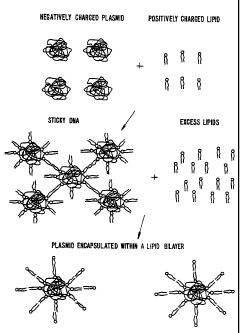
of formation, Figure 3 depicts a detergent dialysis approach to the formation
of the
plasmid-lipid particles. With reference to Figure 3, a plasmid or other large
nucleic acid
is contacted with a detergent solution of cationic lipids to form a coated
plasmid
complex. These coated plasmids can aggregate and precipitate. However, the
presence
of a detergent reduces this aggregation and allows the coated plasmids to
react with
excess lipids (typically, non-cationic lipids) to form particles in which the
plasmid is
encapsulated in a lipid bilayer. As noted above, these particles differ from
the more
classical liposomes both in size (liposomes being typically 200-400 nm) in
that there is
little or no aqueous medium encapsulated by the particle's lipid bilayer. The
methods
CA 02222328 2010-02-05
24
described below for the formation of plasmid-lipid particles using organic
solvents follow
a similar scheme.
In some embodiments, the particles are formed using detergent dialysis.
Thus, the present invention provides a method for the preparation of serum-
stable
plasmid-lipid particles, comprising:
(a) combining a plasmid with cationic lipids in a detergent solution to form a
coated plasmid-lipid complex;
(b) contacting non-cationic lipids with the coated plasmid-lipid complex to
form a detergent solution comprising a plasmid-lipid complex and
non-cationic lipids; and
(c) dialyzing the detergent solution of step (b) to provide a solution of
serum-
stable plasmid-lipid particles, wherein the plasmid is encapsulated in a
lipid bilayer and the particles are serum-stable and have a size of from
about 50 to about 150 nm.
An initial solution of coated plasmid-lipid complexes is formed by combining
the plasmid
with the cationic lipids in a detergent solution. -
In these embodiments, the detergent solution is preferably an aqueous
solution of a neutral detergent having a critical micelle concentration- of 15-
300 mM,
more preferably 20-50 mM. Examples of suitable detergents include, for
example,
N,N'-((octanoylimino)-bis-(trimethylene))-bis-(D-gluconamide) (BIGCHAP); BRIJ
35;
Deoxy-BIGCHAP; dodecylpoly(ethylene glycol) ether; Tween 20TM; Tween 40TM,
Tween
60TM; Tween 80TM; Tween 85TM; Mega 8; Mega 9; Zwittergent 3-08; Zwittergent
3-10;
Triton X-405TM; hexyl-, heptyl-, octyl- and nonyl-(3-D-glucopyranoside; and
heptylthioglucopyranoside; with octyl (3-D-glucopyranoside and Tween-20TH
being the most
preferred. The concentration of detergent in the detergent solution is
typically about 100
mM to about 2 M, preferably from about 200 mM to about. 1.5 M.
The cationic lipids and plasmid will typically be combined to produce a
charge ratio (+/-) of about 1: 1 to about 20: 1, preferably in a ratio of
about 1: 1 to about
12: 1, and more preferably in a ratio of abo,'t 2:1 to about 6: 1.
Additionally, the overall
concentration of plasmid in solution will typically be from about 25 g/mL to
about I
mg/mL, preferably from about 25 g/mL to about 200 gg/mL, and more preferably
from
about 50 g/mL to about 100 g/mL. The combination of plasmids and cationic
lipids in
detergent solution is kept, typically at room temperature, for a period of
time which is
sufficient for the coated complexes to form. Alternatively, the plasmids and
cationic
CA 02222328 2010-02-05
lipids can be combined in the detergent solution and warmed to temperatures of
up to
about 37 C. For plasmids which are particularly sensitive to temperature, the
coated
complexes can be formed at lower temperatures, typically down to about 4 C.
The detergent solution of the coated plasmid-lipid complexes is then
5 contacted with non-cationic lipids to provide a detergent solution of
plasmid-lipid
complexes and non-cationic lipids. The non-cationic lipids which are useful in
this step
include, diacylphosphatidylcholine, diacylphosphatidylethanolamine, ceramide,
sphingomyelin, cephalin, cardiolipin, and cerebrosides. In preferred
embodiments, the
non-cationic lipids are diacylphosphatidylcholine,
diacylphosphatidylethanolamine,
10 ceramide or sphingomyelin. The acyl groups in these lipids are preferably
acyl groups
derived from fatty acids having C,,; C24 carbon chains. More preferably the
acyl groups
are lauroyl, myristoyl, palmitoyl, stearoyl or oleoyl. In particularly
preferred
embodiments, the non-cationic lipid will be 1,2-.sn-
dioleoylphosphatidylethanolamine
(DOPE), palmitoyl oleoyl phosphatidylcholine (POPC)'or egg phosphatidylcholine
15 (EPC). In .the most preferred embodiments, the plasmid-lipid particles will
be fusogenic
particles with enhanced properties in vivo and the non-cationic lipid will be
DOPE. In
other preferred embodiments, the non-cationic lipids will further comprise
polyethylene
glycol-based polymers such as PEG 2000, PEG 5000 and polyethylene glycol
conjugated
to ceramides, as described in U.S. Patent No. 5,820,873.
The amount of non-cationic lipid which is used in the present methods is
typically about 2 to about 20 mg of total lipids to 50 g of plasmid.
Preferably the
amount of total lipid is from about 5 to about 10 Ong per 50 g of plasmid.
Following formation of the detergent solution of plasmid-lipid complexes
and non-cationic lipids, the detergent is removed, preferably by dialysis. The
removal of
the detergent results in the formation of a lipid-bilayer which surrounds the
plasmid
providing serum-stable plasmid-lipid particles which have a size of from about
50 nm to
about 150 nm. The particles thus formed do not aggregate and are optionally
sized to
achieve a uniform particle size.
The serumstable plasmid-lipid particles can be sized by any of the
methods available for sizing liposomes. The sizing may be conducted in order
to achieve
a desired size range and relatively narrow distribution of particle sizes.
Several techniques are available for sizing the particles to a desired size.
One sizing method, used for liposomes and equally applicable to the present
particles is
CA 02222328 1997-11-25
WO 96/40964 PCTIUS96/09949
26
described in U.S. Pat. No. 4,737,323, incorporated herein by reference.
Sonicating a
particle suspension either by bath or probe sonication produces a progressive
size
reduction down to particles of less than about 50 nm in size. Homogenization
is another
method which relies on shearing energy to fragment larger particles into
smaller ones.
In a typical homogenization procedure, particles are recirculated through a
standard
emulsion homogenizer until selected particle sizes, typically between about 60
and 80
nm, are observed. In both methods, the particle size distribution can be
monitored by
conventional laser-beam particle size discrimination, or QELS.
Extrusion of the particles through a small-pore polycarbonate membrane or
an asymmetric ceramic membrane is also an effective method for reducing
particle sizes
to a relatively well-defined size distribution. Typically, the suspension is
cycled through
the membrane one or more times until the desired particle size distribution is
achieved.
The particles may be extruded through successively smaller-pore membranes, to
achieve
a gradual reduction in size.
In another group of embodiments, the present invention provides a method
for the preparation of serum-stable plasmid-lipid particles, comprising;
(a) preparing a mixture comprising cationic lipids and non-cationic lipids in
an
organic solvent;
(b) contacting an aqueous solution of nucleic acid with said mixture in step
(a)
to provide a clear single phase; and
(c) removing said organic solvent to provide a suspension of plasmid-lipid
particles, wherein said plasmid is encapsulated in a lipid bilayer, and said
particles are stable in senim and have a size of from about 50 to about 150
nm.
The plasmids (or nucleic acids), cationic lipids and non-cationic lipids
which are useful in this group of embodiments are as described for the
detergent dialysis
methods above.
The selection of an organic solvent will typically involve consideration of
solvent polarity and the ease with which the solvent can be removed at the
later stages of
particle formation. The organic solvent, which is also used as a solubilizing
agent, is in
an amount sufficient to provide a clear single phase mixture of plasmid and
lipids.
Suitable solvents include chloroform, dichloromethane, diethylether,
cyclohexane,
cyclopentane, benzene, toluene, nw hanol, or other aliphatic alcohols such as
propanol,
isopropanol, butanol, tent-butanol, iso-butanol, pentanol and hexanol.
Combinations of
CA 02222328 2010-02-05
27
two or more solvents may also be used in the present invention.
Contacting the plasmid with the organic solution of cationic and non-
cationic lipids is accomplished by mixing together a first solution of
plasmid, which is
typically an aqueous solution and a second organic solution of the lipids. One
of skill in
the art will understand that this mixing can take place by any number.of
methods, for
example by mechanical means such as by using vortex mixers.
After the plasmid has been contacted with the organic solution of lipids,
the organic solvent is removed, thus forming an aqueous suspension of serum-
stable
plasmid-lipid particles. The methods used to remove the organic solvent will
typically
involve evaporation at reduced pressures or blowing a stream of inert gas
(e.g., nitrogen
or argon) across the mixture.
The serum-stable plasmid-lipid particles thus formed will typically be sized
from about 50 nm to 150 nm. To achieve further size reduction or homogeneity
of size
in the particles, sizing can be conducted as described above.
In other embodiments, the methods will further comprise adding nonlipid
polycations which are useful to effect the transformation of cells using the
present
compositions. Examples of suitable nonlipid polycations include,
hexadimethrine
bromide (sold under the brandname POLYBRENE , from Aldrich Chemical Co.,
Milwaukee, Wisconsin, USA) or other salts of heaxadimethrine. Other suitable
polycations include, for example, salts of poly-L-ornithine, poly-L-arginine,
poly-L-
lysine, poly-D-lysine, polyallylamine and polyethylene] mine.
In other embodiments, the polyoxyethylene conjugates which are used in
the plasmid-lipid particles of the present invention can he prepared by
combining the
conjugating group (i.e. phosphatidic acid or phosphatidylethanolamine) with an
appropriately functionalized polyoxyethylene derivative. For example,
phosphatidylethanolamine can be combined with polyoxyethylene bis(p-
toluenesulfonate)
to provide a phosphatidylethanolamine-polyoxyethylene conjugate. See, Woodle,
et at.,
Biochim. Biophys. Acta 1105:193-200 (1992),
In certain embodiments, the formation of the lipid-nucleic acid complexes
can be carried out either in a monophase system (e.g., a Bligh and Dyer
monophase or
similar mixture of aqueous and organic solvents) or in a two phase system with
suitable
mixing.
When formation of the complexes is carried out in a monophase system,
the cationic lipids and nucleic acids are each dissolved in a volume of the
monophase
CA 02222328 1997-11-25
WO 96/40964 PCT/US96/09949
28
mixture. Combination of the two solutions provides a single mixture in which
the
complexes form. Alternatively, the complexes can form in two-phase mixtures in
which
the cationic lipids bind to the nucleic acid (which is present in the aqueous
phase), and
"pull" it in to the organic phase.
Without intending to be bound by any particular theory of formation,
Figure 1 provides a model for the binding of monocationic lipids to DNA which
results
in the formation of a hydrophobic (organic-soluble) lipid-nucleic acid
complex. In this
figure, cationic lipids first bind to the DNA to form a complex in which the
DNA is
uncondensed. This complex is soluble in the organic phase or in a monophase
and the
DNA remains uncondensed. Upon the addition of other lipids and removal of
solvent,
and hydration, the complexes form particles (described in more detail below).
In another embodiment, the present invention provides a method for the
preparation of lipid-nucleic acid particles, comprising:
(a) contacting nucleic acids with a solution comprising non-cationic lipids
and
a detergent to form a nucleic acid-lipid mixture;
(b) contacting cationic lipids with the nucleic acid-lipid mixture to
neutralize a
portion of the negative charge of the nucleic acids and form a charge-
neutralized mixture
of nucleic acids and lipids; and
(c) removing the detergent from the charge-neutralized mixture to provide the
lipid-nucleic acid particles in which the nucleic acids are protected from
degradation.
Without intending to be limited by any particular aspect of the illustration,
Figure 30 provides a depiction of one method of forming the particles using
detergent
dialysis. In this figure, DNA in an aqueous detergent solution (OGP) is
combined with
non-cationic lipids. (ESM) in an aqueous detergent solution and allowed to
anneal for
about 30 min. A previously sonicated mixture of cationic lipid (DODAC) in
detergent is
added and the resulting mixture is dialyzed for 3 days to remove detergent and
thereby
form lipid-nucleic acid particles. One of skill in the art will understand
that for the
kinetic formation of such particles, the order of addition of cationic lipids
and
non-cationic lipids could be reversed, or the lipids could be added
simultaneously. In
addition, it is possible to cover the nucleic acid with multivalent cations,
such that it now
hinds anions.
In one group of embodiments, the solution of non-cationic lipids and
detergent is an aqueous solution. Contacting the nucleic acids with the
solution of non-
cationic lipids and detergent is typically accomplished by mixing together a
first solution
CA 02222328 2010-02-05
29
of nucleic acids and a second solution of the lipids and detergent. One of
skill in the art
will understand that this mixing can take place by any number of methods, for
example
by mechanical means such as by using vortex mixers. Preferably, the nucleic
acid
solution is also a detergent solution. The amount of non-cationic lipid which
is used in
the present method is typically determined based on the amount of cationic
lipid used,
and is typically of from about 0.2 to 5 times the amount of cationic lipid,
preferably
about 0.5 to 2 times the amount of cationic lipid used.
The nucleic acid-lipid mixture thus formed is contacted with cation-ic lipids
to neutralize a portion of the negative charge which is associated with the
nucleic acids
(or other polyanionic materials) present. The amount of cationic lipids used
will
typically be sufficient to neutralize at least 50% of the negative charge of
the nucleic
acid. Preferably, the negative charge will be at least 70% neutralized, more
preferably
at least 90% neutralized. Cationic lipids which are useful in the present
invention,
include, for example, DODAC, DOTMA, DDAB, DOTAP, DC-Chol and DMRIE.
These lipids and related analogs have been described in
U.S. Patent Nos. 5,208,036, 5,264,618, 5,279,833, 5,283,185, and 5,753,613.
Additionally, a number of commercial
preparations of cationic lipids are available and can be used in the present
invention.
These include, for example, LIPOFECTIN (commercially available cationic
liposomes
comprising DOTMA and DOPE, from GIBCO/BRL, Grand Island, New York, USA);
LIPOFECTAMINE (commercially available cationic liposomes comprising DOSPA and
DOPE, from GIBCO/BRL); and TRANSFECTAM (commercially available cationic
lipids comprising DOGS in ethanol from Promega Corp., Madison, Wisconsin,
USA).
Contacting the cationic lipids with the nucleic acid-lipid mixture can be
accomplished by any of a number of techniques, preferably by mixing together a
solution
of the cationic lipid and a solution containing the nucleic acid-lipid
mixture. Upon
mixing the two solutions (or contacting in any other manner), a portion of the
negative
charge associated with the nucleic acid is neutralized. Nevertheless, the
nucleic acid
remains in an uncondensed state and acquires hydrophilic characteristics.
After the cationic lipids have been contacted with the nucleic acid-lipid
mixture, the detergent (or combination of detergent and organic solvent) is
removed, thus
forming the lipid-nucleic acid particles. The methods used to remove the
detergent will
typically involve dialysis. When organic solvents are present, removal is
typically
accomplished by evaporation at reduced pressures or by blowing a stream of
inert gas
CA 02222328 1997-11-25
WO 96/40964 PCT/US96/09949
(e. g., nitrogen or argon) across the mixture.
The particles thus formed will typically be sized from about 100 nm to
several microns. To achieve further size reduction or homogeneity of size in
the
particles, the lipid-nucleic acid particles can be sonicated, filtered or
subjected to other
5 sizing techniques which are used in liposomal formulations and are known to
those of
skill in the art.
In other embodiments, the methods will further comprise adding nonlipid
polycations which are useful to effect the lipofection of cells using the
present
compositions. Examples of suitable nonlipid polycations include,
hexadimethrine
10 bromide (sold under the brandname POLYBRENE , from Aldrich Chemical Co.,
Milwaukee, Wisconsin, USA) or other salts of hexadimethrine. Other suitable
polycations include, for example, salts of poly-L-ornithine, poly-L-arginine,
poly-L-
lysine, poly-D-lysine, polyallylamine and polyethyleneimine. Addition of these
salts is
preferably after the particles have been formed.
15 In another aspect, the present invention provides methods for the
preparation of lipid-nucleic acid particles, comprising:
(a) contacting an amount of cationic lipids with nucleic acids in a solution;
the
solution comprising of from about 15-35% water and about 65-85% organic
solvent and
the amount of cationic lipids being sufficient to produce a +/- charge ratio
of from about
20 0.85 to about 2.0, to provide a hydrophobic, charge-neutralized lipid-
nucleic acid
complex;
(b) contacting the hydrophobic, charge-neutralized lipid-nucleic acid complex
in solution with non-cationic lipids, to provide a lipid-nucleic acid mixture;
and
(c) removing the organic solvents from the lipid-nucleic acid mixture to
25 provide lipid-nucleic acid particles in which the nucleic acids are
protected from
degradation.
The nucleic acids, non-cationic lipids, cationic lipids and organic solvents
which are useful in this aspect of the invention are the same as those
described for the
methods above which used detergents. In one group of embodiments, the solution
of
30 step (a) is a monophase. In another group of embodiments, the solution of
step (a) is
two-phase.
In preferred embodiments, the cationic lipids are DODAC, DDAB,
DOTMA, DOSPA, DMRIE, DOGS or combinations thereof. In other preferred
embodiments, the non-cationic lipids are ESM, DOPE, polyethylene glycol-based
CA 02222328 1997-11-25
WO 96/40964 PCT/US96/09949
31
polymers (e.g., PEG 2000, PEG 5000, PEG-modified phospholipids or PEG-modified
ceramides) or combinations thereof. In still other preferred embodiments, the
organic
solvents are methanol, chloroform, methylene chloride, ethanol, diethyl ether
or
combinations thereof.
In a particularly preferred embodiment, the nucleic acid is a plasmid; the
cationic lipid is DODAC, DDAB, DOTMA, DOSPA, DMRIE, DOGS or combinations
thereof; the non-cationic lipid is ESM, DOPE, polyethylene glycol-based
polymers or
combinations thereof; and the organic solvent is methanol, chloroform,
methylene
chloride, ethanol, diethyl ether or combinations thereof.
As above, contacting the nucleic acids with the cationic lipids is typically
accomplished by mixing together a first solution of nucleic acids and a second
solution of
the lipids, preferably by mechanical means such as by using vortex mixers. The
resulting mixture contains complexes as described for one aspect of the
invention above.
These complexes are then converted to particles by the addition of non-
cationic lipids and
the removal of the organic solvent. The addition of the non-cationic lipids is
typically
accomplished by simply adding a solution of the non-cationic lipids to the
mixture
containing the complexes. A reverse addition can also be used. Subsequent
removal of
organic solvents can he accomplished by methods known to those of skill in the
art and
also described above.
The amount of non-cationic lipids which is used in this aspect of the
invention is typically an amount of from about 0.2 to 5 times the amount (on a
mole
basis) of cationic lipids which was used to provide the charge-neutralized
lipid-nucleic
acid complex. Preferably, the amount is from 0.5 to 2 times the amount of
cationic
lipids used.
In yet another aspect, the present invention provides lipid-nucleic acid
particles which are prepared by the methods described above. In these
embodiments, the
lipid-nucleic acid particles are either net charge neutral or carry an overall
charge which
provides the particles with greater gene lipofection activity. Preferably, the
nucleic acid
component of the particles is a nucleic acid which encodes a desired protein
or blocks the
production of an undesired protein. In particularly preferred embodiments, the
nucleic
acid is a plasmid, the non-cationic lipid is egg sphingomyelin and the
catio:ic lipid is
DODAC.
As noted above, the lipid-nucleic acid particles are useful for the
lipofection of cells, either in vitro or in vivo. Accordingly, the present
invention
CA 02222328 1997-11-25
WO 96/40964 PCT/US96/09949
32
provides, in yet another aspect, a method for introducing a nucleic acid into
a cell,
comprising;
(a) preparing a lipid-nucleic acid particle according to the methods above;
and
(b) contacting the cell with the lipid-nucleic acid particle for a period of
time
sufficient to introduce the nucleic acid into the cell.
Although discussed in more detail below, preferred embodiments are those
in which the lipid-nucleic acid particle comprises a plasmid, DODAC and ESM.
Unlike viral-based gene therapy vectors which can only incorporate a
relatively small non-viral nucleic acid sequence into the viral genome because
of size
limitations for packaging virion particles, the lipid-nucleic acid complexes
of the prtesent
invention may be used to transfer large (e.g., 50-5,000 kilobase) exogenous
nucleic acids
into cells. This aspect of lipofection is particularly advantageous since many
genes
which may be targets for gene therapy span over 100 kilobases (e.g., amyloid
precursor
protein (APP) gene, Huntington's chorea gene) and large homologous targeting
constructs or transgenes may be required for therapy.
Cells can be lipofected with an exogenous nucleic acid at high efficiency
and with cell type specificity by contacting the cells with a receptor-
recognition
transfection complex comprising: (1) an exogenous nucleic acid, (2) a receptor-
ligand
protein ("rip") which is covalently linked to a polycation, and (3) a cationic
or neutral
lipid. It has been found that a combination of a polycation-linked receptor-
recognition
protein and a suitable cationic (or neutral) lipid can be used to transfect
nucleic acids,
and that the combination retains cell type targeting specificity conferred by
the receptor-
recognition protein and also exhibits high efficiency transfection conferred,
in part, by
the inclusion of a cationic lipid, neutral lipid, or lipopolyamine.
The exogenous nucleic acid is typically dsDNA, ssDNA, ssRNA, dsRNA;
most typically the exogenous nuclei.' acid is dsDNA such as a cloned DNA
sequence in a
cloning vector such as a plasmid or viral genome. Multiple species of
exogenous nucleic
acid may be combined in a transfection complex, such as for co-transfection of
unlinked
nucleic acid sequences or to accomplish in vivo homologous recombination
shuffling.
Frequently, the exogenous nucleic acid(s) are not capable of autonomous
replication in
cells which incorporate the transfection complex, and are either transiently
expressed or
are stably integrated into a host cell chromosome by homologous recombination
or
nonhomologous integration. Often at least one selectable marker (e.g., a neon
expression
cassette) is included in the exogenous nucleic acid(s) to facilitate selection
of cells which
CA 02222328 1997-11-25
WO 96/40964 PCT/US96/09949
33
have incorporated the exogenous nucleic acid(s). Typically, an exogenous
nucleic acid
comprises a structural gene encoding a polypepticle to be expressed in a
target cell which
has incorporated the exogenous nucleic acid, and the structural gene usually
is operably
linked to appropriate cis-acting regulatory elements (e.g., promoter,
enhancer,
polyadenylation site). Although gene therapy may be performed in a variety of
ways, a
typical receptor-recognition lipofection complex comprises a nucleic acid
which
comprises at least one transcriptional unit.
The lipid nucleic acid particles of the invention can be designed to contain,
in addition to the species of nucleic acid, a receptor-recognition molecule
(rim), such as
a protein. The rim can be covalently bound to lipids that comprise the nucleic
acid-lipid
particle. Its presence on the particle increases the efficiency aand
specificity with the
particle contacts and enters target cells. For example, a suitable rim is a
non-
immunoglobulin protein that binds to a cell surface receptor of a target cell
which
mediates internalization of a transfection complex comprising the rlm-
polycation
conjugate by, for example, the process of endocytosis and/or membrane fusion.
Additional suitable rim species typically are naturally-occurring
physiological ligands
which comprise a polypeptide portion (e.g., adhesion molecules such as ICAM-
1, ICAM-
2, ELAM-1, VCAM-1). Viral proteins (e.g., spike glycoproteins) which bind to
viral
receptors on eukaryotic cells and mediate virus internalization may also be
used as rim
species for forming rim-polycation conjugates. Examples also include viral
glycoproteins
which attach to cell surface receptors and lead to internalization and/or
membrane fusion
include the gB, gC, gD, gE, gH, and gl virion glycoproteins of HSV-I, and
gp120 of
HIV- 1.
Fragments and analogs of naturally-occurring proteins may be used as well
as full-length mature proteins as rim species in forming transfection
complexes of the
invention. For example, fragments, analogs, and fusion proteins comprising a
portion of
an adhesion molecule or virion attachment protein which mediates attachment to
a target
cell may be used as rim species without other portions of the naturally-
occurring full-
length protein that are not essential for cell attachment and/or membrane
fusion. Thus,
for example, a cytoplasmic tail peptide portion of a virion glycoprotein
usually may be
omitted and the resultant protein may still serve as a suitable rim..
The rim selected will vary with the particular target cell type. For specific
targeting to hepatocytes, asialoglycoproteins (galactose-terminal) are
preferred as rim
species. Examples of asialoglycoproteins include asialoorosomucoid,
asialofetuin, and
CA 02222328 1997-11-25
WO 96/40964 PCT/US96/09949
34
desialylated vesicular stomatitis virus virion proteins. These can be formed
by chemical
or enzymatic desialylation of those glycoproteins that possess terminal sialic
acid and
penultimate galactose residues. Alternatively, rim species suitable for
forming
lipofection complexes that selectively target hepatocytes may be created by
coupling
lactose or other galactose-terminal carbohydrates (e.g., arabinogalactan) to
non-galactose-
bearing proteins by reductive lactosamination. Other useful galactose-terminal
carbohydrates for hepatocyte targeting include carbohydrate trees obtained
from natural
glycoproteins, especially tri- and tetra-antennary structures that contain
either terminal
galactose residues or that can be enzymatically treated to expose terminal
galactose
residues. For targeting macrophages, endothelial cells, or lymphocytes, rim
species
comprising mannose or mannose-6-phosphate, or complex carbohydrates comprising
these terminal carbohydrate structures may be used.
Since a variety of different cell surface receptors exist on the surfaces of
mammalian cells, cell-specific targeting of nucleic acids to nonhepatic cells
can involve
lipofection complexes that comprise various rim species. For example,
transferrin can be
used as a suitable rim for forming receptor-recognition transfection complexes
to cells
expressing transferrin receptors. Other receptor ligands such as polypeptide
hormones
(e.g., growth hormone, PDGF, FGF, EGF, insulin, IL-2, IL-4, etc.) may be used
to
localize receptor-recognition transfection complexes to cells expressing the
cognate
receptor.
The nucleic acid-lipid particles may comprise multiple rim species.
Frequently, an agent having membrane fusion activity (e. g., influenza virus
hemagglutinin, HSV- l gB and gD) is used as an rim for forming rlm-polycation
complexes, either alone or in combination with other rim species, typically
with those
which lack membrane fusion activity.
These transfection methods generally comprise the steps of: (1) forming a
nucleic acid-lipid-rlmm particle consisting essentially of an exogenous
nucleic acid, a
polycation conjugate consisting essentially of a polycation linked to a non-
immunoglobulin receptor-recognition molecule that binds to a predetermined
cell surface
receptor, and a lipid component consisting essentially of a neutral or
cationic lipid
(optionally including a quaternary ammonium detergent and/or a lipopolvamine),
and (2)
contacting cells expressing the predetermined cell surface receptor with a
composition
comprising the receptor-recognition transfection complex under physiological
transfection
conditions which permit uptake of the exogenous nucleic acid into said cells.
In
CA 02222328 1997-11-25
WO 96/40964 PCT/US96/09949
alternative embodiments, the rim is attached to the polycation by covalent
linkage,
frequently by covalent linkage through a crosslinking agent or by peptide
linkage.
Overall particle charge is an important property of the particles, as it may
affect particle clearance from the blood. Particles with prolonged circulation
half-lives
5 are typically desirable for therapeutic and diagnostic uses. For instance,
particles which
can be maintained from 8, 12, or up to 24 hours in the bloodstream are
particularly
preferred. Negatively charged liposomes and particles are typically, taken up
more
rapidly by the reticuloendothelial system (Juliano, Biochem. Biophys. Res.
Common.
63:651 (1975)) and thus have shorter half-lives in the bloodstream.
CA 02222328 1997-11-25
WO 96/40964 PCT/US96/09949
36
C. Pharmaceutical Preparations
The nucleic acid-lipid particles of the present invention can be
administered either alone or in mixture with a physiologically-acceptable
carrier (such as
physiological saline or phosphate buffer) selected in accordance with the
route of
administration and standard pharmaceutical practice. Generally, normal saline
will be
employed as the pharmaceutically acceptable carrier. Other suitable carriers
include,
e.g., water, buffered water, 0.4% saline, 0.3% glycine, and the like,
including
glycoproteins for enhanced stability, such as albumin, lipoprotein, globulin,
etc.
The pharmaceutical carrier is generally added following particle formation.
Thus, after the particle is formed, the particle can be diluted into
pharmaceutically
acceptable carriers such as normal saline.
The concentration of particles in the pharmaceutical formulations can vary
widely, i.e., from less than about 0.05%, usually at or at least about 2-5% to
as much as
10 to 30% by weight and will be selected primarily by fluid volumes,
viscosities, etc., in
accordance with the particular mode of administration selected. For example,
the
concentration may be increased to lower the fluid load associated with
treatment. This
may be particularly desirable in patients having atherosclerosis-associated
congestive
heart failure or severe hypertension. Alternatively, particles composed of
irritating lipids
may be diluted to low concentrations to lessen inflammation at the site of
administration.
It is often desirable to include polyethylene glycol (PEG), PEG-ceramide, or
modified
(e.g., ganglioside.GM,-modified) lipids to the particles. Addition of such
components
prevents particle aggregation and provides a means for increasing circulation
lifetime and
increasing the delivery of the lipid-nucleic acid particles to the target
tissues. Typically,
the concentration of the PEG, PEG-ceramide or GM,-modified lipids in the
particle will
be about 1-15%.
The pharmaceutical compositions may be sterilized by conventional, well
known sterilization techniques. Aqueous solutions can be packaged for use or
filtered
under aseptic conditions and lyophilized, the lyophilized preparation being
combined with
a sterile aqueous solution prior to administration. The compositions can
contain
pharmaceutically acceptable auxiliary substances as required to approximate
physiological
conditions, such as pH adjusting and buffering agents, tonicity adjusting
agents and the
like, for example, sodium acetate, sodium lactate, sodium chloride, potassium
chloride,
and calcium chloride. Additionally, the particle suspension may include lipid-
protective
CA 02222328 1997-11-25
WO 96/40964 PCT/US96/09949
37
agents which protect lipids against free-radical and lipid-peroxidative
damages on
storage. Lipophilic free-radical quenchers, such as alphatocopherol and water-
soluble
iron-specific chelators, such as ferrioxamine, are suitable.
In another example of their use, lipid-nucleic acid particles can be
incorporated into a broad range of topical dosage forms including but not
limited to gels,
oils, emulsions and the like. For instance, the suspension containing the
nucleic acid-
lipid particles can be formulated and administered as topical creams, pastes,
ointments,
gels, lotions and the like.
The present invention also provides lipid-nucleic acid particles in kit form.
The kit will typically be comprised of a container which is compartmentalized
for
holding the various elements of the kit. The kit will contain the compositions
of the
present inventions, preferably in dehydrated form, with instructions for their
rehydration
and administration. In still other embodiments, the particles and/or
compositions
comprising the particles will have a targeting moiety attached to the surface
of the
particle. Methods of attaching targeting moieties (e.g., antibodies, proteins)
to lipids
(such as those used in the present particles) are known to those of skill in
the art.
D. Administration of Lipid-Nucleic Acid Particle Formulations
The serum-stable nuch is acid-lipid particles of the present invention are
useful for the introduction of nucleic acids into cells. Accordingly, the
present invention
also provides methods for introducing a nucleic acids (e.g., a plasmid) into a
cell. The
methods are carried out in vitro or in vivo by first forming the particles as
described
above, then contacting the particles with the cells for a period of time
sufficient for
transfection to occur.
The nucleic acid-lipid particles of the present invention can be adsorbed to
almost any cell type with which they are mixed or contacted. Once adsorbed,
the
particles can either be endocytosed by a portion of the cells, exchange lipids
with cell
membranes, or fuse with the cells. Transfer or incorporation of the nucleic
acid portion
of the particle can take place via any one of these pathways. In particular,
when fusion
takes place, the particle membrane is integrated into the cell membrane and
the contents
of the particle combine with the intracellular fluid.
CA 02222328 2010-02-05
38
1. In vitro gene transfer
For in vitro applications, the delivery of nucleic acids can be to any cell
grown in culture, whether of plant or animal origin, vertebrate or
invertebrate, and of
any tissue or type. In preferred embodiments, the cells will be animal cells,
more
preferably mammalian cells, and most preferably human cells.
Contact between the cells and the lipid-nucleic acid particles, when carried
out in vitro, takes place in a biologically compatible medium. The
concentration of
particles varies widely depending-on the particular application, but is
generally between
about I mol and about 10 mmol. Treatment of the cells with the nucleic acid-
lipid
particles is generally carried out at physiological temperatures (about 37 C)
for periods
of time of from about 1 to 48 hours, preferably of from about 2 to 4 hours.
In one group of preferred embodiments, a lipid-nucleic acid particle
suspension is added to 60-80% confluent plated cells having-a cell density of
from about
103 to about 105 cells/mL, more preferably about 2 X 10' cells/mL. The
concentration
of the suspension added to the cells is preferably of from about 0.01 to 0.2
ug/mL, more
preferably about 0. 1 g/mL.
2. In vivo gene transfer
Alternatively, the compositions of the present invention can also be used
for the in vivo gene transfer, using methods, which are known to those of
skill in the art.
In particular, Zhu, et at., Science 261:209-211 (1993), incorporated herein by
reference,
describes the intravenous delivery of cytomegalovirus (CMV)-chloramphenicol
acetyltransferase (CAT) expression plasmid using DOTMA-DOPE complexes. Hyde,
et
al., Nature 362:250-256 (1993), incorporated herein by reference, describes
the delivery
of the cystic fibrosis transmembrane conductance regulator (CFTR) gene to
epithelia of
the. airway and to alveoli in the lung of mice, using liposomes. Brigham, et
al., Am. J.
Med. Sci. 298:278-281 (1989), describes the in vivo
transfection of lungs of mice with a functioning prokaryotic gene encoding the
intracellular enzyme chloramphenicol acetyltransferase (CAT).
For in vivo administration, the pharmaceutical compositions are preferably
administered parenterally, i.e., intraarticularly, intravenously,
intraperitoneally,
subcutaneously, or intramuscularly. More preferably, the pharmaceutical
compositions
are administered intravenously or intraperitoneally by a bolus injection. For
example,
see Stadler, et al., U.S. Patent No. 5,286,634.
CA 02222328 2010-02-05
39
Intracellular nucleic acid delivery has also been discussed in Straubringer,
et
al., METHODS IN ENZYMOLOGY, Academic Press, New York. 101:512-527 (1983);
Mannino, et al., Biotechniques 6:682-690 (1988); Nicolau, ct al., Crit. Rev.
Ther. Drug
Carrier Syst. 6:239-271 (1989), and Behr, Acc. Chem. Res. 26:274-278 (1993).
Still
other methods of administering lipid-based therapeutics are described in, for
example,
Rahman et al., U.S. Patent No. 3,993,754; Sears, U.S. Patent No. 4,145,410;
Papahadjopoulos et al., U.S. Patent No. 4,235,871; Schneider, U.S. Patent No.
4,224,179; Lenk et al., U.S. Patent No. 4,522,803; and Fountain et al., U.S.
Patent No.
4,588,578.
In certain embodiments, the pharmaceutical preparations may be contacted
with the target tissue by direct application of the preparation to the tissue.
The
application may be made by topical, "open" or "closed" procedures. By
"topical", it is
meant the direct application of the pharmaceutical preparation to a tissue
exposed to the
environment, such as the skin, oropharynx, external auditory canal, and the
like.
"Open" procedures are those procedures which include incising the skin of a
patient and
directly visualizing the underlying tissue to which the pharmaceutical
preparations are
applied. This is generally accomplished by a surgical procedure, such as a
thoracotomy
to access the lungs, abdominal laparotomy to access abdominal viscera, or
other direct
surgical approach to the target tissue. "Closed" procedures are, invasive
procedures in
which the internal target tissues are not directly visualized, but accessed
via inserting
instruments through small wounds in the skin. For example, the preparations
may be
administered to the peritoneum by needle lavage. Likewise, the pharmaceutical
preparations may be administered to the meninges or spinal cord by infusion
during a
lumbar puncture followed by appropriate positioning of the patient as commonly
practiced for spinal anesthesia or metrazamide imaging of the spinal cord.
Alternatively,
the preparations may be administered through endoscopic devices.
The lipid-nucleic acid particles can also be administered in an aerosol
inhaled into the lungs (see, Brigham, et al., Am. J. Sci. 298(4):278-281
(1989)) or by
direct injection at the site of disease (Culver, HUMAN GENE. THERAPY, MaryAnn
Liebert, Inc., Publishers, New York. pp.70-71 (1994)).
The methods of the present invention may be practiced in a variety of
hosts. Preferred hosts include mammalian species, such as humans, non-human
primates, dogs, cats, cattle, horses, sheep, and the like.
The amount of particles administered will depend upon the the ratio of
CA 02222328 1997-11-25
WO 96/40964 PCT/US96/09949
nucleic acid to lipid; the particular nucleic acid used, the disease state
being diagnosed;
the age, weight, and condition of the patient and the judgement of the
clinician; but will
generally be between about 0.01 and about 50 mg per kilogram of body weight;
preferably between about 0.1 and about 5 mg/kg of body weight or about 108-101
5 particles per injection.
3. Insertion of Functional Copy of a Gene
Some methods of gene therapy serve to compensate for a defect in an
endogenous gene by integrating a functional copy of the gene into the host
chromosome.
The inserted gene replicates with the host DNA and is expressed at a level to
compensate
10 for the defective gene. Diseases amendable to treatment by this approach
are often
characterized by recessive mutations. That is, both copies of an endogenous
gene must
be defective for symptoms to appear. Such diseases include cystic fibrosis,
sickle cell
anemia, /3-thalassemia, phenylketonuria, galactosemia, Wilson's disease,
hemochromatosis, severe combined immunodeficiency disease, alpha- l-anti
trypsin
15 deficiency, albinism, alkaptonuria, lysosomal storage diseases, Ehlers-
Danlos syndrome,
hemophilia, glucose-6-phosphate dehydrogenase deficiency, agammaglobulimenia,
diabetes insipidus, Lesch-Nyhan syndrome, muscular dystrophy, Wiskott-Aldrich
syndrome, Fabry's disease, fragile X-syndrome, and the like. Other recessive
mutations
are known in the art, and the use of the methods of the present invention to
treat them is
20 contemplated herein.
There are several methods for introducing an exogenous functional gene to
compensate for the above genetic defects. In one approach, cells are removed
from a
patient suffering from the disease and contacted with a lipid-vector complex
in vitro.
Cells should be removed from a tissue type in which disease symptoms are
manifested.
25 If the cells are capable of replication, and the vector used includes a
selective marker,
cells having internalized and expressed the marker can be selected.
Particularly if
selection is not performed, it is important that the frequency of gene
transfer into cells be
high, for example, at least about 1, 5, 10, 25 or 50% of cells.
After integration of the vector into the cellular genome, and optionally,
30 selection, cells are reintroduced into the patient. In this application,
and others discussed
below (except site-specific recombination to correct dominant mutations), it
is not
necessary that the gene supplied by the lipid-nucleic acid particle be
delivered to the
same site as is occupied by the defective gene for which it is compensating.
CA 02222328 1997-11-25
WO 96/40964 PCT/US96/09949
41
Alternatively, the lipid-vector complex can be introduced directly into a
patient as a pharmaceutical composition. The complex is delivered to the
tissue(s)
affected by the genetic disorder being treated in a therapeutically effective
dose. In this
and other methods, a therapeutically effective dose is an amount sufficient to
cure, or at
least partially arrest, the symptoms of the disease and its complications.
Effective doses
of the compositions of the present invention, for the treatment of the above
described
conditions will vary depending upon many different factors, including means of
administration, target site, physiological state of the patient, and other
medicants
administered. Thus, treatment dosages will need to be titrated to optimize
safety and
efficacy. Doses ranging from about 10 ng to l g, 100 ng to 100 mg, I pg to 10
mg, or
30-300 g DNA per patient are typical. Routes of administration include oral,
nasal,
gastric, intravenous, intradermal and intramuscular.
The nucleic acid-lipid complexes can also be used to transfect embryonic
stem cells or zygotes to achieve germline alterations. See Jaenisch, Science,
240,
1468-1474 (1988); Gordon et al. (1984) Methods Enzymol. 101, 414; Hogan et
al.,
Manipulation of the Mouse Embryo: A Laboratory Manual, C.S.H.L. N.Y. (1986);
and
Hammer et al. (1985) Nature 315, 680; Gandolfi et al. (1987) J. Reprod. Fert.
81,
23-28; Rexroad et al. (1988) J. Anim. Sci. 66, 947-953 and Eyestone et al.
(1989) J.
Reprod. Fert. 85, 715-720; Camous et al. (1984) J. Reprod. Fert. 72, 779-785;
Heyman
et al. (1987) Theriogenology 27, 5968. However, these methods are presently
more
suitable for veterinary applications that human treatment due to ethical and
regulatory
constraints in manipulating human embryos.
As an example, cystic fibrosis (CF) is a usually fatal recessive genetic
disease, having a high incidence in Caucasian populations. The gene
responsible for this
disease was isolated by Riordan et al, Science 245, 1059-1065 (1989). It
encodes a
protein called the cystic fibrosis transmembrane conductance regulator (CFTR)
which is
involved in the transfer of chloride ions (C1-) through epithelial cell
membranes.
Mutations in the gene cause defects of Cl- secretion in epithelial cells
leading to the
various clinical manifestations. Although CF has a number of symptoms
including
thickened exocrine gland secretions, pancreatic deficiency, intestinal
blockage and
malabsorption of fat, the most serious factor affecting mortality is chronic
lung disease.
Accordingly, to treat a CF patient, a vector containing a coding sequence for
a functional
CFTR gene product can be complexed with lipid, and optionally, a
pharmaceutical
excipient and introduced into the patient via nasal administration so that the
vector-lipid
CA 02222328 1997-11-25
WO 96/40964 PCT/US96/09949
42
composition reaches the lungs. The dose of vector-lipid complex is preferably
about 10x-
10)0 particles.
As another example, defects in the a or -y globin genes (see McDonagh &
Nienhuis in Hematology of Infancy and Childhood (eds. Nathan & Oski, Saunders,
PA,
1992) at pp. 783-879) can be compensated for by ex vivo treatment of
hemopoietic stem
cells with an nucleic acid-lipid complex containing a functional copy of the
gene. The
gene integrates into the stem cells which are then reintroduced into the
patient. Defects
in the gene responsible for Fanconi Anemia Complement Group C can be treated
by an
analogous strategy (see Walsh et al., J. Clin. Invest. 94, 1440-1448 (1994)).
Other applications include the introduction of a functional copy of a tumor
suppressor gene into cancerous cell or cells at risk of becoming cancerous.
Individuals
having defects in one or both copies of an endogenous tumor suppressor gene
are
particularly at risk of developing cancers. For example, Li-Fraumeni syndrome
is a
hereditary condition in which individuals receive mutant p53 alleles,
resulting in the early
onset of various cancers (Harris, Science 262, 1980-1981 (1993) Frebourg et
al., PNAS
89, 6413-6417 (1992); Malkin et al., Science 250, 1233 (1990)). Expression of
a tumor
suppressor gene in a cancerous cell or a cell at risk of becoming cancerous is
effective to
prevent, arrest and/or reverse cellular proliferation and other manifestations
of the
cancerous state. Suitable tumor suppressor genes for use in the invention
include p53
(Buchman et al., Gene 70, 245-252 (1988)), APC, DCC, Rb, WT1, and NFI (Marx,
Science 260, 751-752 (1993); Marshall, Cell 64, 313-326 (1991)). Lipid-nucleic
acid
complexes bearing a functional copy of a tumor suppressor gene are usually
administered
in vivo by the route most proximal to the intended site of action. For
example, skin
cancers can be treated by topical administration and leukemia by intravenous
administration.
4. Suppression of Gene Expression
Methods of gene therapy using the nucleic acid-lipid complexes of the
invention can also be used for prophylactic or therapeutic treatment of
patients or cells,
infected with or at risk of being infected with, a pathogenic microorganism,
such as
HIV. The effectiveness of antisense molecules in blocking target gene
functions of
impeding virus replication has been demonstrated in a number of different
systems
(Friedman et al., Nature 335, 452-54 (1988), Malim et al., Cell 58, 205-14
(1989) &
Trono at al., Cell 59, 113-20 (1989)). The vector used includes a DNA segment
CA 02222328 1997-11-25
WO 96/40964 PCT/US96/09949
43
encoding an antisense transcript, which is complementary to a segment of the
genome
from the pathogenic microorganism. The segment should preferably play an
essential
role in the lifecycle of the microorganism, and should also be unique to the
microorganism (or at least absent from the genome of the natural genome of a
patient
undergoing therapy). For example, suitable sites for inhibition on the HIV
virus includes
TAR, REV or nef (Chatterjee et al., Science 258, 1485-1488 (1992)). Rev is a
regulatory RNA binding protein that facilitates the export of unspliced HIV
pre mRNA
from the nucleus. Malim et al., Nature 338, 254 (1989). Tat is thought to be a
transcriptional activator that functions by binding a recognition sequence in
5' flanking
mRNA. Karn & Graeble, Trends Genet. 8, 365 (1992). The nucleic acid-lipid
complex
is introduced into leukocytes or hemopoietic stem cells, either ex vivo or by
intravenous
injection in a therapeutically effective dose. The treatment can be
administered
prophylactically to HIV- persons, or to persons already infected with HIV.
Analogous methods are used for suppressing expression of endogenous
recipient cell genes encoding adhesion proteins. Suppression of adhesion
protein
expression in useful in aborting undesirable inflammatory responses. Adhesion
proteins
that can be suppressed by antisense segments present in seelcted vectors
include
integrins, selectins, and immunoglobulin (Ig) superfamily members (see
Springer, Nature
346, 425-433 (1990). Osborn, Cell 62, 3 (1990); Hynes, Cell 69, 11 (1992)).
Integrins
are heterodimeric transmembrane glycoproteins consisting of an a chain (120-
180 kDa)
and a fl chain (90-110 kDa), generally having short cytoplasmic domains. The
three
known integrins, LFA-l, Mac-1 and P150,95, have different alpha subunits,
designated
CD I I a, CD 11 b and CD I I c, and a common beta subunit designated CD 18.
LFA-1
(aJ32) is expressed on lymphocytes, granulocyte and monocytes, and binds
predominantly to an Ig-family member counter-receptor termed ICAM- I (and
perhaps to
a lesser extent ICAM-2). ICAM-1 is expressed on many cells, including
leukocytes and
endothelial cells, and is up-regulated on vascular endothelium by cytokines
such as TNF
and IL-I. Mac-1 (aM02) is distributed on neutrophils and monocytes, and also
binds to
ICAM-1 (and possibly ICAM-2). The third 02 integrin, P150,95 (ax(32), is also
found
on neutrophils and monocytes. The selectins consist of L-selectin, E-selectin
and P-
selectin.
5. Cells to be transformed
The compositions and methods of the present invention are used to treat a
CA 02222328 1997-11-25
WO 96/40964 PCT/US96/09949
44
wide variety of cell types, in vivo and in vitro. Among those most often
targeted for
gene therapy are hematopoietic precursor (stem) cells. Other cells include
those of
which a proportion of the targeted cells are nondividing or slow dividing.
These include,
for example, fibroblasts, keratinocytes, endothelial cells, skeletal and
smooth muscle
cells, osteoblasts, neurons, quiescent lymphocytes, terminally differentiated
cells, slow or
non-cycling primary cells, parenchymal cells, lymphoid cells, epithelial
cells, bone cells,
etc. The methods and compositions can be employed with cells of a wide variety
of
vertebrates, including nmatnmals, and especially those of veterinary
importance, e.g,
canine, feline, equine, bovine, ovine, caprine, rodent, lagomorph, swine,
etc., in
addition to human cell populations.
To the extent that tissue culture of cells may be required, it is well known
in the art. Freshney (1994) (Culture of Animal Cells, a Manual of Basic
Technique,
third edition Wiley-Liss, New York), Kuchler of al. (1977) Biochemical Methods
in Cell
Culture and Virology, Kuchler, R.J., Dowden, Hutchinson and Ross, Inc., and
the
references cited therein provides a general guide to the culture of cells.
Cultured cell
systems often will he in the form of monolayers of cells, although cell
suspensions are
also used.
Gene therapy relies on the efficient delivery of therapeutic genes to target
cells. Most of the somatic cells that have been targeted for gene therapy,
e.g.,
hematopoietic cells, skin fibroblasts and keratinocytes, hepatocytes,
endothelial cells,
muscle cells and lymphocytes, are normally non-dividing. Retroviral vectors,
which are
the most widely used vectors for gene therapy, unfortunately require cell
division for
effective transduction (Miller et al., Mot. Cell. Biol. 10:4239-4242 (1990)).
This is also
true with other gene therapy vectors such as the adeno-associated vectors
(Russell et al.,
Proc. Natl. Acad. Sci. USA 91: 8915-8919 (1994); Alexander et al., J. Virol.
68: 8282-
8287 (1994); Srivastrava, Blood Cells 20: 531-538 (1994)). Recently, HIV-based
vectors
has been reported to transfect non-dividing cells (CITE) Nonetheless, the
majority of
stem cells, a preferred target for many gene therapy treatments, are normally
not
proliferating. Thus, the efficiency of transduction is often relatively low,
and the gene
product may not be expressed in therapeutically or prophylactically effective
amounts.
This has led investigators to develop techniques such as stimulating the stem
cells to
proliferate priot to or during gene transfer (e.g., by treatment with growth
factors)
pretreatment with 5-fluorouracil, infection in the presence of cytokines, and
extending the
vector infection period to increase the likelihood that stem cells are
dividing during
CA 02222328 1997-11-25
WO 96/40964 PCTIUS96/09949
infection, but these have met with limited success.
6. Detection of foreign nucleic acids
After a given cell is transduced with a nucleic acid constrict that encodes
a gene of interest, it is important to detect which cells or cell lines
express the gene
5 product and to assess the level of expression of the gene product in
engineered cells.
This requires the detection of nucleic acids that encode the gene products.
Nucleic acids and proteins are detected and quantified herein by any of a
number of means well known to those of skill in the art. These include
analytic
biochemical methods such as spectrophotometry, radiography, electrophoresis,
capillary
10 electrophoresis, high performance liquid chromatography (HPLC), thin layer
chromatography (TLC), hyperdiffusion chromatography, and the like, and various
immunological methods such as fluid or gel precipitin reactions,
immunodiffusion (single
or double), immunoelectrophoresis, radioimmunoassays (RIAs), enzyme-linked
immunosorbent assays (ELISAs), iminunofluorescent assays, and the like. The
detection
15 of nucleic acids proceeds by well known methods such as Southern analysis,
northern
analysis, gel electrophoresis, PCR, radiolabeling, scintillation counting, and
affinity
chromatography.
The selection of a nucleic acid hybridization format is not critical. A
variety of nucleic acid hybridization formats are known to those skilled in
the art. For
20 example, common formats include sandwich assays and competition or
displacement
assays. Hybridization techniques are generally described in "Nucleic Acid
Hybridization,
A Practical Approach," Ed. Hames, B.D. and Higgins, S.J., IRL Press, 1985.
The sensitivity of the hybridization assays may be enhanced through use of
a nucleic acid amplification system which multiplies the target nucleic acid
being
25 detected. In vitro amplification techniques suitable for amplifying
sequences for use as
molecular probes or for generating nucleic acid fragments for subsequent
subcloning are
known. Examples of techniques sufficient to direct persons of skill through
such in vitro
amplification methods, including the polymerase chain reaction (PCR) the
ligase chain
reaction (LCR), Q(3-replicase amplification and other RNA polymerase mediated
30 techniques (e.g., NASBA) are found in Berger, Sambrook, and Ausubel, as
well as
Mullis et al. (1987), U.S. Patent No. 4,683,202; PCR Protocols A Guide to
Methods and
Applications (Innis et al. eds) Academic Press Inc. San Diego, CA (1990)
(Innis);
CA 02222328 1997-11-25
WO 96/40964 PCT/US96/09949
46
Arnheim & Levinson (October 1, 1990), C&EN 36-47; The Journal Of NIH Research
(1991), 3: 81-94; (Kwoh et al. (1989), Proc. Natl. Acad. Sci. USA, 86: 1173;
Guatelli et
al. (1990), Proc. Natl. Acad. Sci. USA, 87: 1874; Lomeli et al. (1989), J.
Clin. Chem.,
35: 1826; Landegren et al. (1988), Science, 241: 1077-1080; Van Brunt (1990),
Biotechnology, 8: 291-294; Wu and Wallace (1989), Gene, 4: 560; Barringer et
al.
(1990), Gene, 89: 117, and Sooknanan and Malek (1995), Biotechnology, 13: 563-
564.
Improved methods of cloning in vitro amplified nucleic acids are described in
Wallace et
al., U.S. Pat. No. 5,426,039. Other methods recently described in the art are
the
nucleic acid sequence based amplification (NASBATM, Cangene, Mississauga,
Ontario)
and Q Beta Replicase systems. These systems can be used to directly identify
mutants
where the PCR or LCR primers are designed to be extended or ligated only when
a
select sequence is present. Alternatively, the select sequences can be
generally amplified
using, for example, nonspecific PCR primers and the amplified target region
later probed
for a specific sequence indicative of a mutation.
Oligonucleotides for use as probes, e.g., in in vitro amplification methods,
for use as gene probes, or as inhibitor components are typically synthesized
chemically
according to the solid phase phosphoramidite triester method described by
Beaucage and
Caruthers (1981), Tetrahedron Letts., 22(20): 1859-1862, e.g., using an
automated
synthesizer, as described in Needham-VanDevanter et al. (1984), Nucleic Acids
Res., 12:
6159-6168. Purification of oligonucleotides, where necessary, is typically
performed by
either native acrylamide gel electrophoresis or by anion-exchange HPLC as
described in
Pearson and Regnier (1983), J. Chrom., 255: 137-149. The sequence of the
synthetic
oligonucleotides can be verified using the chemical degradation method of
Maxam and
Gilbert (1980) in Grossman and Moldave (eds.) Academic Press, New York,
Methods in
Enzymology, 65: 499-560.
An alternative means for determining the level of expression of the gene is
in situ hybridization. In situ hybridization assays are well known and are
generally
described in Angerer -et al. (1987), Methods Enzymol. , 152: 649-660. In an in
situ
hybridization assay cells are fixed to a solid support, typically a glass
slide. If DNA is
to be probed, the cells are denatured with heat or alkali. The cells are then
contacted
with a hybridization solution at a moderate temperature to permit annealing of
specific
probes that are labelled. The probes are preferably labelled with
radioisotopes or
fluorescent reporters.
CA 02222328 1997-11-25
WO 96/40964 PCT/US96/09949
47
7. Detection of foreign gene products
The expression of the gene of interest to produce a product may be
detected or quantified by a variety of methods. Preferred methods involve the
use of
specific antibodies.
Methods of producing polyclonal and monoclonal antibodies are known to
those of skill in the art. See, e.g., Coligan (1991), CURRENT PROTOCOLS IN
IMMUNOLOGY, Wiley/Greene, NY; and Harlow and Lane (1989), ANTIBODIES: A
LABORATORY MANUAL, Cold Spring Harbor Press, NY; Stites et al. (eds.) BASIC
AND
CLINICAL IMMUNOLOGY (4th ed.) Lange Medical Publications, Los Altos, CA, and
references cited therein; Goding (1986), MONOCLONAL ANTIBODIES: PRINCIPLES AND
PRACTICE (2d ed.) Academic Press, New York, NY; and Kohler and Milstein
(1975),
Nature, 256: 495-497. Such techniques include antibody preparation by
selection of
antibodies from libraries of recombinant antibodies in phage or similar
vectors. See,
Huse et al. (1989), Science, 246: 1275-128 1; and Ward c't al. (1989), Nature,
341:
544-546. Specific monoclonal and polyclonal antibodies and antisera will
usually bind
with a Ka of at least about .1 mM, more usually at least about I AM,
preferably at least
about .1 AM or better, and most typically and preferably, .01 AM or better.
The presence of a desired polypeptide (including peptide, transcript, or
enzymatic digestion product) in a sample may be detected and quantified using
Western
blot analysis. The technique generally comprises separating sample products by
gel
electrophoresis on the basis of molecular weight, transferring the separated
proteins to a
suitable solid support, (such as a nitrocellulose filter, a nylon filter, or
derivatized nylon
filter), and incubating the sample with labeling antibodies that specifically
bind to the
analyte protein. The labeling antibodies specifically bind to analyte on the
solid support.
These antibodies are directly labeled, or alternatively are subsequently
detected using
labeling agents such as antibodies (e.g., labeled sheep anti-mouse antibodies
where the
antibody to an analyte is a marine antibody) that specifically bind to the
labeling
antibody.
ViI. Exam Imes
The following examples are offered solely for the purposes of illustration,
CA 02222328 1997-11-25
WO 96/40964 PCT/US96/09949
48
and are intended neither to limit nor to define the invention. In each of
these examples,
the term "DNA" or "plasmid" refers to the plasmid pCMV (pCMV4-CAT).
A. Materials
Transfecting agents Lipofectin and Lipofectamine were purchased from
Gibco/BRL (Grand Island, New York, USA). Transfectam Reagent was purchased
from
Promega Corp. (Madison, Wisconsin, USA). The monocationic lipid DDAB, calcium
chloride, L-lysine (free base), poly L-lysine hydrobromide (Avg. MW 52,000), n-
octyl
-D-glucopyranoside (OGP) and DNase I were obtained from Sigma Chemical Company
(St. Louis, Missouri, USA). TO-PRO-1 (thiazole orange monomer) was obtained
from
Molecular Probes Inc., Eugene, Oregon, USA. The plasmid pCMV (GenBank
accession
# U02451) encoding E. coli -galactosidase (-gal), a 7.2 kb plasmid DNA
reporter gene,
was obtained from Clontech Laboratories, Palo Alto, California, USA. /(3-gal
DNA was
propagated and purified using standard techniques (Sambrook et al., Molecular
Cloning,
A Laboratory Manual, Second Ed., Cold Spring Harbor, New York (1989)). Egg
sphingomyclin (SM) and 1,2-dioleoyl-sn-Glycero-3-phosphoethanolamine (DOPE)
were
purchased from Avanti Polar Lipids (Alabaster, AL). N-N-diolcoyl-N,N-
dimethylammonium chloride (DOl)AC) was synthesized and supplied by Steven
Ansell of
INEX Pharmaceuticals Corp. (Vancouver, B.C.). TO-PRO-t was purchased from
Molecular Probes Inc. (Eugene, OR). Dialysis membrane (SPECTRA/POR, mwco:
12.000-14,000) was purchased from Fisher Scientific (Ottawa, ON). All other
chemicals
used were reagent grade and all solvents used were HPLC grade. Radiolabeled
DNA
was used as a tracer and was generated by incorporating '3H-dUTP into the
plasmid
during bacterial growth, resulting in specific activities of -50,000 dpm/g of
DNA. All
other chemicals used in these Examples were of reagent grade and all solvents
used were
HPLC grade. Sterile distilled water was used throughout all experiments. All
materials
were used without further purification.
B. Methods
Bligh and Dyer Extraction Procedure
Non-cationic lipids, cationic lipids and DNA were solubilized in
chloroform: methanol: water (1:2.1:1) prior to mixing. This mixture of
solvents and water
is equivalent to that used in the preparation of a Bligh and Dyer monophase
(Bligh and
CA 02222328 1997-11-25
WO 96/40964 PCT/US96/09949
49
Dyer, Can. J. Biochein. Physiol. 37:91-97 (1959)). Typically, DNA was added to
achieve a final concentration of 10 g/mL in solution while lipid was added at
various
concentrations. Trace quantities of 3H-plasmid DNA were added such that 2000
to 4000
dpm were present per 10 g unlabelled DNA. The reaction mixtures were incubated
at
room temperature for 30 min in a total volume of I mL. Subsequently the Bligh
and
Dyer monophase was partitioned into a two phase system by the addition of
water and
chloroform (250 L each). The samples were mixed by vortexing and the
separation of the
lower organic and upper aqueous phases was facilitated by centrifugation at
2000 rpm for
5 min at room temperature. The aqueous phase was removed and retained for
scintillation counting. The solvent phase was dried using a stream of nitrogen
gas, and
the resulting film was resuspended in SOLVABLE solubilizing agent (Dupont NEN,
Boston, Massachusetts, USA) and incubated at 501 C for 1 hour. This last step
was
necessary to solubilize the dried DNA/lipid complex since the addition of the
scintillation
cocktail alone was not sufficient to dissociate the complex. PICOFLUOR
scintillant
(Canberra Packard, Meriden, Connecticut, USA) was added to all samples and the
radioactivity (3H-DNA) was measured using a Packard TR 1900 Scintillation
Counter
(Canberra Packard).
Assays evaluating the stability of charge-neutralized, lipid-nucleic acid
complexes were done in the presence of varying concentration of NaCl and OGP.
Briefly, cationic lipid-nucleic acid complexes were prepared under conditions
where
100% of the plasmid was expected to be recovered in the organic phase. NaCl or
OGP
was then added to the monophase system and incubations carried out at room
temperature
for 15 min. Bligh and Dyer extractions were performed as described above.
The binding of calcium, .L-lysine, and poly-L-lysine to the plasmid was
evaluated using a
modification of the above procedure. These nonlipid cationic materials were
dissolved at
various concentrations in sterile distilled water and incubated with the
plasmid (10 g/mL
final concentration in water) at room temperature for 30 min in a final volume
of 250 L.
Reaction volumes were adjusted to I mL with chloroform: methanol (1:2.1) to
produce a
monophase. Bligh and Dyer extractions were then performed as described.
Dye Intercalation Assay
The fluorochrome TO-PRO-1 was used to evaluate the state of
condensation of the plasmid in the charge-neutralized lipid-nucleic acid
complex.
TO-PRO-1 was used in this study due to its stable intercalation into the
plasmid as well
CA 02222328 1997-11-25
WO 96/40964 PCT/US96/09949
as the high sensitivity in the fluorescence detection compared with the more
common
intercalator ethidium bromide (see, Hirons, et al., Cytometry 15:129-140
(1994)).
Plasmid was dissolved in either the Bligh and Dyer monophase or in 100 mM OGP.
Poly-L-lysine or DODAC were each added to 10 g plasmid at a 1: 1 charge ratio.
5 Agarose Gel Electrophoresis
Complexes involving plasmid and poly-L-lysine were formed at a nucleic
acid concentration of 10 g/mL and a 1: 1 charge ratio in the presence of 100
mM OGP.
Complexes involving the cationic lipid DODAC and plasmid were formed at a
plasmid
concentration of 10 g/mL and increasing concentrations of DODAC (10 to 320
10 nmoles/mL). The mixtures were incubated at room temperature for 30 min
prior to
loading onto a 0.8% agarose gel. Electrophoresis was carried out in TBE buffer
according to standard techniques (Sambrook, et al., Molecular Cloning: A
Laboratory
Manual, Second Edition, Cold Spring Harbor, New York (1989)). Nucleic acids
were
visualized after staining the gel with ethidium bromide (0.5 g/mL, 20 min) by
15 photography with UV transillumination.
DNAse I Assay
To evaluate the protective effect of cationic lipids on DNA, the complexes
formed in the presence of OGP were incubated with DNase I. Preformed
charge-neutralized lipid-nucleic acid complexes (plasmid/DODAC; 1: 1 charge
ratio) were
20 mixed with DNase I at a concentration where plasmid alone was susceptible
to
degradation at 37 C for 10 min. The reactions were stopped by the addition of
25 mM
EDTA and the samples were extracted using the Bligh and Dyer procedure in the
presence of 150 mM NaCl. Under these conditions the charge-neutralized lipid-
nucleic
acid complexes dissociate and plasmid can be efficiently recovered in the
aqueous
25 fraction. This DNA was precipitated with 1/10th volume of 3 M sodium
acetate (pH 5.2)
and 2.5 volumes of 95% EtOH and recovered by centrifugation at 14,000 g for 30
min at
4C. The DNA pellet was resuspended in sterile distilled water and subjected to
electrophoresis on a 0.8% agarose gel.
EXAMPLE 1
30 This example illustrates the encapsulation of a plasmid in a lipid particle
CA 02222328 2010-02-05
51
using either a reverse-phase method or a detergent dialysis method.
Reverse Phase Method
pCMV4-CAT plasmid was encapsulated in a lipid particle which was
constructed using about 10 mg or 20 mg of lipid. The encapsulation method
involved a
modification of the classical reverse phase method for entrapment. Generally,
1.050 ml
of chloroform: methanol in a 1:2-1 mole % ratio was added to a lipid film
containing 2
l of 14C-cholesteryl hexadecyl ether (6.66 pcl/pCi). This was followed by the
addition of
220 g1 H2O and 33 l 3H-pCMVCAT plasmid (158,000 dpm/ l; 1.5 mg/nil). This
combination provided a clear single phase. The chloroform and most of the
methanol
were removed under a stream of nitrogen while vortexing. In some cases, the
resulting
250 l suspension of encapsulated plasmid was diluted with 1 in] of H2O and
extruded 5
times through one 400 nm filter followed by extrusion 5 times through one 200
nm filter.
The resulting vesicle size was approximately 150 to 200 nm in diameter.
Liposome sizes
before extrusion varied greatly depending on the lipid composition.
Detergent Dialysis Method
pCMVCAT was incubated with DODAC at various DODAC
concentrations in 100 l of 1 M n-octyl-B-D-glucopyranoside and 400 l of H2O
for 30
min at room temperature. The resulting plasmid:DODAC mixture was added to a
suspension of approximately 10 _mg of lipid containing 1 l, "C-cholesteryi
hexadecyl
ether; 6.66 l / /Ci in 100 l of 1 M n-octyl-(3-D-glucopyranoside. The
suspension was
dialysed against HBS at pH 7.4 overnight. The resulting encapsulated plasmid
could be
used without further sizing.
EXAMPLE 2
This example illustrates the level of plasmid "protection" from the external
medium using anion exchange chromatography.
The extent of encapsulation or protection of the plasmid from the external
medium was assessed by anion exchange chromatography as follows: a 50 l
aliquot of
each sample was eluted on a DEAE SepharoserM CL-6B column and the fractions
were
assessed for both 3H-plasmid and '4C-lipid by scintillation counting. Any
exposed
negative charges, such as those present on DNA molecules will bind to the
anion
CA 02222328 1997-11-25
WO 96/40964 PCT/US96/09949
52
exchange column and will not elute with the 14C-lipid. DNA which has its
negative
charge, "protected" or non-exposed will not bind to the anion exchange resin
and will
elute with the 14C-lipid. Alternatively, plasmid DNA was measured using the
indicator
dye, PicoGreen .
Reverse Phase Method
Figure 4 presents the results describing the relationship between the
amount of DODAC present in the formulation and the encapsulation efficiency
for
POPC:TDODAC:PEG-Cer-C20(20 mg total lipid) compositions after extrusion
through a
400 nm filter and a 200 nm filter as measured by anion exchange
chromatography. Lipid
was composed of 10% PEG-Cer-Cn, and the remaining percentage was attributable
to
POPC and DODAC. An increase in percent plasmid recovered was observed
corresponding to an increase in DODAC concentration. No plasmid was recovered
in
the absence of DODAC while, at a DODAC concentration of 1.5 mole%, 90% of the
plasmid was recovered after extrusion through a 200 nm filter. Nearly 100% of
the
plasmid recovered from extrusion through a 200 mn filter was recovered by
anion
exchange chromatography (Figure 5) suggesting that all of the recovered
plasmid was
encapsulated. This corresponded to an overall encapsulation efficiency of
about 70%.
Lipid recoveries after extrusion and anion exchange chromatography were 90%
after
extrusion through a 400 nm filter and 70% after extrusion through a 200 nm
filter (see
Figure 6). Of the 70% lipid recovered after extrusion through a 200 nm fitter,
nearly
100% was recovered after anion exchange chromatography (Figure 7). Lipid and
plasmid recovery after extrusion and anion exchange chromatography were nearly
identical. Table l illustrates the encapsulation efficiencies using several
different lipid
compositions. It is quite evident that a wide range of lipid compositions may
be used. It
is also interesting to note that PEG-Cer floes not appear to be necessary in
many of these
lipid compositions.
CA 02222328 1997-11-25
WO 96/40964 PCT/US96/09949
53
Table 1. Some examples of plasmid DNA encapsulation by the modified reverse
phase
method. Data are shown only when at least 70% of the lipid was recovered from
anion
exchange chromatography.
Reverse Phase
Lipid composition % encapsulation
EPC-DODAC (98.9:1.1) 98%
DOPE:EPC:DODAC (10:88.9:1.1) 24%
DOPE:EPC:DODAC (20:78.9:1,1) 13%
DOPE:EPC:DODAC (40:58.9:1,1) 16%
DOPE:EPC:DODAC-(10:85.5:4.5) 78%
DOPE:EPC:DODAC (15:80.5:4.5) 67%
DOPE:SM:EPC:DODAC (15:40:40:5) 53%
DOPE:SM:EPC:DODAC (20:37.5:37.5:5) 37%
DOPE: EPC: DODAC: PEG-Cer-C8 (25:64:1:10) 83%
DOPE:EPC:DODAC:PEG-Cer-C8 (50:39:1:10) 90%
Dialysis Method
Figure 8 presents the results describing tile relationship between the amount
of DODAC present in the formulation and the encapsulation efficiency for
DOPE: DODAC:PEG-Cer-C2(, (84:6:10) as measured by anion exchange
chromatography.
Table 2 illustrates the encapsulation efficiencies using several different
lipid
compositions. It is quite evident that a wide range of lipid compositions may
be used. It
is interesting to note that PEG-Cer appears to be necessary in these lipid
compositions.
CA 02222328 1997-11-25
WO 96/40964 PCT/US96/09949
54
Table 2. Some examples of plasmid DNA encapsulation by the detergent dialysis
method. Data are shown only when at least 70% of the lipid was recovered from
anion
exchange chromatography.
Detergent dialysis
Lipid composition % encapsulation amount of DNA comments
DOPE:DODAC:PEG-CER-C11 64% 50 pg -400 dialyzed
(79:6:15) pg/10 pmole against 150
lipid mM NaCl
DOPE:DODAC:PEG-CER-C14 60% 50 pg -400 dialysed
(84:6:10) pg/10 pmole against 150
lipid mM NaCl
DOPE:DODAC:PEG-CER-C2,, 52% 50 pg -400 dialysed
(84:6:10) pg/10 pmole against 150
lipid mM NaCl
DOPE:DODAC:EPC:PEG-CER-C11 20% 50 pg -400 dialysed
(59:6:20:15) pg/10 pmole against 150
lipid mM NaCl
DOPE:DODAC:DOPC:PEG-CER- 36% 50 pg -400 dialysed
C14 (74:6:10:10) pg/10 pmole against 150
lipid mM NaCl
DOPE:DODAC:DOPC:PEG-CER- 17% 50 pg -400 dialysed
C14 (64:6:20:10) pg/10 pmole against 150
lipid mM NaCl
DOPE:DODAC:EPC:Chol:PEG- 57% 50 Mg -400 dialysed
CER-C14 (41:9:20:20:10) pg/10 pmole against 150
lipid mM NaCl
DOPE:DODAC:EPC:Chol:PEG- 50% 50 pg -400 dialysed
CER-C14 (51:9:20:10:10) pg/l0 pmole against 150
lipid mM NaCl
DOPE:DODAC:PEG-C14 22.5% 50 pg -400 dialysed
(80:10:10) pg/10 pmole against 150
lipid - mM NaCl
DOPE:DODAC:PEG-Cõ 22.7% 50 pg -400 dialysed
(79:11:10) pg/10 pmole against 300
lipid mM NaCl*
DOPE:DODAC:PEG-Cp 57% 50 pg/10 pmole dialysed
(89.4:0.6:10) lipid against 5 mM
NaCl*
DOPE:DODAC:PEG-CER-C1, 51% 50 pg/10 pmole dialysed
*87:3:10) lipid against 50 mM
NaC1*
See Example 6.
EXAMPLE 3
This example illustrates the serum stability achieved using plasmid:lipid
particles prepared by the methods of Example 1.
To establish the serum stability of the plasmid-lipid particles aliquots of
the
particle mixtures prepared according to both the reverse phase and dialysis
method of
CA 02222328 1997-11-25
WO 96/40964 PCT/US96/09949
Example I were incubated in 80% mouse serum (Cedar Lane) for 30 min at 37 C.
Prior to incubation, the lipid associated plasmid was eluted on a DEAE
Sepharose CL-6B
column to remove unencapsulated plasmid. Following incubation, an aliquot of
the
incubation mixture was eluted in HBS on a Sepharose CL-4B column.
5 As a control, 1.5 mg of free 3H-pCMVCAT was eluted on a Sepharose
CL-4B column in HBS, pH 7.4 (see Figure 9A). For comparison, 1.5 mg of free 'H-
-
pCMVCAT was incubated in 500 l of mouse serum at 37" C for 30 min and eluted
in
the same manner (Figure 9B). Note that in Figure 9A, the free plasmid eluted
in the
void volume of the column while, in Figure 9B, the plasmid incubated in serum
eluted in
10 the included volume suggesting that the plasmid had been digested by serum
enzymes.
Serum stability of plasmid-lipid particles prepared by reverse phase
The stability of plasmid-lipid particles was assessed by incubation of a 50 l
aliquot in 500 141 of mouse serum (Cedar Lane) for 15 min at 37 C. A 500 gl
aliquot of
the incubation mixture was eluted in HBS on a Sepharose CL-4B column (Figure
10).
15 Comigration of the plasmid and lipid in the void volume strongly suggests
that no
plasmid degradation has occurred. Any serum-degraded plasmid or lipid should
have
been detected as a peak at around fraction 35 (see control results in Figure
9B).
Serum stability of plasmid-lipid particles prepared by dialysis
A 50 l aliquot of a particle suspension was incubated in 500 l of mouse
20 serum at 37 C for 30 min and eluted on a Sepharose CL-4B column as
described above.
Figure I 1 shows the elution profile of the sample after incubation in serum.
As can be
seen in Figure I1A, 94% of the plasmid is recovered in the void volume
suggesting that
essentially all of the plasmid recovered from anion exchange chromatography
was
encapsulated.
25 To demonstrate that this experiment reflects encapsulation and not
inhibition
of serum nucleases by lipids in the formulation, an encapsulated plasmid DNA
formulation which had not been treated by anion exchange chromatography was
incubated in mouse serum for 30 min (Figure I IB). 47% of the encapsulated
plasmid
DNA was eluted in the included volume while 53% was eluted in the void volume
of the
30 column. The trapping efficiency as measured by anion exchange
chromatography was
55%.
CA 02222328 1997-11-25
WO 96/40964 PCTIUS96/09949
56
EXAMPLE 4
This example illustrates the in vitro resistance of the plasmid:lipid
particles to
DNase I digestion. Complexes formed by the addition of DOPE: DODAC (50:50)
vesicles to plasmid DNA were compared to the encapsulated formulation
(DOPE:DODAC:PEG-Cer-C14i 84:6:10). The samples were incubated in Dnase I,
amplified by PCR (Polymerase Chain Reaction) and run on an agarose gel. The
DNA
bands were visualized with ethidium bromide. The complexes were not stable in
the
DNase I (Figure 12A) in the absence of detergent (lane 9) w (&e the
encapsulated plasmid
(Figure 12B) was stable (lane 9).
EXAMPLE 5
This example illustrates the dependence of plasmid concentration on
encapsulation efficiency.
pCMVCAT plasmid was encapsulated in the lipid particles by detergent
dialysis as described in Example 1. Encapsulation was approximately 50% - 60%
for all
concentrations tested (Figure 13). Encapsulation efficiency was independent of
plasmid
concentration over the range studied.
EXAMPLE 6
This example illustrates the dependence of optimal DODAC concentration for
entrapment on NaCl concentration.
We found that the optimum DODAC concentration for entrapment was not
only dependent oil the lipid composition but also was dependent on the NaCl
concentration of the dialysis buffer. pCMVCAT plasmid was encapsulated in the
lipid
particles by detergent dialysis as described in Example 1. Figure (14) shows
the
optimum DODAC concentration for encapsulation of pCMVCAT plasmid at different
NaCl concentrations. Note that the amount of DODAC required in the membrane
could
be controlled in a predictable manner simply by changing the NaCl
concentration during
dialysis.
CA 02222328 1997-11-25
WO 96/40964 PCT/US96/09949
57
EXAMPLE, 7
This example illustrates the size distribution of plasmid-lipid particles as
measured by quasielastic light scattering using a Nicomp Submicron Particle
Sizer
(Model 370).
Detergent Dialysis
Plasmid-lipid particles were prepared by detergent dialysis as described in
Example 1. The lipid composition was DOPE: DODAC:PEO-Cer-C211. The particles
were sized using a Nicomp Submicron Particle Sizer. Figure 15 shows the volume
weighted measurement while Figure 16 shows the number weighted measurement.
EXAMPLE 8
This example illustrates the size distribution and structure of the plasmid-
lipid
particles as measured by cryoelectron microscopy.
Cryoelectron microscopy is a relatively nonperturbing technique which
routinely has been used to study liposome shape. Liposomes are visible in the
vitreous
ice layer due to the relatively electron-dense phosphate head groups. The same
would
apply to DNA since it consists of many phosphate groups.
Cryoelectron microscopy was performed as described previously (Chakrabarti
et al., 1992; Wheeler et al., 1994). Vesicles containing plasmid DNA were
enriched
from the formulation by differential centrifugation. A 500 I aliquot of the
formulation
was centrifuged in a microultracentrifuge for 90 min at 60,000 x g. The
supernatant was
decanted and the pellet was resuspended in 100 nil of HBS. A drop of the
suspension
was placed on a 700 mesh gold grid, blotted from behind with Whatman No. 50
filter
paper to form a thin film and vitrified by plunging into liquid ethane cooled
with liquid
nitrogen in a Reichart Jung Universal Cryo Fixation system (Reichart Corp.).
The grid
was transferred to a Zeiss 10C STEM electron microscope equipped with a Gatan
126
cold stage. The stage and anticontaminator were kept at 120K and 115k,
respectively,
with liquid nitrogen. Regions of thin vitreous ice were observed with an
acceleration
voltage of 60 kV.
Figure 17A is a cryoelectron microscopy picture of an encapsulated plasmid
DNA formulation. For this preparation, 400 dug of plasmid DNA was used. The
lipid
composition was DOPE:DODAC:PEG-Cer-C14 (84:6: 10). The small arrows denote
empty liposomes approximately 100 nm in diameter. These are compared to the
lipid
CA 02222328 1997-11-25
WO 96/40964 PCTIUS96/09949
58
particles containing electron-dense centers (large arrows). These electron-
dense centers
presumably correspond to plasmid DNA. These structures were not seen in
formulations
made in the absence of DNA Figure 17B.
EXAMPLE 9
This example illustrates the blood clearance of the plasmid:lipid particles in
mice.
Reverse Phase
Encapsulated plasmid blood clearance was tested in three ICR mire as a
function of percent recovered dose over time. Percent recovery of free 'H-
plasmid was
plotted over a similar time course as a control (see Figure 18). The
encapsulated
plasmid exhibits a clearance rate which is much slower than that of'H-plasmid.
Additionally, the plasmid:lipid ratio does not change significantly over the
time course of
the experiment confirming that the plasmid clearance rate is associated with
the clearance
rate of the lipid carrier itself.
Detergent Dialysis
Fusogenic particles of pCMVCAT encapsulated in DOPE:DODAC:PEG-
Cer-C14_or DOPE: DODAC:PEG-Cer-C2õ (84:6:10 mole%) were prepared as follows;
pCMVCAT (50 g)(42 of l of,H-pCMVCAT; 108 (Ipm/ l, 1.19 mg/ml)
was incubated with DODAC in 100 l of 1 M OGP and 400 l of water for 30 min
at
room temperature. This DNA:DODAC complex mixture was added to a suspension of
DOPE-PEG-Cer-C14 or DOPE:PEG-Cer-C2õ and the particles were constructed as
described in Example 1 (detergent dialysis). The plasmid:lipid particles for
blood
clearance studies contained 0.75 l of 14C-cholesteryl hexadecyl ether (CHE)
(6.66
l/IcCi) in I 00 Icl of I M OGP and 400 l of water.
Clearance of pCMVCAT encapsulated in DOPE:DODAC:PEG-Cer-C14 and
DOPE: DODAC. PEC-Cer-C,0(84:6:10).
External "encapsulated" DNA was removed by anion exchange
chromatography using DEAE Sepharose CL-6B prior to injection into mice.
Encapsulation efficiencies were approximately 42% for the systems containing
PEG-Cer-
CA 02222328 1997-11-25
WO 96/40964 PCTIUS96/09949
59
CZõ and 60% for the systems containing PEG-Cer-Cer14.
Three groups of three female ICR mice (20-25 g) were injected with 200 l
of DNA-encapsulated with the plasmid:lipid particle. One group of mice was
sacrificed
and blood was taken at each of three time points (1, 2 and 5 hours). The
plasma was
separated from whole blood by centrifugation in 0.5 ml EDTA coated Tainer
tubes. A
200 l aliquot of the plasma from each mouse was assayed for ;H-DNA and "C-
lipid by
scintillation counting.
Figure 19A shows the Clearance of DNA encapsulated in a particle
composed of DOPE:DODAC:PEG-Cer-C20 (84:6:10). The DNA and lipid were cleared
much less rapidly from the circulation than when the DOPE: DOD AC:PEG-Cer-C14
composition was used. Nearly 50% of the lipid and DNA are present after 2
hour. A
significant amount of DNA and lipid were still present after 5 hours. The
amount of
DNA and lipid injected was 1.8 g and 853 g, respectively.
Figure 19B shows the clearance of DNA encapsulated in particle composed
of DOPE:DODAC:PEG-Cer14 (84:6: mole%). Both DNA and lipid are cleared rapidly
from the circulation with only about 20% of the lipid and 10% of the DNA
present in the
plasma after I hour. The amount of DNA and lipid injected was 2.7 g and 912
g,
respectively.
EXAMPLE 10
This example illustrates the in vitro transfection of BHK cells grown in
tissue
culture.
EXAMPLE 11
This example illustrates the in vivo transfection of tissues in mice.
In vivo transfection in lung, liver and spleen
Three groups of four ICR mice were injected via tail vein with pCMVCAT
encapsulated in lipid particles composed of DOPE:DODAC:PEG-Cer-C14 (84:6:10)
or
DOPE: DODAC:PEG-Cer-C,,,, prepared as described in Example 7, The mice were
sacrificed after 2, 4 and 8 days and the lung, liver and spleen were assayed
for CAT
activity according to a modification of Deigh, Anal. Biochena., 156:251-256
(1986). The
CA 02222328 1997-11-25
WO 96/40964 PCT/US96/09949
amount of plasmid injected was 2.6 lAg for the particles containing PEG-Cer-
C14 and 1.5
14g for the particles containing PEG-Cer-CZ,,.
Figure 20 shows the results of in vivo transfection achieved in the lung. As
can be seen from this figure, treatment with DOPE:DODAC:PEG-Cer-C14 resulted
in
5 transfection (based on CAT activity) up to 4 days. DOPE: DOD AC: PEG-Cer-
C,,, while
resulting in overall lower levels of CAT activity), provided relatively
constant levels of
enzyme activity over 8 days.
Figure 21 shows the results of transfection achieved in the liver. For both
formulations, transfection (and CAT activity) reached a maximum at 4 days.
10 Figure 22 shows the results of transfection achieved in the spleen wherein
the
maximum transfection was found for both formulations to occur after 2 days.
Examples 12-18 illustrate the formation and characterization of charge-
neutralized lipid-nucleic acid intermediate complexes, in which the nucleic
acid adopts
hydrophobic character. In each of these examples, the term "DNA" or "plasmid"
refers
15 to the plasmid pCMVJ3. Examples 19 and 20 illustrate the preparation and
characterization of lipid-nucleic acid particles which are suitable for
transfection of cells.
Examples 21-23 illustrate the serum stability and transfecting ability of
these lipid-nucleic
acid particles.
EXAMPLE 12
20 This example provides a comparison of cationic lipids and non-cationic
lipids
in effecting the formation of hydrophobic charge-neutralized lipid-nucleic
acid
complexes.
L1POFECTIN consists of sonicated unilamellar vesicles composed of DOTMA
and DOPE (50:50 mole ratio, see, Feigner, et al., Proc. Natl. Acad. Sci, USA
84:7413-
25 7417 (1987)). The liposomes are prepared in water and are provided at a
total lipid
concentration of 1 mg/mL. DNA (10 ug) was mixed with the liposomes in water,
as
described below in Example 2, to provide from 0 to 160 nmoles total lipid.
Each of the
mixtures was extracted using the Bligh and Dyer procedure. Surprisingly, in
the
presence of LIPOFECTIN , there was a concentration dependent reduction in DNA
30 recovered from the aqueous phase (see Figure 23). Addition of 80 nmoles of
total lipid
to 10 Itg DNA resulted in greater than 95% loss of DNA from the aqueous phase.
This
CA 02222328 1997-11-25
WO 96/40964 PCTIUS96/09949
61
effect could not be achieved using iiposomes prepared from egg
phosphatidylcholine/DOPE (50:50 mole ratio). Thus, the hydrophobic complex
which
forms and is drawn into the organic phase is a result of the cationic lipid
present in the
complex.
EXAMPLE 13
This example provides a comparison of several cationic lipids in forming
hydrophobic, charge-neutralized lipid-nucleic acid complexes which partition
into organic
solvents.
Purified monovalent cationic lipids (DOTMA, DDAB and DODAC) were
each added to DNA in a Bligh and Dyer monophase solvent system. The resulting
mixtures were each partitioned into two phases by the addition of water and
chloroform.
Plasmid DNA levels were determined in the aqueous and organic phases as
described
above. The results are presented in Figure 24, and are consistent with the
results
presented in Figure 23. In particular, there was found to be a cationic lipid
dependent
loss of DNA from the aqueous phase (Figure 24A). There was no visible evidence
of
precipitated material at the aqueous/organic interface and quantification of
the DNA in
samples collected to include the interface did not account for appreciable DNA
levels
(results not shown). The DNA was found to be quantitatively transferred to the
organic
phase (Figure 24B). Additionally, greater than 95% of the DNA in the monophase
could
be recovered in the organic phase when 40 nmoles monovalent cationic lipid was
added.
This value is identical to results presented in Figure 23 in which 80 nmoles
of
LIPOFECTIIV (50 mol % DOTMA) resulted in the complete loss of DNA from the
aqueous phase. The results presented in Figure 24 indicate that the three
different
monovalent cationic lipids behave in a similar fashion under the conditions
used.
CA 02222328 1997-11-25
WO 96/40964 PCT/US96/09949
62
EXAMPLE 14
This example illustrates the influence of multivalent cationic lipids and
cationic nonlipid species on DNA partitioning into organic solvents.
LIPOFECTAMINE (DOSPA:DOPE, 75:25 mol ratio), and TRANSFECTAM
(100% DOGS) were added to DNA (10 g) as preformed liposomes, as described in
Example 13. The liposomes contain headgroups derived from spermine and exhibit
positive charges of 5 and 4, respectively at pH < 7. As expected,
significantly lower
amounts of these lipids (calculated on the basis of moles) are required to
mediate DNA
partitioning into the organic phase (see Figure 24). Complete partitioning of
the DNA
into the organic phase was achieved after addition of approximately 10 nmoles
DOSPA
and DOGS.
Previous studies have demonstrated that DNA condenses into small toroid or
rod shaped structures when the DNA phosphate charge is at least 90%
neutralized (see
Wilson, et at., Biochemistry 18:2192-2196 (1979). The data presented in Figure
24 was
therefore expressed as a function of cation/phosphate charge ratio (Figure 25A
and 25B).
For comparison, results obtained after the addition of the nonlipid-based
monovalent
(lysine), divalent (calcium) and multivalent (poly-L-lysine) cations are
included (Figures
25C and 5D). The results shown in Figure 25 demonstrate that for monovalent
cationic
lipids, greater than 99% of the DNA partitioned into the organic phase when a
+/-
charge ratio > 1 was achieved. Similar results were observed when the
polyvalent lipids
DOSPA and DOGS were used, although a slightly greater charge ratio was
required to
mediate efficient DNA transfer. However, DNA partitioning into the organic
phase did
not occur as a result of simple charge neutralization. When the DNA was mixed
with
the nonlipid cations, at charge ratios up to and in excess of 4, the majority
of the DNA
was invariably recovered in the aqueous phase.
EXAMPLE 15
This example illustrates that the hydrophobic, charge-neutralized lipid-
nucleic
acid complexes formed as described in Examples 12-14 provide the nucleic acid
in an
uncondensed (unprotected) configuration.
Evaluation of the hydrophobic, charge-neutralized lipid-nucleic acid
complexes was carried out by assessing the ability of a small fluorescent
probe to bind to
CA 02222328 1997-11-25
WO 96/40964 PCT/US96/09949
63
the nucleic acid in the complex. This evaluation is similar to an approach
using ethidium
bromide (see Gershon, et al., Biochemistry 32:7143-7151 (1993)). TO-PRO-1 is a
more
sensitive, membrane impermeable, nucleic acid intercalating dye and therefore,
provides
a more stringent test of DNA binding. DNA was mixed with either a monovalent
cationic lipid or poly-L-lysine in the Bligh and Dyer monophase (Figure 26A).
TO-
PRO-1 was then added to a final concentration of 1 yM and fluorescence was
measured
at 533 nm (probe excitation at 509 nm). In the absence of DNA no fluorescence
was
observed. However, when plasmid DNA was added (10 g/mL) there was a > 600
fold
increase in fluorescence at 533 nm. When TO-PRO-1 was added to the DNA/poly-I-
lysine mixture, no fluorescence was observed. This is consistent with the
existence of
the DNA in a condensed state due to charge neutralization. In dramatic
contrast,
addition of TO-PRO-1 to a hydrophobic charge-neutralized lipid-nucleic acid
complex
(plasmid/DODAC complex), TO-PRO-1 binding was not excluded. This result is
consistent with the concept that DNA within the hydrophobic complex does not
exist as a
condensed structure. Figure 26B shows that similar results were obtained when
TO-
PRO-1 was added to plasmid DNA mixed with either poly-L-lysine or the cationic
lipid
DODAC in the presence of 100 mM OGP, a nonionic detergent.
EXAMPLE 16
This example illustrates the stability of the hydrophobic, charge-neutralized
lipid-nucleic acid complex in detergent solutions (Figure 27) and instability
in the
presence of added salts (Figure 28).
Plasmid DNA (10 g) was mixed with 40 nmoles of DODAC in a Bligh and
Dyer monophase as described in Example 12. OGP was added to achieve
concentration
up to 20 mM (20 moles in 1 mL) prior to separating the sample into two phases.
This
concentration was the maximum amount which could be added from a 2 M stock
solution
of OGP without disrupting the nionophase system. Regardless of the OGP
concentration,
greater than 99% of the DNA partitioned into the organic phase, demonstrating
the
stability of the hydrophobic, charge-neutralized complexes.
The effect of increasing concentrations of NaCl on the stability of the
hydrophobic, charge-neutralized lipid-nucleic acid complex was also evaluated.
As
illustrated in Figure 28, monovalent cationic lipid binding to DNA was
completely
inhibited in the presence of I mole NaCl. At this level, Na' is present in a
25 molar
CA 02222328 1997-11-25
WO 96/40964 PCT/tJS96/09949
64
excess relative to the amount of cationic lipid added. As expected, the
complex between
DNA and the polyvalent.lipid DOSPA was more stable in the presence of NaCl. In
fact,
addition of Na' in a 300-fold molar excess relative to DOSPA did not cause
partitioning
of the charge-neutralize lipid-nucleic acid complex into the aqueous phase.
EXAMPLE 17
This example illustrates the influence of cationic lipid binding on DNA
migration by agarose gel electrophoresis.
Figure 29A shows the gel mobility characteristics of the charge-neutralized
lipid-nucleic acid complexes made in the presence of OGP compared to that of
the poly-
L-lysine condensed DNA control. Lane 2 shows that the nonlipid-based DNA/poly-
L-
lysine complexes exhibit significantly reduced mobility in an agarose gel.
This result is
consistent with studies which have demonstrated that DNA condensed with
cationic
liposomes adopt a macromolecular structure that does not move within an
applied electric
field (see, Bertling, et al., Biotechnol. Appl. Biochem. 13:390-405 (1991)).
This effect
may be a consequence of charge neutralization and/or increases in molecular
size. In
contrast, when DNA is mixed with cationic lipids under conditions of Example
12, there
is no indication that the migration of DNA has been altered (see Figure 29A,
lanes 3-5).
These studies provide further evidence suggesting that cationic lipid binding
to DNA
using the methods of the present invention does not result in the condensation
of DNA.
Changes in DNA mobility were observed, however, when the cationic lipid
concentration
was increase beyond cationic lipid to DNA phosphate charge ratios of 2 (see
lanes 6 to
8). For example, addition of 320 nmoles of DODAC resulted in a decrease in DNA
migrating into the gel and a small proportion of the DNA migrating near the
top of the
gel. This indicates that condensation of DNA can be achieved with excess
cationic
lipids.
CA 02222328 1997-11-25
WO 96/40964 PCT/US96/09949
EXAMPLE 18
This example illustrates the ability of cationic lipids to protect plasmid DNA
from enzymatic digestion.
To determine the ability of cationic lipids to protect plasmid DNA from
5 enzymatic digestion, DNase I mediated degradation of the lipid-nucleic acid
complex
(plasmid/DODAC complex prepared as described above) was also evaluated using
agarose gel electrophoresis (see Figure 29B). In these experiments, plasmid in
OGP
solution was mixed with a sufficient amount of DNase I to generate small DNA
fragments after a 10 min incubation at 37 C (lane 2). Lane 1 shows undigested
plasmid
10 as a control. Using identical conditions, the complexes (plasmid complexed
with the
monocationic lipid DODAC) was not protected against the enzymatic activity of
DNase I
(lane 4). DNA extracted from the complex in the absence of DNase I (lane 3)
shows
intact DNA. This provides further evidence that the nucleic acids in the lipid-
nucleic
acid complexes is in an uncondensed state and is susceptible to degradation.
15 EXAMPLE 19
This example illustrates the preparation of lipid-nucleic acid particles of
/3-gal, DODAC and ESM.
Cationic lipid DODAC, non-cationic lipid ESM, and nucleic acid a-gal
plasmid were formulated using a detergent dialysis method according to the
"strategy of
20 reverse order" (see Figure 30) as follows:
Individual solutions of DNA (10 g in 200 L of 200 mM aqueous OGP),
DODAC (160 nmoles in 400 L OGP) and ESM (160 nmoles in 400 1L OGP) were
prepared. The ESM and DODAC solutions were each sonicated at low power at 10-
20
pulses. The DNA solution was then added to the ESM solution and the mixture
was
25 allowed to incubate for 0.5 hr at room temperature. The DODAC solution was
added
slowly to the DNA/ESM mixture while vortexing the mixture at low speed. The
resultant mixture (l mL) was placed in a SPECTRA/POR, mwco: 12-14,000 dialysis
tube (Fisher Scientific) and dialyzed against six changes of 2 L of distilled
sterile water
over 36 hours. Size distribution of the complexes formed was determined using
30 quasielastic light scattering (QELS) technique (Nicomp 370 particle sizer
operating at a
wavelength of 632.8 nm). Figure 31 shows that two populations of particles
were
CA 02222328 1997-11-25
WO 96/40964 PCT/US96/09949
66
observed, one group sized from 50 to 150 nm and the second sized 500 to 1000
nm.
The relative numbers of each depended on the type of non-cationic lipid(s)
used, the
amount and concentration of the two lipid components, and the DNA/lipid ratio.
About
20-40% of the relative volume of the mixture were the smaller sized complexes
which
accounted for over 90% of the total particle number.
EXAMPLE 20
This example illustrates the state of condensation of the DNA in the lipid-
nucleic acid particle.
The fluorochrome (TO-PRO-1) was used to evaluate the state of condensation
of the DNA in the lipid-nucleic acid particle. A 200 pL aliquot of the lipid-
nucleic acid
particle (containing 2 g plasmid DNA prepared with the protocol given in
Example 19)
was diluted to 1 mL with 100 mM OGP. TO-PRO-1 was added to make a final
concentration of 1 M. To measure fluorescence, spectrotluorometric
measurements
were performed using a Luminescence Spectrometer 50B (Perkin Elmer Ltd.,
Buckinghamshire, England) with an excitation wavelength of 509 nm and an
emission
wavelength of 533 nm. The results are presented in Figure 32 in which the
values are
expressed as arbitrary fluorescence units. As Figure 32 illustrates, plasmid
DNA in
lipid-nucleic acid complexes containing DODAC/ESM is condensed or protected by
the
lipid component. Moreover, the detergent (OGP) can dissolve the complex to
uncondense the DNA (see Figure 32).
DNA in lipid-nucleic acid particles containing DODAC/DOPE is partially
accessible to TO-PRO-1 at a lipid/DNA charge ratio (+/-) of 4:1, however, at
8:1 DNA
is completely protected by the lipid component. This result suggests that the
nucleic acid
(DNA) is partially condensed at the lower charge ratio and fully condensed at
the higher
ratio (Figure 32).
EXAMPLE 21
This example demonstrates the stability of lipid-nucleic acid particles in
phosphate-buffered saline and in serum containing media.
A lipid-nucleic acid particle formulation was prepared according to the
procedure described in Example 19. Portions of the formulation (using either
ESM or
CA 02222328 1997-11-25
WO 96/40964 PCT/US96/09949
67
DOPE as the neutral lipid) were combined with PBS (140 mM NaCl, 10 mM Na,HPO4)
or serum-containing medium and incubated for two hours at 37 C. The resulting
complexes were isolated and examined for any changes in QELS size results or
transfection efficiency. No difference was found for any of the formulations,
indicating
that the complexes were not disrupted by either sodium or serum components.
One
portion which was incubated with PBS for 10 days still showed very good
transfection
efficiency.
EXAMPLE 22
This example illustrates the protection of DNA against DNase I which is
afforded by the lipid-nucleic acid particles.
A lipid-nucleic acid particle formulation of 10 g DNA, 160 nmoles DODAC
and 160 nmoles ESM in 1 mL total volume was prepared according to the method
described in Example 19. The susceptibility of the DNA in this formulation to
degradation by DNase I was evaluated by mixing the formulation with DNase I in
the
presence of OGP (1:1 charge ratio). The level of DNase I was equivalent to
that which
degrades uncomplexed DNA within 10 minutes at 37 C. The reactions were stopped
after 10 min by the addition- of 25 mM EDTA. DNA was extracted using the Bligh
and
Dyer extraction procedure in the presence of 150 mM NaCl. Under these
conditions the
cationic lipid/DNA complex dissociates and the resulting DNA can be
efficiently
recovered from the aqueous fraction. This DNA was precipitated with 1/10th
volume of
3M sodium acetate (pH 5.2) and 2.5 volumes of 95% ethanol and recovered by
centrifugation at 14,000 g for 30 min at 4 C. The DNA pellet was resuspended
in
sterile distilled water and subjected to electrophoresis on a 0.8% agarose gel
(Gibco,
BRL). The results are shown in Figure 33. As Figure 33 indicates, complexes
containing ESM provide protection of DNA from DNase I degradation.
A
CA 02222328 1997-11-25
WO 96/40964 PCT/US96/09949
68
EXAMPLE 23
This example illustrates the in vitro transfection of CHO or B16 cell lines
using lipid-nucleic acid particles prepared by the method of Example 19.
In vitro transfection was performed using a 96-well cell culture plate
(Costar,
Cambridge, Massachusetts, USA) containing 50% confluent growth of either
Chinese
Hamster Ovary (CHO) or murine melanoma (B16) cell lines. Appropriate amounts
(about 6-50 L) of the lipid-nucleic acid particle formulation (10 g DNA/mL)
were
premixed with medium containing 10% serum to a final volume of 150 L. The
medium
surrounding the cells was removed using a needle syringe and replaced with the
lipid-nucleic acid particles in 10% serum-containing medium. The cells and
complex
were incubated for a further 48 hours at 37 C. The transfection efficiency was
evaluated using fl-gal stain or an enzyme activity assay. Results are
presented in Figure
34.
The transfection study showed excellent transfection efficiency with
ESM-containing complexes and with DOPE-containing complexes (not shown). A
cationic lipid to DNA charge ratio of 3:1 to 4:1 gave the best in vitro
transfection
results.
EXAMPLE 24
This example illustrates the properties of nucleic acid-lipid particles
prepared
as described below in the presence of 100 mM (A) or 20 mM (B) n-octyl
Q-D-glucopyranoside (OGP).
The protocol involves the preparation of solutions of pCMV(3 DNA in OGP
and lipid-detergent mixed micelles. DODAC and the neutral lipid were dissolved
in the
same concentration of OGP used to dilute the DNA solution. To ensure that the
lipids
were completely dissolved, the mixtures were heated to 50 C for 5 min and
vortexed
vigorously. Individual solutions were prepared with or without neutral lipid.
When
there was no neutral lipid involved, the DNA was added to the DODAC solution
followed by gentle vortexing and then incubated at room temperature for 30
min. When
the neutral lipid was present, the detergent solution containing DNA was mixed
with the
detergent solution containing the neutral lipid. This mixture was incubated
for 30 min at
room, temperature and (lien added to detergent solution containing the
cationic lipid
CA 02222328 1997-11-25
WO 96/40964 PCTIUS96/09949
69
DODAC. To remove detergent mixtures were transferred to dialysis bags and
dialyzed
against six changes of sterile water over 72 hrs. The volume of each sample
was less
than I mL.
nucleic acid-lipid particle formation was evaluated by measuring changes in
90 light scattering intensity at 600 nm (slit width of 2.5 nm). This
wavelength was
used because light scattering from detergent micelles alone was negligible,
therefore, the
formation of nucleic acid-lipid particles could be monitored. This technique
was also
used to assess the ability of OGP to solubilize preformed liposomes of DODAC
or SM.
Multilamellar liposomes were prepared at a final lipid concentration of 1.0 mM
by
hydrating powdered lipid in distilled water at 60 C. The lipid suspensions
were
sonicated for 5 min (100 watts, 90% duty cycle, at 20 kHz) using a probe
sonicator
(Sonilier Cell Disrupter 350, Branson Sonic Power Co., Danbury, CN) to produce
homogeneous suspension. For the lipid dissolution measurement,- an aliquot of
the lipid
suspension was diluted with distilled water to a final lipid concentration of
0.2-1.0 mM.
This lipid suspension was titrated with 200 mM -OGP and mixed well by
pipetting. Light
scattering intensity was measured at room temperature using a Luminescence
Spectrometer 50B (Perkin Elmer).
In 100 mM OGP there were no significant changes in solution turbidity
observed when DNA was added to DODAC/OGP mixed micelle solution in the
presence
and absence of SM. After 3 hrs of dialysis the solutions became turbid and
light
scattering increased, a reflection of increased particle size and/or
aggregation. After 4.5
hrs., a decrease in light scattering was observed for systems prepared in the
absence of
SM, a result of the formation of larger visible aggregates. When the samples
were
prepared in 20 mM OGP (Figure 35B), a concentration close to the critical
micelle
concentration of OGP in the absence of added lipids, light scattering
increased at the time
when DNA was added to DODAC/OGP micelles. This increase in turbidity is
indicative
of spontaneous particle formation spheroidal mixed micelles. It is therefore
unlikely that
lipid vesicles form under conditions where the detergent concentration is
equal or greater
than 20 mM. nucleic acid-lipid particle formation in the presence of 20 mM OGP
is
likely not due to DNA-mediated aggregation of cationic liposomes. We believe
that
nucleic acid-lipid particle formation is the result of the hydrophobic lipid-
DNA complex
adopting a structure that minimizes lipid acyl chain contact with water.
The physical characteristics of the nucleic acid-lipid particles, formed
either
spontaneously or following detergent removal, are summarized in Table 3 and
Figure 37.
CA 02222328 1997-11-25
WO 96/40964 PCT/US96/09949
The parameters evaluated include (1) particle size as estimated by QELS and
electron
microscopy, (ii) the observed degree of aggregation/ flocculation, and (iii)
an assessment
of TO-PRO-1 binding, an intercalating agent that fluoresces when bound to DNA.
Since it is believed that nucleic acid-lipid particle formation is dependent
on
5 cationic lipid binding to DNA, particle characteristics were assessed under
conditions
where the cationic lipid to anionic phosphate charge ratio was varied from 1:
1 to 8: 1.
Under conditions where particle formation occurred following detergent removal
(i.e.
lipid and DNA mixtures prepared in 100 Mm OGP) the resulting particles were
large
(>2000 nm) and aggregated (Table 3). This tendency to aggregate was dependent
on
10 the charge ratio.
After nucleic acid-lipid particle formation, the DNA assumed a structure that
was not accessible to TO-PRO-1 intercalation, suggesting that the DNA was
condensed.
It should be noted that a condensation index of = 1.0 is equivalent to that
obtained when
DNA is condensed by the addition of polyiysine (Reimer et al., 1995).
15 When low detergent concentrations (20 mM OGP) were used to promote
spontaneous nucleic acid-lipid particle formation there was no significant
change in
particle size or aggregation state as a function of detergent removal, except
at charge
ratios of 1:1 and 1.5:1 (Table 32), where significant increases in particle
size were
observed.
20 As shown in Figure 37A, QELS data indicated that for samples prepared
using the 2:1 charge ratio, the particles were homogeneous and fit a Gaussian
analysis
with a mean diameter of 59 38 nm.
This result is comparable with observations made using negative stain
electron microscopy (Figure 37B). nucleic acid-lipid particles were evaluated
by electron
25 microscopy (EM) using two methods. First, the samples were prepared for
negative
stain EM by placing a drop of a concentrated nucleic acid-lipid particle
formulation (3
mM lipid) onto a formvar coated nickel grid. After I min the sample was
carefully
drawn away using filter paper and stained with a 2.5% ammonium molybdate
solution.
The stained samples were immediately examined and photographed using a Carl
Zeiss
30 EM 10CR electron microscope operated at 80 Kv. Second, nucleic acid-lipid
particles
were prepared for freeze-fracture EM, where a sample of concentrated nucleic
acid-lipid
particle formulation (15 mM lipid) was mixed with glycerol (25% v/v), frozen
in a freon
slush, and subjected to freeze-fracture employing a Balzers BAF 400D
apparatus.
Micrographs were obtained using a JEOL Model JEM- 1200EX electron microscope.
CA 02222328 1997-11-25
WO 96/40964 PCT/1JS96/09949
71
Data obtained from freeze-fracture electron microscopic analysis of the
particles (Figure 37C) indicated that, regardless of sample concentration (up
to 15 mM
total lipid), there were only a few regions on the freeze-fracture replica
that exhibited
fracture surfaces typical of membrane bilayer structures. Instead, numerous
bumps were
detected on the replica. This is consistent with the suggestion that particles
rather than
liposomes were formed using the procedures described here.
DNA was accessible to TO-PRO-1 following spontaneous particle formation
and condensation indices of less than 0.05 were typically measured prior to
detergent
removal. This result was unexpected and suggests that particle formation is
not a
indicator of whether DNA is condensed. After detergent removal, TO-PRO-1
intercalation was not observed and the resulting DNA condensation indices were
high
(~ 1.0) (Table 3).
CA 02222328 1997-11-25
WO 96/40964 PCT/US96/09949
72
Table 3. Characteristics of lipid-DNA particles formed with pCMVO/DODAC/SM
prepared using 20 mM and 100 mM OGP before and after dialysis.
mean diameter SD(nm)"
before after aggregation condensation
(cation/anion,) dialysis dialysis state' index"
100 mM OGP
1:1 ND* >2000 ++ 0.759
2:1 ND >2000 + 0.927
4:1 ND >2000 + 0.974
8:1 ND >2000 ++ 0.991
mM OGP
1:1 71.2 37.0 192 110 - 0.875
1.5:1 63.1 33.8 119 76 -- 0.985
15 2:1 60.8 33.3 58.6 37.8 -- 0.991
4:1 56.7 32.0 55.9 32.6 -- 0.994
8:1 64.6 33.4 66.4 35.4 -- 0.989
The charge ratio of cationic lipids to DNA phosphate groups.
" Mean diameter was measured using QELS techniques as described in the
20 Methods. The instrument used to evaluate particle size is accurate only
under conditions where the mean particle size is less than 1.0 um. The
aggregation state of the formulations after dialysis was evaluated
qualitatively through visual inspection of the samples and scored as
follows: ++ large aggregates that settle out of solution within 5 min
after sample mixing; + small to medium aggregates present but the solution
retains a uniform "milky" appearance; - no obvious aggregates unless
viewed by microscopy-, -- no aggregates and homogeneous as assessed by
QELS.
" DNA condensation index, a reflection of TO-PRO-1 binding to DNA in the
presence and absence of lipid binding, was determined as described in the
Methods.
NID: not detectable because particles were not formed.
DNA Stability. Assay
To evaluate the protective effect of lipids on DNA, 100 l of the
formulations containing I g pCMVI3 DNA were incubated with 0.67 unit of DNase
I at
37 C for 20 min in the presence of buffer (0.05 Ni Tris-HCI pH 8.0, 0.01 M
MgSO4i
0. 1 mM dithiothreitol) or 20 mM OGP. The enzymatic reactions were stopped by
the
addition of 5 l of 0.5M EDTA and 3 l of 5M NaCI. DNA was extracted using a
modified Bligh and Dyer extraction procedure (Reimer e/ al., 1995). Under
these
conditions, lipid and DNA dissociated and the resulting DNA was efficiently
recovered
in the aqueous phase. DNA was precipitated with one-tenth volume of sodium
acetate
(pH 5.2) and 2.5 volumes of 95% ethanol at -20 C for 30 min and recovered by
centrifugation at 12,000 x g for 30 min at 4 C in a microcentrifuge
(Eppendorf). The
CA 02222328 1997-11-25
WO 96/40964 PCT/US96/09949
73
DNA pellet was resuspended in 10 l sterile distilled water and subjected to
electrophoresis on a 0.8% agarose gel in TBE buffer (89 mM Tris-Borate, 2 mM
EDTA,
pH 8.0).
To further define the characteristics of the nucleic acid-lipid particles
produced as a consequence of DNA-cationic lipid complex formation, we
evaluated
whether the DNA in the nucleic acid-lipid particles was protected against the
endonttclease activity of DNase I. This is an important characteristic since
we are
developing these systems for in vitro and in vivo DNA transfer. The results
presented in
Figure 38A show that after detergent removal, DNA within the particle remained
intact
in the presence of DNase I (lanes 5 and 7). Interestingly, DNA within
particles that had
formed spontaneously in the presence of 20 mM OGP remained intact in the
presence of
DNase I even in the absence of detergent removal (Figure 38B, lane 5).
EXAMPLE 25
This example illustrates that the nucleic acid-lipid particles prepared as
described in Example 24 are useful as plasmid delivery systems in vitro.
CHO cells (American Type Tissue Culture, Rockville, MD) were plated at
2x10⁴ cells per well in a 96 well culture plate (Costar, Cambridge, MA) in
(aMEM
supplemented with 5% Fetal Bovine Serum (FBS). The cells were grown for 24 hrs
in a
37 C 5% CO2 incubator and were 40-50% confluent at the time of transfection.
Media
was removed from cells prior to addition of 100 l of diluted nucleic acid-
lipid particle
formulations prepared from 25 l nucleic acid-lipid particles formulation
containing
0.3-1.2 g DNA and 75 l of (aMEM supplemented with 10% FBS. Cells were
incubated at 37 C for 4 hrs, prior to the addition of 100 l of aMEM (10% FBS)
containing 100 g/ml, gentamicin sulphate. The cells were further incubated at
37 C for
two days and then assayed for 0-galactosidase activity. Media was removed from
each
well and 301tl of a cell lysis buffer (0.1 % Triton X-100, 250 mM Na,HPO4, pH
8.0) was
added. Subsequently, 50 l of bovine serum albumin (0.5% in phosphate buffer,
pH
8.0) was added to each well followed by the addition of 150 l of chlorophenol
red
galactopyranoside (CPRG, 1- mg/mL in 60 mM Na7HPO4, 1 mM MgSO4, 10 mM KC 1,
50 mM a-mercaptoethanol). Absorbance at 590 nm was read on a Titertek
Multiscan
Type 310C microtiter plate reader (Flow Laboratories, Mississauga, ONT) at
various
times and the resulting optical densities were converted to mU f3-
galactosidase using a
CA 02222328 1997-11-25
WO 96/40964 PCT/US96/09949
74
standard curve obtained for each plate. All assays were evaluated in at least
3 wells per
plate and the values are reported as means standard deviations.
Chinese Hamster Ovary (CHO) cell transfection studies using nucleic acid-
lipid pirticles prepared using 100 mM OGP are presented in figure 39 and are
evaluated
by enzyme production (Q-galactosidase activity) as the end product of gene
transfer.
Only those systems that were > 2000 nm were effective at transfecting cells.
The
transfection efficiency for these systems increased as the cationic lipid to
DNA nucleotide
phosphate (charge) ratio increased from 1: 1 to 4:1 (Figure 39A). Unlike
results with
preformed liposome-DNA aggregates (see, e.g., Jarnagin et al., 1992); however,
transfection was not affected by the presence of serum.
Particle-induced cell toxicity data are shown in Figure 39B as a reduction in
enzyme activity/well with increasing amounts of nucleic acid-lipid particle
formulation.
A significant difference between preformed liposome-DNA aggregates and
the nucleic acid-lipid particles concerns the use of DOPE as a helper lipid
required for
optimal :ransfection (Feigner & Ringold, 1989; Smith et al., 1993; Farhood et
al.,
1995). As shown in Figure 39C, large particles prepared using the detergent
dialysis
procedure with DODAC and SM were more effective in transfecting CHO cells in
vitro
than particles prepared using DODAC and DOPE.
EXAMPLE 26
This example illustrates the encapsulation of plasmid DNA in a lipid vesicles
by the detergent dialysis method using different cationic lipids. The dialysis
method is as
described previously for DODAC (EXAMPLE 1). The amount of plasmid entrapped
with different mol % of the various cationic lipids was determined by DEAE
Sepharose
chromatography (described in EXAMPLE 2). The entrapment efficiency was similar
for
all cationic lipids tested with approximately 50 to 60% of plasmid DNA. The
cationic
lipid concentration required in the formulation for optimal plasmid
encapsulation was 6.5
% for DOTMA, DSDAC and DODMA-AN in Figure 41(a); 8% DODAC and DMRIE
in 41(b); DCchol in 41(c).
EXAMPLE 27
This example demonstrates the stability of plasmid containing vesicles
CA 02222328 1997-11-25
WO 96/40964 PCT/US96/09949
prepared with different cationic lipids. The serum stability and protection of
the plasmid
form serum nucleases was determined by the method described in EXAMPLE 3.
Stability and protection was similar for all preparations obtained with the
different
cationic lipids. As examples the elution profile for preparations containing
DODAC
5 Figure 42(a); DOTMA, Figure 42(b), and DSDAC, Figure 42(c) are given after
incubation in mouse serum for 30 min at 37 C.
EXAMPLE 28
This example demonstrates the encapsulation of plasmid DNA with the
ionizable lipid AL-1 (pK, = 6.6) by the dialysis method (as described in
EXAMPLE 1).
to AL-I is positively charged at acidic pH and neutral at pH > 7. Different
concentrations
of AL-I were used in the lipid formulations at pH 4.8 and 7.5 respectively.
The amount
of encapsulated DNA was determined using the PicoGreen assay. Non-encapsulated
DNA was removed first by anion exchange chromatography and the entrapped DNA
determined with PicoGreen after solubilization of the lipid vesicles in
detergent.
15 Encapsulation of plasmid DNA using DODAC is shown as comparison. At pH 4.8
maximal encapsulation of approximately 75% of plasmid DNA was achieved with 8%
AL-1 similar to the DODAC formulation at pH 7.5. However, no DNA entrapment
was
obtained with AL-1 at pH 7.5. Figure 43. This clearly demonstrates the
requirement of
positively charged lipids for DNA entrapment.
20 EXAMPLE 29
This example shows the stability of the plasmid containing vesicles formed
with AL-1 at pH 4.8 and the protection of the entrapped DNA from degradation
by
serum nucleases at pH 7.5. 'H-DNA and 14C-CHE (cholesteryl hexadecyl ether)
were
used to follow the DNA and lipid respectively. The vesicles formed with AL-1
at Ph 4.8
25 were incubated in mouse serum for 1.5 hr at 37 C at pH 7.5. The non-
encapsulated
DNA was not removed in the preparations used for serum incubation. After
incubation
in serum the vesicles were separated on a Sepharose CL6B column. Lipid and DNA
were detected by radioactivity in the different fractions. Figure 44.
Approximately 60%
of the DNA was protected from serum nucleases. When vesicles formed with AL-1
at
30 pH 7.5 were incubated in serum virtually all the DNA was degraded and
eluted as
CA 02222328 2010-02-05
76
fragments separated from lipids. Figure 45.
EXAMPLE 30
Example 30 demonstrates the effect of the PEG-ceramide concentration on
the encapsulation efficiency by the dialysis method with 7.5 % DODAC and DOPE.
The non entrapped DNA in the various formulations with different PEG-C14
concentrations was separated by DEAE Sepharose CL6B chromatography. DNA and
lipid recovered are shown as a function of %PEG-C14. Best entrapment was
obtained
with 10 mol % PEG-C 14. Figure 46. However, a more recent experiment showed
optimum entrapment in the range of 10 to 15 mol% (data not shown).
VII. Conclusion
As discussed above, the present invention comprises novel lipid-nucleic acid
complexes and methods of making them. In a number of embodiments, hydrophobic
DNA intermediates can he isolated and the DNA exists in a non-condensed form
as
measured by dye binding and DNase I sensitivity. These complexes can be used
in the
preparation of other lipid-nucleic acid particles.
In further embodiments, the invention provides methods for preparing
serum-stable nucleic acid-lipid particles which are useful for the
transfection of cells,
both in vitro and in vivo.
The methods described for the preparation and uses of the various nucleic
acid particles can be used with essentially any nucleic acid which can exist
in a lipophilic
state when complexed with an appropriate cationic lipid. Examples of some
constructs
include those encoding adenosine deaminase, the low density lipoprotein
receptor for
familial hypercholesterolemia, the CFTR gene for cystic fibrosis,
galactocerebrosidase
for Gaucher's disease, and dystrophin or utrophin into muscle cells for
Duchenne's
muscular dystrophy.
Although the foregoing invention has been described in some detail by way of
illustration and example for purposes of clarity of understanding, it will be
obvious that
certain changes and modifications may be practiced within the scope of the
appended
claims.