Note: Descriptions are shown in the official language in which they were submitted.
CA 02231471 1998-03-06
I
WO 97/10778 PCT~US96/13833
TITLE OF THE INYENTION
.
A DELIVERY SYSTEM FOR INTRALUMINAL VASCULAR GRAFTS
.
FIELD OF INVENTION
... .
s This invention relates to the field of delivery systems useful
for the delivery and implant of intraluminal vascular grafts.
~ .
BACKGROUND OF THE INVENTION
I
Intraluminal vascular grafts ;~re fitted into the lumen of living
blood vessels when it is desired to provide such a vessel with a new
o luminal surface for purposes of treating various vascular problems.
These grafts are conventionally de-livered using balloon catheters and
guidewires. Once located as desired to provide the new vessel lining
at the correct site, the intralumirlal graft is deployed by inflation
of the balloon portion of the balloon catheter to cause the
intraluminal graft to deploy sufficiently to force it against the
lumen of the living vessel, thereby providing the vessel with a new
luminal surface. One shortcoming of this conventional method is due
-to the relatively short length of the balloons employed, requiring
that the intraluminal graft be deployed in length segments by
deflating the balloon after deploying a segment, moving the balloon to
the next segment and reinflating the balloon. This is done repeatedly
; until the entire length of the intraluminal graft has been adequately
deployed. One or both ends of the intraluminal graft are secured to
~I the blood vessel by the use of stents or by sutures. In some
2'3 instances it may be acceptable to secure only the proximal end of the
, graft with either a stent, or sutures. A securing stent may be
deployed simultaneously with ballocn deployment of the end of the
intraluminal graft, or alternatively the stent may be deployed
= ~subsequent to deployment of the intraluminal graft.
,. :
= = . = .
=
CA 02231471 1998-03-06
~ ~VO97/10778 PCT~US96/13833
SUMMARY OF THE INVENTION
The present invention relates to an intraluminal delivery system
for vascular grafts. An intraluminal vascular graft is defined herein
as any vascular graft which is used to provide a new luminal surface
for another conduit, with the new luminal surface located coaxially
within that conduit. While the term conduits herein primarily
describes living blood vessels, it is also intended to include other
living body conduits. The term conduits is also considered to include
prosthetic vascular grafts, stents including covered stents, and
combinations thereof. The delivery system allows for simple and
effective delivery of an intraluminal vascular graft to a desired
location in the vascular system of a living body, and for deployment
of the intraluminal graft as appropriate to fit the luminal surface of
the conduit at the desired location. After deployment of the
intraluminal graft, the delivery system is easily removed. The
;ntraluminal graft may then be secured to the conduit by conventional
surgical means such as by sutures. Alternatively, the system may be
configured to include a stent located at one or both ends of the
intraluminal graft with the stent placed coaxially between the balloon
and the end of the intraluminal graft, whereby the stent and the end
of the intraluminal graft are simultaneously deployed causing
simultaneous deployment and attachment of the end of the intraluminal
graft to the luminal surface of the conduit.
The system comprises a guidewire having a hollow, bullet-shaped
distal end, a balloon catheter and separate inflation means for the
balloon catheter and for deployment of an intraluminal graft.
Deployment as used herein describes the process of causing an
intraluminal graft to fit coaxially in close contact with the luminal
surface of the conduit within which the graft has been placed, with
little or no wrinkling of the intraluminal graft. Deployment may
involve the circumferential distension of the graft or may involve
unfolding of a graft previously folded into a compact volume for
insertion. The hollow, bullet-shaped distal end encloses the balloon
and the distal end of the intraluminal graft, allowing for easy
insertion of the delivery system into the vascular system. The
guidewire is located within a lumen of the catheter shaft of the
CA 02231471 1998-03-06
- ~ WO 97/io778 PCT~US96/13833
,
-3-
ba~loon catheter to allow axial mo~ement of the hollow, bullet-shaped
end with respect to the balloon anci the intral umi nal graft. Balloon
inflation means such as a syringe iis fitted to the proximal end of the
balloon catheter to accomplish inflation of the balloon located at the
!~ distal end. Separate inflation mea~ns such as a second syringe is
provided for deployment of the intraluminal graft.
In use, the assembled system along with an intralum~inal graft is
introduced into the vascular system at a convenient site by
conventional means such as a catheter introducer. The delivery system
iS inserted further into the vascular system until the desired
location for the intraluminal graft is reached, which may be verified
by conventional imaging techniques such as angiography in that
portions of the system may be made to be radiopaque. Once properly
located, the hollow, bullet-shaped tip is extended distally beyond the
balloon by axial movement of the guidewire, after which the balloon is
inflated causing deployment of the distal end of the intraluminal
- graft. The balloon is adequately inflated to cause the end of the
~ntraluminal graft to be secured against the lumen of the conduit in
~ which it is located and thereby sealed to the lumen. The intraluminal
graft is then held captive between the balloon at the distal end and
its attachment to a seal fitting located at the proximal end of the
graft. The means for deploying the intraluminal graft is then
activated, introducing a volume of an inflating medium, preferably a
' liquid such as saline into the interior of the tubular intraluminal
~raft between its ends adequate to cause deployment of the
intraluminal graft, thereby bringing it into contact with the lumen of
the living vessel. The pressure within both the balloon and the
intraluminal graft is then released, leaving the intraluminal graft
deployed outwardly against the lumen of the conduit. For a surgically
transected conduit, the proximal end of the intraluminal graft is
transected even with the transected end of the conduit. Again using
the guidewire, the hollow, bullet-shaped end is moved in a proximal
~- direction to enclose the deflated balloon, after which the delivery
system is withdrawn leaving the intr~luminal graft behind. The
35 - proximal end and optionally the distal end of the intraluminal graft
-are then secured using sutures if such an attachment is desired.
Alternatively, the proximal end and optionally the distal end of the
,
CA 02231471 1998-03-06
W O 97/10778 PCTAJS96/13833
--4--
intraluminal graft may be secured using expandable stents, which offer
the advantage of accomplishing attachment of the intraluminal graft
via transluminal placement. An attaching stent may be deployed during
inflation of the balloon portion of the delivery system, or
alternatively may be separately deployed to attach the intraluminal
graft subsequent to its delivery and deployment. According to either
of the above attachment methods, it may be acceptable to leave the
distal end of the intraluminal graft without attachment.
A primary advantage of the intraluminal graft delivery system of
lo the present invention is that it does not require a protective tubular
sheath to enclose the full length of the intraluminal graft during
insertion. The use of such a sheath has multiple disadvantages. For
example the presence of a sheath requires that the catheter shaft have
adequate length to allow the sheath to be moved proximally for the
equivalent of the full length of the intraluminal graft in order to
free the length of the intraluminal graft for deployment. The
presence of a sheath also increases the diameter of the delivery
system for the full length of the intraluminal graft and thereby
increases the bending resistance of the delivery system, causing it to
be vulnerable to kinking and making it more difficult to navigate
through tortuous pathways. The protective sheath can also be
difficult to remove by sliding it proximally from over the
intraluminal graft, which poses a risk of improper placement, or
damage to the graft.
Still another advantage of the delivery system of the present
invention is that it reduces the risk of back-filling of blood between
the exterior surface of the intraluminal graft and the luminal surface
of the conduit. The bullet-shaped tip also prevents the entry of
blood into the lumen of the intraluminal graft until deployment is
complete. Further, pressure may be applied to the balloon while it
and the intraluminal ~raft are encased by the hollow tip, causing the
intraluminal graft to be immobilized with respect to the tip and the
balloon. This bullet-shaped tip may also be used advantageously with
any angioplasty balloon catheter, whereby following inflation and
deflation of such a catheter balloon, the tip may be moved proximally
to enclose the balloon, thereby reducing its maximum transverse
diameter to a minimum and consequently reducing the amount of drag
CA 02231471 1998-03-06
_W O 97/10778 - -PCT~US96/13833
._5_
* -*
caused by the balloon during subsequent withdrawal of the catheter.
The tip may also be used as a deflation aid to an inflated balloon.
While the delivery system of the present invention is intended
primarily for use within the vascular system of a living body, it is
apparent that the system may be used within any body conduit which may
be provided with a new lining. Further, the system and method of the
present invention are also anticipated to be useful for providing a
new interior surface lining to various pipes, tubes and vessels used
in various mechanical or industrial applications.
.
BRIEF DESCRIPTION OF THE DRAWINGS
-
Figure 1 is a perspective view of the delivery system of the
present invention.
Figures 2-6 are longitudinal cross sections sequentially
describing the delivery system of Figure 1 during use.
1'5 Figure 7 is a cut away perspec:tive view of an alternative
embodiment of the delivery system clescribed by Figures 1-6
incorporating a stent between the balloon and the intraluminal
vascular graft.
Figure 8 is a perspective view of an alternative embodiment of
2C) the delivery system incorporating a balloon at each end of the
intraluminal graft;
Figures 9-11 are longitudinal cross sections sequentially
describing the delivery system of Figure 8 during use.
I
.
DETAILED DESCRIPTION OF THE INVENTION
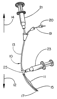
Figure 1 is a perspective view of the delivery system 10 of the
; present invention. The delivery system 10 with its various components
has a distal end 12 and a proximal end 14, as does the conduit 31
within which the delivery system is used. An intraluminal graft 11 is
~ fitted coaxially over the distal end 12 of a balloon catheter 13. The
balloon 15 is located within the distal end 12 of the intraluminal
- graft 11; both the balloon 15 and distal end 12 of the intraluminal
~' ,
I =
CA 02231471 1998-03-06
W O 97/10778 PCT~US96/13833
graft 11 are enclosed within a hollow, bullet-shaped end 17 which is
axially movable with respect to the balloon catheter 13 by a guidewire
19 connected to the hollow, bullet-shaped end 17. Guidewire 19
extends through guidewire lumen of the balloon catheter 13 within
catheter shaft 13A and is operable by relative movement at the
proximal end 14 of the balloon catheter 13 using the torquing device
20 as a handle.
Balloon 15 is connected via a second lumen of the balloon
catheter 13 to a means for balloon inflation 21 such as the syringe
0 shown by Figure 1. This second lumen is preferably located coaxially
around the first lumen. After the hollow, bullet-shaped end 17 has
been moved axially by use of the guidewire 19 so as to no longer
enclose the balloon, inflation of balloon 15 causes circumferential
distension of the distal end 12 of intraluminal graft 11.
Proximal end 14 of intraluminal graft 11 is sealed to the
exterior surface of balloon catheter 13 by seal fitting 25, which is
in turn connected to a means for graft deployment 23 such as the
syringe shown by Figure 1.
Figure 2 describes in longitudinal cross section the initial
configuration of delivery system 10 as it appears during insertion
into a conduit 31 for delivery and deployment of intraluminal graft
11. The axial discontinuities in the cross section indicate that the
lengths between the portions shown by the figures may be any length
desired. In the case depicted, the proximal end 12 of conduit 31 has
been surgically transected to provide access and to al~ow for
attachment of intraluminal graft 11 to the proximal end 12 of conduit
31 by sutures, stent or other suitable means.
Balloon catheter 13 is provided with at least two lumens. The
guidewire is contained within a separate guidewire lumen while a
second lumen is provided to connect means for balloon inflation 21
with balloon 15. Figure 2 depicts these lumens in coaxial
relationship with the guidewire lumen enclosed by catheter shaft 13A
and the lumen connecting balloon 15 and means for balloon inflation 21
enclosed by catheter shaft 13B. While a coaxial relationship is
CA 02231471 1998-03-06
~-W O 97/10778 PCTAUS96/13833
-- - V 7-
-: ~epicted, any geometric relationship may be used which provides the at least two lumens.
Figure 2 and subsequent figures depict balloon 15 connected to
the distal end 12 of catheter shaft 13B in end-to-end fashion at
location 16. It is apparent that an alternative connection may be
made by overlapping the proximal end of balloon 15 with catheter shaft
13B.
During insertion, balloon 15 is coaxially enclosed by hollow,
bullet-shaped end 17 which is connected to and axially movable by
guidewire 19. End 17 comprises a bullet-shaped tip 37 and tubular
portion 39. While Figure 2 describes that tip 37 is made of metal for
easy visualization and tubular portion 39 is of a plastic which is
preferably a lubricous plastic such as PTFE, it is apparent that end
17 may be made as a one-piece construction from a single material
which may be radiopaque if desired.
. Seal fitting 25 seals the proximal end 14 of the intraluminal
; graft 11 to the exterior surface of balloon catheter 13 and provides
for connection to means for graft deployment 23. Seal fitting 25
~ lncludes sealing means 27 such as the seal shown by Figure 2 and
attaching means 2g such as the ring shown for sealingly connecting the
proximal end 14 of intraluminal graft 11 to seal fitting 25. All
sealing functions provided by seal fitting 25 must withstand the
pressure from the means for graft deployment 23 during deployment of
the intraluminal graft 11.
2!~ When located as desired, the hollow, bullet-shaped end 17 is
moved axially in a distal direction 12 by guidewire 19 with activation
of guidewire 1g provided at the proximal end 14 of balloon catheter
13. This is described by Figure 3.. After end 17 is clear of balloon
. 15, balloon 15 is inflated as shown by Figure 4 by pressure from the
means for balloon inflation 21. Irlflation of balloon 15 is continued
until balloon 15 has increased in diameter adequately to deploy the
distal end 12 of intraluminal graf1; 11 enough to bring it into good
contact with the lumen of conduit '11. This causes the distal end of
the intraluminal graft 11 to become sealed against the luminal surface
3'~ of conduit 31 which then allows pressure to be applied to the interior
of the remainder of intraluminal gr-aft 11 via means for graft
distension 23. This pressure is applied until the intraluminal graft
.
CA 02231471 1998-03-06
W O 97/10778 PCTAJS96/13833
11 is fully deployed along its length into contact with the lumen of
conduit 31 as described by Figure 5.
As shown by Figure 6, once the intraluminal graft 11 has been
fully deployed, the pressure applied to the interior of the balloon 15
is released by withdrawing the inflating medium or by sliding hollow
tip 17 over the balloon 15, causing it to compact.
Proximal end 14 of intraluminal graft 11 is transec~ted even with
the previously transected proximal end 12 of conduit 31 as shown by
edges 41, thus enabling subsequent attachment of the proximal end 14
lo of intraluminal graft 11 to conduit 31 by sutures, a stent or other
suitable means.
Also as shown by Figure 6, withdrawal of the delivery system 10
following deployment of intraluminal graft 11 i s accomplished by
axially moving hollow, bullet-shaped end 17 back over deflated balloon
15, thereby again enclosing balloon 15 and minimizing the diameter of
the deflated balloon. Enclosing balloon 15 in such a manner with a
tubular, lubricous cover as provided by end 17 allows for easy removal
of delivery system 10 with minimum drag.
Figure 7 describes a cutaway perspective view of the use of a
balloon expandable stent in conjunction with the delivery system 10.
The view shown is sequentially equivalent to the longitudinal cross
sectional view of Figure 4 which describes inflation of the balloon 15
and deployment of the distal end of the intraluminal graft 11. To
create the delivery system shown by Figure 7, stent 71 is fitted
coaxially over balloon 15 and coaxially within the distal end 12 of
intraluminal graft 1I prior to insertion of the delivery system 10
into the vascular system of a living body. The distal ends 12 of the
stent 71 and the intraluminal graft 11 extend to the distal end of
balloon 15, which is not apparent from the cutaway perspective view of
Figure 7. Following insertion to a desired location within the
vascular system, inflation of balloon 15 results in simultaneous
deployment of the distal end 12 of intraluminal graft 11 and stent 71,
so that when the distal end 12 of intraluminal graft 11 has been
deployed sufficiently to come into circumferential contact with the
lumen of conduit 31, it is simultaneously attached thereto by the
balloon-expanded stent 71.
CA 02231471 1998-03-06
=i~ wo 97/10778 PCT~US96/13833
While this procedure describes securing of the intraluminal graft
by the use of balloon-expandable stents, it is apparent that other
types of stents may be used, such as, for example, self-expanding
~stents.
c, Figure 8 describes an alternative embodiment wherein seal fitting
25 is replaced by a second balloon catheter 83 having a balloon 81
located at the proximal end of the intraluminal graft. The use of the
additional balloon at the proximal end of the intraluminal graft
~ allows the entire procedure to be a~ccomplished transluminally without
lo requiring a surgical cut-down and avoids surgical transection of the
conduit being repaired. Balloon 81 is provided with its own separate
means for inflation 85 at the proxilnal end 14 of the delivery system
10. Graft 11 is deployed via means for graft deployment 24 at the
proximal end of the delivery system 10. Seal fitting 22 seals the
15, proximal end of the intraluminal graft to the exterior surface of
balloon catheter 83. Alternatively (not shown by the Figures) balloon
81 may be inflated simultaneously with balloon 15 using a common means
for balloon inflation. Preferably, balloon catheter 83 is slidably
coaxial with balloon catheter 13 whereby balloon 81 may be moved
20~ axially along balloon catheter 13 to allow the intraluminal graft 11
~ to be cut to any desired length and allow balloon 81 to be located at
the proximal end of intraluminal graft 11 regardless of the length of
that graft.
; As described by Figures 8-11, catheter 83 is provided with an
25~ inner lumen which enables deployment of intraluminal graft 11 and is
enclosed by catheter shaft 83A, and with an outer lumen which allows
inflation of balloon 81, the outer lumen being enclosed by catheter
shaft 83B.
Figure 8 and subsequent figures depict balloon 81 connected to
the distal end 12 of catheter shaft 83B in end-to-end fashion at
location 82. It is apparent that an alternative connection may be
made by overlapping the proximal end of balloon 81 with catheter shaft
83B.
Figures 9, 10 and 11 are longitudinal cross sections sequentially
describing the delivery system 10 of Figure 8 during use. Figure 9
describes this embodiment after insertion into a vascular system and
after the bullet-shaped tip 17 has been extended distally by guidewire
-
CA 02231471 1998-03-06
W O 97/10778 PCT~US96/13833
--10--
19 beyond the end of balloon 15. Proximal end 14 of intraluminal
graft 11 coaxially covers the second balloon 81. As shown by Figure
10, the next step involves inflation of both balloons 15 and 81.
Preferably balloon 15 at the distal end of the intraluminal graft 11
is inflated first, thereby securing that end of intraluminal graft 11
to the wall of the conduit 31. Second balloon 81 is then inflated,
securing the proximal end 14 of intraluminal graft 11 tQ the wall of
the conduit 31. Following inflation of both balloons 15 and 81 the
portion of the intraluminal graft 11 between the distal and proximal
lo ends previously secured by the balloons is deployed by activating
means for graft deployment 24 as described by Figure 11. After the
entire length of the intraluminal graft 11 has been deployed against
the wall of the conduit 31, balloons 15 and 81 are deflated, the
bullet-shaped tip 17 is moved proximally back into place over balloon
15 and the entire delivery system 10 is withdrawn.
ExamPle
This example describes the construction of an embodiment of the
present invention.
One end along the longitudinal axis of a female tee luer lock
fitting (part number H-06359-47, supplied by Cole Parmer, Niles, IL)
having a 4 mm inner diameter was fitted with a self sealing injection
site (Injection Site with Luer Lock manufactured by Baxter Healthcare
Corporation, Deerfield, IL). A hole was created through the injection
site, and the balloon end of a model 12TL0806F Fogarty Thru-Lumen
Embolectomy Catheter manufactured by Baxter Healthcare Corporation
(Irvine, CA) was passed through this hole, situating the female tee
luer lock fitting with one uncovered end facing toward the distal end
of the catheter, another uncovered end perpendicular to the catheter
shaft, and the third end (fitted with the injection site) facing the
proximal end of the catheter. The catheter was then fitted with a
0.64 mm diameter Ultra-Select Nitinol Guidewire manufactured by
Microvena (White Bear Lake, MN). This guidewire was modified to the
extent that the flexible end was removed, and a stainless steel
bullet-shaped tip having a 3.2 mm outer diameter was welded onto the
end of the wire. This bullet-shaped tip had a 0.76 mm diameter hole
bored along its longitudinal axis, and was stepped at one end to an
CA 02231471 1998-03-06
,- W O 97/10778 PCTAUS96/13833
outer diameter of about 2.9 mm so that a 2.3 cm long piece of PTFE
tubing having and inner diameter of 2.9 mm and an outer diameter of
3.2 mm could be pressed onto the stepped end of the bullet-shaped tip.
This resulted in one end of the guidewire having a securely affixed
bullet-shaped tip including a short hollow section. When the
guidewire was fully inserted into its lumen in the embolectomy
catheter, the hollow section of thle bullet-shaped tip coaxially
covered the balloon portion of the embolectomy catheter. The torquing
device provided with the guidewire was placed on the wire
approximately 3 cm away from the elld of the threaded fitting attached
to the proximal end of the catheter shaft. This placement of the
~torquing device enabled the device to be used to slide the guidewire
and the attached bullet-shaped tip distally, such that the hollow
section of the tip was no longer coaxially covering the balloon
portion of the catheter.
At this point the delivery sy~;tem was ready to have an
intraluminal vascular graft installled onto it. The graft, having an
inside diameter of 3 mm and a wall thickness of about 0.13 mm was slid
~ over the bullet-shaped tip, coaxially fitting it over the catheter
shaft. The torquing device was used to slide the guidewire and the
attached bullet shaped tip distally, such that the hollow portion of
; the tip was no longer encasing the balloon portion of the catheter.
The distal end of the graft was plalced such that it coincided with the
proximal edge of the distal radiopa,que balloon marker band. The
2~, hollow portion of the bullet-shapedi tip was then slid proximally,
encasing both the most distal portion of the vascular graft as well as
- the balloon portion of the catheter. The female tee luer lock fitting
; and attached injection site were then slid distally along the shaft of
the catheter to the proximal end of the intraluminal graft, and this
end of the intraluminal graft was then ligated onto the open end of
the female tee luer lock fitting facing the balloon portion of the
catheter. The delivery system, now fitted with an intraluminal graft,
was ready for use.
~ While particular embodiments of the present invention have been
illustrated and described herein, the present invention should not be
limited to such illustrations and descriptions. It should be apparent
that changes and modifications may be incorporated and embodied as
. ~
. . .
CA 02231471 1998-03-06
~ W O 97/10778 PCTrUS96/13833
-12-
part of the present invention within the scope of the following
claims.