Note: Descriptions are shown in the official language in which they were submitted.
CA 02231593 1998-03-10 ,
WO 97/03614 PCT/US96/10937
DEVICE AND METHOD FOR REINFORCING SURGICAL STAPLES
' FIELD OF THE INVENTION
~ The present invention relates to surgical staple devices and
methods for reinforcing the seams formed by such devices.
BACKGROUND OF THE INVENTION
One of the more commercially successful innovations in surgical
procedures in recent years is the development of surgical
stapler
devices. These devices are designed to simultaneously cut
and seal an
extended segment of tissue in a patient, vastly reducing
the time and
risks of such procedures. Typically, a surgical stapler
comprises two
stapler arms, one containing two or more lines of multiple
staples and
a second containing corresponding means to bend each of
the staples
into a closed position. For most applications, a surgical
blade is
included in the device to quickly sever tissue between the
lines of
staples. Those stapler devices employing a cutting blade
are referred
to as "anastomotic staplers" and those used without a cutting
blade
are referred to as "non-anastomotic staplers."
In the operation of a typical anastomotic stapler, the two
stapler arms are positioned around tissue to be cut and
then locked
firmly together. In one motion the surgeon then actuates
the stapler
device, which simultaneously installs two or more lines
of staples
through the tissue and cuts a line down the middle of the
staple
lines. In this manner, the physician can quickly cut and
seal up to
about 8 cm of tissue at a time. This procedure is much faster
than
using a conventional process of cutting with scissors or
a scalpel and
then laboriously sealing the incision with sutures. As a
result,
patient care is dramatically improved by minimizing bleed
time from
the surgical site and significantly increasing the speed
with which an
operation can be completed.
For most procedures, the use of bare staples, with the staples
in
direct contact with the patient's tissue, is generally acceptable.
CA 02231593 2000-OS-26
_2_
The integrity of the tissue itself will normally serve to prevent the
staples from tearing out of the tissue and compromising the seart~
before healing has occurred. However, in certain circumstances the
tissue that is being sealed is too fragile to securely hold the
staples in place. In these instances, the tissue will tend to rip at
or near the staple lines, slowing healing and possibly leading to
serious complications.
One area where fragile tissue is of particular concern is the use
of stapler devices in lung tissue, and especially lung tissue that is
affected by emphysema or similar condition. Diseased lung tissue is
very fragile and, in extreme cases, will readily tear through
unprotected staple lines. With the growing use of surgical staplers
in operations on diseased lung tissues such as bullectomies and volume
reduction procedures, it has become increasingly important to develop
some reliable means to protect fragile tissue from tissue tears due to
surgical staples or surgical stapling procedures.
One product that attempts to correct these problems i * PERI-
STRIPS staple line reinforcement sleeves available from Bio-
~ascular,Inc. of Saint Paul, MN. This product is specified for use in
lung resection procedures in order to buttress the staple lines and
help prevent air leakage that can occur through staple holes. The.
sleeves are of tri-component construction, comprising (1) a thin strip
of processed bovine pericardial tissue attached with (2) suture to (3)
a section of polyethylene backing material to form a tubular sleeve.
These tri-component sleeves are slid over each of the arms of a
surgical stapler, with the bovine pericardial tissue carefully
positioned on the operative faces of each of the stapler arms.
During an operation, a surgeon staples and cuts through both the
bovine pericardial tissue and the patient's lung tissue in order to
perform the lung resection procedure. Once the staples are in place,
the surgeon must then cut the suture lines holding the bovine
pericardial strips in place and remove the polyethylene backing
material and sutures.
While the PERI-STRIPS sleeves offer improvement in preventing
lung tissue tearing, this product has numerous deficiencies. First,
the use of bovine pericardial tissue creates numerous handling
problems and costs. This natural tissue must be stored in
TRADEMARK
CA 02231593 1998-03-10
WO 97/03614 PCT/US96/10937
-3-
preservatives (e. g., propylene oxide) before use and the preservatives
must be carefully removed through a saline so~ution wash prior to use.
This is viewed as a needless waste of personnel time and
effort prior
to use of the sleeves. Even after cleaning, the PERI-STRIPS
sleeves
are required to be kept moist at all times prior to use.
These demanding handling characteristics make it very difficult
to quickly employ the PERI-STRIPS sleeves. As a result,
it is common
that the surgeon will have to waste some of these strips
during each
operation in order to assure that an adequate number will
always~be
prepared and ready. Since the PERI-STRIPS sleeves are quite
expensive, usually constituting one of the most expensive
single
implements used in a typical lung resection procedure, the
need to
prepare extra sleeves that may not be used is not a trivial
matter.
Another problem with the PERI-STRIPS sleeves is that they
are of
multiple component construction. The surgeon must exercise
particular
care that the sleeves are properly aligned prior to stapler
actuation
and that staples are driven through only the bovine pericardial
tissue. Since the polyethylene backing material is not approved
for
human implantation, it is crucial that only the bovine pericardial
tissue is attached to the staple lines and that all of the
backing
material is removed.
Other concerns with the PERI-STRIPS sleeves include: the
need to
employ scissors or a scalpel to cut the sutures holding
the two
materials together; inconsistent product performance due
to normal
differences in natural animal tissues; difficulties in cutting
through
the bovine pericardial tissue; and possible contamination
or
immunological problems where preparation of the PERI-STRIPS
sleeves
has not been properly performed. Despite all of these constraints,
the PERI-STRIPS reinforcement sleeve product remains the
primary
choice of surgeons performing lung resection procedures.
In an effort to address some of these drawbacks, it has
been
attempted to form a staple reinforcement device from an
artificial
implantable material, such as strips of polytetrafluoroethyiene
(PTFE)
cut from vascular grafts or similar devices. The strips
of material
are held to the operative faces of the stapler arms by loops
of suture
wrapped around the stapler arms. Once staples are driven
through the
strip, the surgeon must then cut the suture to free the
device from
CA 02231593 1998-03-10
WO 97/03614 PCT/US96/10937
the surgical site. This technique has not been widely employed due to
difficulties in preparing, mounting. and,using the strips in this
form. Additionally, the use of relatively narrow strips of artificial
implantable material has a centering problem similar to that
encountered with the use of strips of bovine pericardial tissue. In
both cases, the strips must be carefully centered on the operative
face of the surgical arm or proper staple reinforcement will not
occur.
In light of these problems, it is a primary purpose of the
present invention to provide an improved staple line reinforcement
material that will fully protect surgical staple lines while being
easy to prepare and use.
It is still another purpose of the present invention to provide
an improved staple line reinforcement material that is safe and
effective in use.
These and other purposes of the present invention will become
evident from review of the following specification.
SUMMARY OF THE INVENTION
The present invention is an improved device for reinforcing
surgical staples. While the present device may be used for a wide
variety of surgical procedures using surgical staples, it is
particularly suitable for use on fragile tissue, such as lung tissue
in lung resection procedures. The device of the present invention is
considered an important implement in establishing improved seals of
surgical sites, with reduced possibility of tearing or leaks at the
surgical sites through or around surgical staples.
The device of the present invention comprises an essentially
tubular structure made entirely from implantable material. The
preferred device is formed from a porous poiytetrafluoroethylene
(PTFE), and most preferably an expanded PTFE. The device comprises a
sleeve that readily slides over each arm of a surgical stapler device.
The surgeon then seals through both sleeves and the tissue to
accomplish the stapling procedure.
CA 02231593 1998-03-10
WO 97/03614 PCT/US96/10937
-5-
The sleeve of the present invention is far easie~ to prepare
and
use tha~~ previous staple reinforcement devices. The use
of expanded
PTFE allows the reinforcement device of the present invention
to be
used directly out of the package, with no arduous preparation
procedures. This not only saves personnel time, but also
assures that
' only precise number of sleeves that are needed for the procedure
need
to-be-prepared. This saves significant expense over numerous
operations. Moreover, the sleeve of the present invention
can be
easily installed and used without fear that misplacement
might lead to
IO inadequate staple reinforcement or the attachment of non-
implantable
material.
It is particularly preferred that the sleeve of the present
invention includes multiple operative portions or "faces."
This
allows any one of the faces of the sleeve to be centered
over the
operative face of the stapler arm. For instance, a sleeve
with an
essentially rectangular cross-section can provide four different
operative faces that can be centered over the faces of the
stapler
arms. With this construction, the surgical staff can quickly
and
easily mount the sleeves on the stapler arms without fear
that a wrong
face may be oriented to receive the staples.
Ideally, the sleeve of the present invention includes pre-defined
tear lines along the length of the sleeve. This allows the
surgeon to
quickly and easily separate the operative face of the device
from.
excess material after installation. As is disclosed, the
tear lines
can be created by selective modification of the expanded
PTFE material
to allow it to rip more readily along the tear lines, or
merely by
providing scoring along the tear lines.
The device of the present invention can be formed in a number
of
different forms. The device preferably comprises an essentially
tubular sleeve, either a tube of continuous material or one
or more
sheets of material attached to together to form a tube. Although
not
required, for ease in staple installation, the tube ideally
has at
least one flatten face into which staples are introduced.
As has been
noted, the use of one or more flatten faces makes installation
of the
sleeve on the stapler easier and aids in orientation of tear
lines for
easy separation of excess material following installation.
CA 02231593 1998-03-10
WO 97/03614 PCT/IJS96/10937
-6-
BRIEF DESCRIPTION OF THE ORAWIN6S
The operation of the present invention should become apparent
from the following description when considered in conjunction with the
accompanying drawings, in which:
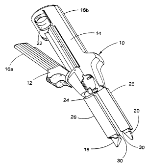
Figure 1 is a perspective view of a surgical stapler having two
surgical staple reinforcement devices of the present invention mounted
on its stapler arms;
Figure 2 is a three-quarter isometric view of one embodiment of a
surgical staple reinforcement device of the present invention;
Figure 3 is a three-quarter isometric view of another embodiment
of a surgical staple reinforcement device of the present invention;
Figure 4 is a three-quarter isometric view of still another
embodiment of a surgical staple reinforcement device of the present
invention;
Figure 5 is a perspective view of two surgical staple
reinforcement devices of the present invention shown attached to
either side of tissue immediately following actuation of the stapler
device, with the reinforcement devices shown partially separated ane
with the stapler not shown for clarity;
Figure 6 is a three-quarter isometric view of yet another
embodiment of a staple reinforcement device of the present invention;
and
Figure 7 is a cross-section view an embodiment of an extrusion
die suitable for production of one embodiment of a reinforcement
device of the present invention.
DETAILED DESCRIPTION OF THE INVENTION
The present invention is an improved device for use in
reinforcing staple lines created by a surgical stapler.
Shown in Figure 1 is a conventional surgical stapler 10. The
stapler 10 comprises two separate halves 12, 14 that can be locked
together. Each of the halves 12 and 14 has its own handle 16a and
16b, respectively, allowing manipulation of the stapler. On the first
half 12 is a first stapler arm 18 that is loaded with one or more rows
CA 02231593 1998-03-10 .-
WO 97/03614 PCT/US96/10937
of surgical staples. A corresponding second stapler arm 20
is on the
second half 14, containing means to bend each of the staples
contained
in the first stapler arm 18 into a closed position. This
means to
bend the staples usually comprises a series of contoured
grooves, each
corresponding to one of the staples contained in the first
stapler arm
18. Finally, one of the halves contains an actuation arm
22 that
fires each of the staples. In an anastomotic stapler device,
the
actuation arm 22 both fires the staples and actuates a cutting
blade
24. The cutting blade 24 is oriented between at least two
rows of
staples, allowing each row of staples to seal on either side
of the
cutting blade simultaneously with the cutting action.
In operation, the two halves 12, 14 of the stapler 10 are
locked
together with each of the stapler arms 18, 20 positioned
on either
side of tissue to be sealed. Once the surgeon assures that
the arms
18, 20 are properly positioned, the actuation arm 22 is moved
forward,
firing the staples and sealing the surgical site. In an anastomotic
stapler device, the staples are fired simultaneously with
the slicing
the tissue with cutting blade 24. The result is a rapid and
accurate
cutting and sealing of a patient's tissue that is much faster
than
previous cutting and suturing techniques.
As has been noted, while commercially available staplers
function
well for most cutting and sealing applications, problems
have been
experienced with the placement of staples in relatively weak
and
fragile tissue, such as the lung tissue of emphysema patients.
The
need for some form of staple reinforcement has been recognized,
but
until the present invention no fully adequate staple reinforcement
device has been available.
In the present invention a staple reinforcement device is
provided that overcomes many of the problems previously experienced
with such devices. A first embodiment of the staple reinforcement
device 26 of the present invention is shown in Figures 1
and 2. This
device 26 comprises a sleeve 28 having at least one face
30 adapted to
receive the rows or lines of surgical staples and at least
one
side/back wall 32 adapted to surround the stapler arms 18,
20 and hold
the device 26 in place. An opening 34 is provided on at least
one end
of the sleeve 28 to allow installation of the sleeve over
the stapler
arms.
CA 02231593 1998-03-10
WO 97/03614 PCT/LTS96/10937
_g_
Unlike previous tubular staple reinforcement devices, the device
of the present invention is formed entirely from an implantable
material. This allows the device to be mounted and used with
substantially less care than previous staple reinforcement devices.
For instance, a slight misalignment of the device will never result in
the accidental attachment of non-implantable material within the
patient or an inadequate amount of reinforcement material protecting
the tissue.
In the embodiment of Figures 1 and 2, the wall 32 comprises
essentially three other operative faces 35, 36, and 37. This
construction allows any one of the faces 30, 35, 36, or 37 to receive
and reinforce the staples in a patient's tissue. As a result, less
care and manipulation is required by the surgical team to mount and
center the sleeve prior to use.
Preferably, the device 26 is constructed from porous
polytetrafluoroethylene (PTFE), and particularly a stretched or
expanded PTFE such as that made in accordance with United States
Patents 3,953,566, 3,962,153, 4,096,227, and 4,187,390, all
incorporated by reference. By heating and rapidly expanding PTFE in
accordance with the teachings of these patents, the resulting material
exhibits exceptional strength in the direction that it has been
expanded.
PTFE, and particularly expanded PTFE, has numerous properties
that make it particularly suitable for use as an implantable material.
First, the material is highly inert, sterilizable, and bio-compatible.
As a result,-it is widely employed as vascular grafts and various
other implantable tube and sheet materials. Further, PTFE has
extremely low coefficient of friction, which allows the material to
slide easily onto and off of the stapler arms I8, 20 as well as being
easily and smoothly cut by the cutting blade 24 and sealed by the
surgical staples. Finally, expanded PTFE material can be selectively
expanded to have exceptional strength where needed to resist staple
pull-out and to have ready severability in the direction of cut of the
device.
The preferred sleeve of expanded PTFE for use with the present
invention is formed in the following manner. A fine powder PTFE resin
is blended with a lubricant, such as odorless mineral spirits, until a
CA 02231593 1998-03-10
WO 97/03614 PCT/US96/10937
_g_
compound is formed. The volume of lubricant used should be sufficient
to lubricate the primary particles of the PTFE resin so as to mini:~ize
the potential of the shearing of the particles prior to extruding.
The compound is then compressed into a billet and extruded, such as
through a ram type extruder, to form a coherent extrudate. A
reduction ratio of 3D:1 to 300:1 may be used (i.e., reduction ratio =
cross-section area of extrusion cylinder divided by the cross-section
of the extrusion die). For most applications a reduction ratio of
75:1 to 150:1 is preferred. The lubricant may then be removed, such
as through volatilization. If desired, the extruded product may then
be further expanded in at least one direction 1.1 to 50 times its
original length. Expansion may be accomplished by passing the dry
coherent extrudate over a series of rotating heated rollers or plates.
A tube can be stretched in a hot oven to maintain its tubular
structure.
Finally, the product should be heat set (also referred to as
"amorphorously locked") to retain the material in its final expanded
condition. This may be accomplished by exposing the material to a
heat of about 327 to 380°C for about 25 seconds to about 4 minutes or
more.
To form a tubular structure for use as the present invention, it
is preferred that the extrusion step occur through a circular, semi-
circular, triangular, rectangular, or other closed ring die so as to
deliver a tubular product. The die should be proportioned so that the
final product will fit snugly over the desired stapler-arms.
Alternatively, the tubular structure can be formed by creating a
sheet or tape of the expanded material and then wrapping the sheet or
tape into a tubular form. This can be accomplished through any
suitable means, such as longitudinally wrapping (i.e., in a
"cigarette" wrap fashion) or helica7ly wrapping (e. g., over a mandrel
or similar structure). The wrapped product may be bonded to itself by
adhesive, heat bonding, mechanical means (e.g., a suture seam) or
similar means to form a sleeve that will attach over the stapler arms.
It should be understood that it is contemplated by the present
invention that small amounts of materials such as adhesives or suture
may be used to bind the tubular structure together without departing
from the intend scope of the present invention.
CA 02231593 1998-03-10
WO 97/03614 PCT/US96/10937
-10-
Without intending to limit the scope of the present invention,
the fin d product preferably comprises an expanaed PTFE structure with
the following range of properties: an expansion/stretch ratio of 2:1
to 6:1 or more (e. g., 10:1); a fibril length of about 2 to 90 micron;
a longitudinal strength of above about 10 kg; a transverse strength of
above about 5 kg; a density of about 0.8 to 1.5 g/cc; and an average
wall thickness of about 0.125 to 2.5 mm.
Each of these properties may be measured in a conventional
manner. Fibril length may be determined by the mean length of the
fibrils extending between nodes of a sample of the expanded PTFE
material measured on a scanning electromicrograph (SEM) of the sample.
Longitudinal and transverse strength may be determined through use of
a tensile strength tester, such as an INSTRON tensile tester available
from Instron Corporation. Density may be determined by dividing the
measured weight of the sample by the computed volume of the sample.
Average wall thickness may be determined through conventional means,
such as through the use of calipers or measurements from SEMs.
Material suitable for use in the present invention is
commercially available in a number of forms. For instance, tubular
structures of expanded PTFE that may be modified for use on surgical
stapler arms are commercially available from W. L. Gore & Associates,
Inc., Flagstaff, AZ, in the form of prosthetic vascular grafts under
the trademark GORE-TEX~. Additionally, sheets and tapes of expanded
PTFE material that may be constructed into the sleeves of the prey
invention are commercially available in a wide variety of farms from a
number of sources, including W. L. Gore & Associates, Inc., Elkton,
MD, under the trademark GORE-TEX~.
Although not preferred, other possible implantable materials that
may be employed with the present invention include: nylon;
polypropylene; polyurethane; silicone; DACRON~ polymer; etc. For some
applications, it may be desirable to use a bio-absorbable implantable
material, such as polyglycolic acid (PGA), polylactic acid (PLA),
polycaprolactone, or natural animal membranes.
It is particularly preferred that the device of the present
invention includes means to allow separation of the attached face of -
the sleeve from the remainder of the sleeve following actuation of the
stapler. This can be accomplished in any one or more of a number of
CA 02231593 1998-03-10
WO 97/03614 PCT/US96/10937
-11-
ways. The tubular structure of the sleeve may be modified
during its
formation to selectively weaken certain areas so that they
will
readily rip longitudina?ly. Where sleeve is being created
by
extrusion, this can be accomplished by modifying the extrusion
die to
reduce the thickness of the sleeve in certain areas to create
tear
lines. For instance, one or more projections may be provided
into the
flow of extrudate passing through the die that will reduce
the
thickness along longitudinal lengths of the tubular structure
being
produced. These longitudinal lengths will thereby be weakened,
allowing the material to more readily separate (or "tear")
along these
lengths. Any structure that will provide for controlled separation
of
material in this manner is referred to herein as a "tear
line."
One example of tear lines is shown in Figure 2. In that
embodiment, the tube being extruded has an essentially rectangular
cross-section, with a wall thickness of about 0.125 to 1.0
mm, with
about 0.375 to 0.8 being a typical thickness. If desired,
the wall
thickness may be increased up to about 2.5 mm for use with
most
current stapler devices. By modifying the corners of the
die
extruding this tube, the wall thickness in corners can be
reduced by
about 25 to 75~, with a preferred reduction being about 65~.
This
produces four tear lines 40a, 40b, 40c, 40d running the length
of the
sleeve. When a transverse tension is applied to the sleeve,
separation of material will readily occur along the tear
lines and the
separation will easily propagate along the length of the
tube to allow
the backing material to be removed from an attached face.
For
example, with the attachment of face 30, separation of backing
material 32 can be accomplished by tearing along tear lines
40a and
40b.
For further ease in separation, small cuts 43 may be provided at
an end of the tear lines 40 to ease in starting the tear propagation.
The cut 43 may be provided by the surgical personnel before or after
actuation of the stapler. Alternatively, the cut 43 may be supplied
on the sleeve by the sleeve manufacturer.
It should be appreciated that the tear lines 40 may be provided
at any desired location on the sleeve to address particular needs.
For example, in the embodiment of Figure Z, two folds are provided
longitudinally on faces 35 and 37. Tear lines 40e and 40f may
CA 02231593 1998-03-10
WO 97/03614 PCT/LTS96/10937
-12-
alternatively or additionally be provided along these folds to provide
different or increased options for separating the sleeve following
installation.
Another method of creating tear lines is to produce the tear
lines following creation of the sleeve. This can be accomplished by
stripping or modifying the sleeve material in the places where tears
are desired, such as through: selective heating or altering of the
sleeve material to create the tear line (e. g., through use of a laser
or heated cutting implement); cutting the sleeve to a prescribed depth
along the desired tear line (e. g., with a cutting blade); mechanically
altering 'the material (e. g., through use of pinch rollers);
selectively weakening the material; etc.
Alternatively, the sleeve may be scored with lines of holes or
similar structures that will provide sufficient weakening to allow
easier separation of remainder portions of the sleeve following
installation. This can be accomplished through a number of means,
such as: creating holes with lasers; punching holes; using a pinch
roller with teeth; etc.; or through some combination of any of the
methods described.
Once tear lines are created, separation of material following
installation can be easily and rapidly accomplished. Shown in Figure
5 is one example of two devices 26a, 26b of the present invention
essentially of the construction shown in Figures 1 and 2. As is
shown, the devices 26a, 26b are attached by staples 43 to two segments
of tissue 44, 46 along faces 30a and 30b. The tissue segments 44, 46
have been cut from one another along incision line 48 using a
anastomotic surgical stapler and sealed by staple lines 50a, 50b, and
50c, 50d, respectively.
Once the stapler has been actuated, cutting and sealing the
tissue, the backing material 32 of each of the sleeves can be
separated from attached faces merely be ripping along tear lines 40a
and 40b. This is normally done with the stapler arms still in place
around the cut site. In the illustration of Figure 5, the surgical
stapler is not shown at the cut site so as not to obscure details
concerning the surgical cut 48 and the placement of the staples 43.
As is shown, once the backing material 32a, 32b is removed, only the
operative faces 30a, 30b of the sleeves are left in place.
CA 02231593 1998-03-10
WO 97/03614 PCT/US96/10937
-13-
The provision of tear lines that readily separate the stapler
from the attached reinforcement material is co~~sidered
to be an
extremely useful attribute of the present invention. Previous
sleeve
' devices required some form of cutting of attachment sutures
or similar
action to release an applied staple reinforcement device
from its
backing material and the stapler itself. This is an extra
step for
the surgeon, but may not be particularly burdensome for
many operative
procedures where there is unobstructed access to the surgical
site.
However, with the growing use of endoscopic surgical procedures,
with their intentionally limited access to the surgical
site, the need
to perform an additional cutting step in order to separate
a stapler
from staple reinforcement material can be quite burdensome.
In fact,
the presence of non-implantabie material attached to the
staple
reinforcement material, such as that present with the PERI-STRIPS
reinforcement materials, raises even more concerns for the
surgeon who
must be assured that all such material is completely removed
from the
endoscopic surgical site before terminating the procedure.
If
multiple staple lines are being installed, this increases
the risks
even more for the surgeon that non-implantable material
may be
accidentally attached to the surgical site. With each of
these
problems, the endoscopic surgeon must address these concerns
with
severely restricted space and tools.
The reinforcement device of the present invention avoids
all of
these problems. First, the fact that the device is made
entirely from
implantable material assures the surgeon that non-implantable
material
will not be accidentally attached to the patient. Second,
the
provision of tear lines allows the surgeon to easily separate
the
stapler from the surgical site with little or no additional
cutting
procedures. In fact, it is preferred that the tear lines
are
proportioned so that the mere action of separating the stapler
arms
from one another will completely cut the tear lines and
allow removal
of the stapler from the surgical site. Excess portions of
the
reinforcement device can then be removed by forceps or similar
method.
Further, particularly for endoscopic procedures, it is
contemplated that means may be provided on the stapler device
to aid
in the extraction of excess reinforcement material following
automatic
reinforcement device separation. For example, the reinforcement
CA 02231593 1998-03-10
W~ 97/03614 PCT/US96/10937
-14-
material may be adhered to the stapler through mechanical means (e. g.,
clips, to~her lines, etc.), pressure sensitive ao~~esive strips, et,:.
In this manner, excess reinforcement material can be withdrawn from
the surgical site automatically along with the stapler.
Figure 6 illustrates two examples of means to adhere a sleeve 68
to a stapler for ease in extraction from a surgical site. The sleeve
68 shown is essentially rectangular and includes an operative face 70
and two tear lines 72a, 72b. That portion of the sleeve opposite the
operative face 70, referred to as a remainder or excess portion 76,
includes both a tether 78 and a self-adhesive strip 80 to assist in
anchoring the sleeve 68 to a stapler arm. The tether 78 is adapted to
attach to the stapler arm, preferably to a clip or similar device
provided thereon, and the adhesive strip is adapted to attach to the
back of the surgical arm. In operation, once the operative face 70 is
attached to the surgical site and the tear lines 72 are separated, the
remainder portion 76 is simply extracted from the surgical site by
removing the surgical stapler arm. It should be understood that
stapler arm attachment methods such as these may be employed alone or
in combination with each other to effectuate remainder portion removal
from a surgical site.
The exact shape and dimensions of the device of the present
invention is a function of the particular constraints of the surgical
apparatus and procedures with which it is to be employed. As such,
the reinforcement device of the present invention may be formed in
virtually any shape or size, including cross-sections comprising a
circle, semi-circle, oval or other oblong shape, triangle, rectangle,
pentagon, hexagon, etc., or some less defined shape. As has been
noted, the face or faces and side/back walls) of the device need not
be entirely planar, and may include folds or other essentially concave
or convex orientations. In fact, folds or concave wall structure may
be useful on some or all of the faces or walls of the device in order
to assure more secure grip of the stapler arms by the sleeve.
While devices of the present invention may be provided in
plethora of different shapes and sizes to fit different types of
surgical stapler arms, it is believed that the device of the present
invention particularly lends itself to use with means to hold the
device on a variety of different stapler arm sizes and shapes. It has
CA 02231593 1998-03-10
WO 97/03614 PCT/US96/10937
-15-
been explained that the walls or faces of the device may
be bent
concave inward (i.e., with z sharp or smooth fold) to provide
improved
gripping action and greater accommodation of different sizes
and
shapes of stapler arms.
For greater security, it may also be possible to secure an
slightly oversized reinforcement device to a stapler arm
using suture,
elastic material, or similar means that will retain the reinforcement
device in place until activation of the stapler. Such means
may be
applied by the surgical team at the time of use, or may be
pre-
installed on the device.
Shown in Figure 3 is one example of how a supplemental attachment
means may be incorporated into the device by the manufacturer.
This
device 26 is again essentially a rectangular sleeve 52 having
four
operative faces 54a, 54b, 54c, 54d. Toward one end of this
device 26,
a constrictive device 56 is provided. When the device is
installed
over a stapler arm, this constrictive device 56 serves to
grip the arm
and assist in holding the sleeve 52 in place. Suitable constrictive
devices for use with the present invention include: essentially
non-
eiastic materials, such as sutures or thin wires; elastic
materials,
such as natural or synthetic rubbers; mechanical or chemical
means to
reduce the cross-section of the sleeve in the area where
gripping is
desired (e. g., forming a fold in the sleeve and then using
clips,
adhesives, etc., to hold the fold in place); etc. Particularly
preferred is a constrictive device that is at least somewhat
elastic,
such as an elastomeric band adhered to the sleeve, allowing
for easy
installation-of the device on a wider variety of stapler
arms and a
surer fit of the sleeve on the arms.
Still another embodiment of a reinforcement device 58 of
the
present invention is shown in Figure 4. In this instance,
the device
58 comprises a semi-cylindrical sleeve 60, having one relatively
planar operative face 62. Perforated tear lines 64a, 64b
are provided
to allow separation of the operative face 62 from backing
material 66.
Again, the entire device 58 is formed from implantable material
to
assure that accidental attachment of undesirable material
does not
occur.
Without intending to limit the scope of the present invention,
the following examples illustrate how it can be made and
used.
CA 02231593 1998-03-10
WO 97/03614 PCT/CTS96/10937
-16-
EXAMPLE 1
A sleeve ~f the present invention was produced in the following
manner.
A fine powder PTFE resin was combined in a blender with an amount
of an odorless mineral spirit (ISOPAR M available from Exxon
Corporation) until a compound was obtained. The volume of mineral
spirit used per gram of fine powder PTFE resin was approximately 0.264
cc/g. The compound was compressed into a billet and extruded through
a die attached to a ram type extruder to form a coherent extrudate. A
reduction ratio of 127:1 was used (reduction ratio = cross section
area of extrusion cylinder divided by the cross section of the
extrusion die).
The die was proportioned to provide finished sleeve having an
essentially rectangular cross section with selectively weakened
corners. A cross section of this die is shown in Figure 7. As can be
seen the die 82 provides a rectangular gap 84 through which the tube
is expanded. The gap has a first thickness of about 0.375 mm along
each of operative faces 86a, 86b, 86c, 86d and a second, thinner,
thickness of about 0.12 mm at each of corners 88a, 88b, 88c, 88d.
Following extrusion, the odorless mineral spirit was volatilized
and removed from the sleeve. Expansion was then performed on the
tubular sleeve at a ratio of 2.18:1 at an expansion rate of about
100096 per second. Expansion was performed in a hot oven at a
temperature of about 300°C. The sleeve was then subjected to an
amorphous locking step by exposing the sleeve to a temperature of
about 350°C for about 70 seconds.
The resulting sleeve had the following properties:
Average fibril length of 2-5 micron
Expansion/stretch ratio of 2.18:1
Longitudinal strength of about 15-20 Kg
Transverse strength of about 5-10 Kg
Operative face thickness of about 0.375 mm
Corner (tear line) thickness of about 0.12 mm
EXAMPLE 2
Sleeves made in accordance with Example 1 were mounted one on
each of two arms of a anastomotic surgical stapler. The stapler was
CA 02231593 1998-03-10
WO 97/03614 PCT/US96/10937
-I7-
then used to perform a lung volume reduction procedure on a test
animal. The sleeves proved easy to mount, and to cut and staple
through. Following attachment of each of two sets of sleeves, the
backing material was easily removed from the attached portions of the
sleeve merely by ripping the sleeves along the tear lines using
forceps to apply transverse tension. Separation occurred easily and
only minimal shredding of the expanded PTFE material occurred along
the tear lines.
After a series of incisions were made in this manner, the entire
lung was submerged in saline solution to test for air leakage at or
around the staples or the staple reinforcement material. No air
leakage could be detected.
The present invention can be used in a host of surgical
procedures. Among the possible usages are: various lung resection
procedures (e. g., blebectomies, lobectomoies, bullectomies, wedge
resections, and lung reduction procedures, such as those used to treat
symptoms of emphysema); treatment of soft tissue injuries and defects
(e. g., abdominal or thoracic wall procedures, gastro-intestinal
procedures), and as a tool in a variety of other surgical procedures
(e.g., reproductive organ repair procedures, etc.). The device may be
used with either anastomotic staplers or non-anastomotic staplers.
Naturally, the device of the present invention may be used in
conjunction with operations on both humans and animals.
It should be appreciated that while the device of the present
invention may be used in pairs, as shown in Figure 5, it is believed
that it may also be beneficial to use it to reinforce only one side of
certain procedures. For example, the device may be installed on only
one side of a surgical seam joining tissue or devices where a weak
material is being attached to a relatively strong material (i.e.,
certain relatively weak tissue or prosthetic devices that may be prone
to tear along staple lines may be attached to relatively strong tissue
or devices that are not so inclined to tear). In these instances, a
device of the present invention can be provided to cover only the
material prone to staple damage. Without compromising seam integrity,
this allows for a thinner overall seam and reduces the amount of
material placed in the patient.
CA 02231593 1998-03-10
WO 97/03614 PCT/LTS96/10937
-18-
It should be noted that various other materials may be added to
the staple reinforcement device of the present invention to provide
additional utility. For example, an antimicrobial or antibiotic agent _
may be coated on and/or filled within the porous structure of the
sleeve to provide assistance in avoiding infection. This is
considered to be particularly useful in various procedures (e. g.,
intestine resections, surgery on trauma injuries (e.g., chest or
abdominal trauma), etc.) where microbial or bacterial infection is
likely. Other useful additives may include adhesives, radio-visible
compounds, clotting agents, agents promoting healing, cancer treating
agents, etc.
While particular embodiments of the present invention have been
illustrated and described herein, the present invention should not b~
limited to such illustrations and descriptions. It should b
that changes and modifications may be incorporated and embodied as
part of the present invention within the scope of the following
claims.