Note: Descriptions are shown in the official language in which they were submitted.
CA 02240920 1998-06-17
HYPODERMIC NEEDLE ASSEMBLY
Field of the Invention
'The present invention generally relates to hypodermic needle devices for
collecting samples of blood or other body tissue. In particular, the present
invention
relates to such devices that conceal the sharp point of the hypodermic needle
following use.
_Backtrround of the Invention
A hypodermic needle entering into a patient's body is invariably contaminated
by the patient's blood and body fluids. Following use of the needle, the
needle
presents a risk to physicians, nurses, and other health care personnel because
the
needle might transmit an infection or disease to such personnel if it were to
accidentally puncture them. Thus, health care personnel are in constant danger
of
contracting infections and diseases, some of which may be deadly. Other
potential
victims of accidental needle punctures include sanitation workers who later
dispose of
garbage containing the hypodermic needles. Often a needle puncture in a
person's
skin is so trivial that it remains unrecognized until the person becomes
seriously ill.
The diseases which may be transmitted by a contaminated hypodermic needle
include
Immune Deficiency Virus, Hepatitis, Rabies, Cure, Encephalitis, and Arbor
viruses.
The outcome of contracting one of these diseases is often fatal because there
are no
known cures for any of these diseases.
The problem of accidental needle punctures is well recognized, and enormous
inventive effort has been devoted to concealing the sharp needle point of
hypodermic
needles. One such effort is described in the present applicant's U. S. Patent
No.
5,338,311, issued August 16, 1994, and 5,514,100, issued May 7, 1996. A
hypodermic needle has many applications in modern medicine. One application is
to
fit the hypodermic needle onto a syringe so that the needle can be inserted
into a
person's body or vein to obtain samples of tissue or blood for examination. To
obtain
multiple samples for different tests, a dou'~le-ended hypodermic needle is
attached to
the syringe barrel. One sharp end of the needle is used to puncture the vein
of the
patient, while the other sharp end projects inside the syringe barrel. A pre-
vacuumed
test tube with a rubber stopper is forced on the needle end inside the barrel.
Puncture
C: 72867(1K8301!.DOC)
CA 02240920 1998-06-17
of the rubber stopper results in the withdrawal of a blood sample into the
test tube
because of the preexisting vacuum. The test tube containing the blood sample
is then
withdrawn, and another test tube intended for a different test is forced into
the barrel
to collect a second sample in an identical manner. The barrel with the
attached needle
is disposed of in its entirety, but during its transport to final destruction
it may cause
accidental needle sticks and transmit diseases.
The double-ended needles pose special problems because retraction of one
sharp end can result in the projection of another sharp end toward the user.
Further,
the needle may be screwed to the end of the barrel, which makes retraction of
the
needle difficult or impossible.
Summary of the Invention
A primary object of the present invention is to provide an improved blood
sample collection assembly that conceals the sharp needle point after its use.
One specific object of this invention is to provide an improved blood sample
collection assembly which provides good structural stability for the mechanism
that is
used to retract the needle after it has been used.
Yet another object of the present invention is to provide such an improved
blood sample collection assembly which facilitates fabrication, and reduces
the cost,
of the assembly.
Still another object of the present invention is to provide such an improved
blood sample collection assembly which facilitates the operation of the
assembly,
particularly during retracting movement of the needle.
Another object of the present invention is to provide such an improved blood
sample collection assembly which improves the acceptability of the assembly by
providing an external appearance which is virtually the same as that of
conventional
hypodermic needle blood sample collection assemblies which do not provide for
needle retraction.
A further object of the invention is to provide such an improved blood sample
collection assembly that operates in a fail-safe manner in that the retraction
mechanism must be actuated by conscious effort, and the retracting movement
automatically stops when both ends of the double-ended needle are concealed.
C: 72867(1K83011.DOC/
CA 02240920 1998-06-17
Other objects and advantages of the invention will become apparent upon
reading the following detailed description and upon reference to the
accompanying
drawings.
In accordance with the present invention, the foregoing objectives are
realized
by providing a hypodermic-needle sample collection device comprising an
elongated,
generally cylindrical barrel forming an aperture at the distal end of the
barrel and
opening into the interior of the barrel; a needle holder mounted for
longitudinal
movement within the barrel, the needle holder including a pin projecting
laterally
therefrom; a hollow needle carned by the needle holder and projecting from the
holder
along the axis of the barrel; and guide means forming longitudinal linear and
spiral
guide surfaces extending along at least a portion of the length of the barrel
for
engaging the pin and moving the needle longitudinally within the barrel in
response to
relative rotational movement between the linear and spiral guide surfaces.
In a preferred embodiment of the invention, the guide means comprises a
guide tube telescoped within the barrel, the opposed walls of the guide tube
and barrel
forming cooperating spiral and longitudinal slots receiving the laterally
projecting pin.
The guide tube and barrel are rotatable relative to each other so that one of
the side
walls of the spiral slot cams the pin along the longitudinal slot when the
guide tube
and barrel are rotated relative to each other. The guide tube preferably forms
the
spiral slot, and the barrel forms the longitudinal slot, with the longitudinal
slot being
open on the interior surface of the barrel and closed on the exterior surface
of the
barrel, and the pin terminating within the longitudinal slot.
Brief Description of the Drawings
FIG. I is a perspective view of a sample collection device embodying the
present invention, with the needle fully extended;
FIG. 2 is the same perspective view shown in FIG. 1, with the needle partially
retracted;
FIG. 3 is an exploded side elevation of the sample collection device shown in
FIGS. 1 and 2;
FIG. 4 is an enlarged longitudinal section of the sample collection device of
FIGs. 1-3 with the needle in its fully extended position, and showing the
needle and
needle holder in full elevation;
C: 72867( 1 K8301!. DOC)
CA 02240920 1998-06-17
4
FIG. 5 is the same view shown in FIG. 4 with the needle in a partially
retracted
position;
FIG. 6 is the same view shown in FIG. 4 with the needle fully retracted;
FIG. 7 is a section taken generally along line 7-7 in FIG. 6;
FIG. 8 is an end elevation of the right-hand end of the sample collection
device
of FIGs. 1-7;
FIG. 9 is an enlarged longitudinal section of a portion of the right-hand end
of
the sample collection device of FIGs. 1-8, with the guide tube shown in full
elevation;
FIG. 10 is an enlarged, exploded side elevation of a modified needle
subassembly and needle holder for use in the sample collection device of FIGS.
1-9.
FIG. 11 is a longitudinal section of a blood sample collection assembly
embodying the present invention;
FIG. 12 is a longitudinal section of the blood sample collection assembly in
FIG. 11 with the vacuum tube partially advanced within the vacuum tube
chamber;
FIG. 13 is a longitudinal section of the blood sample collection assembly in
FIG. 11 with the vacuum tube completely advanced within the vacuum tube
chamber so
that a needle of the assembly pierces the rubber stopper of the vacuum tube;
FIG. 14a is a side plan view, taken orthogonal to the longitudinal sections in
FIGS. 11-13, of the blood sample collection assembly embodying the present
invention;
FIG. 14b is a top plan view of the blood sample collection assembly in FIG.
11;
FIG. 14c is a longitudinal section of the blood sample collection assembly in
FIG.
11 with the needle holder and mounted needles being shown in the forward
position
(solid lines) and the retracted position (dotted lines);
FIG. 15 is a longitudinal section of the blood sample collection assembly in
FIG.
11 with the needle holder and mounted needles in the retracted position;
FIG. 16a is an exploded perspective view of the blood sample collection
assembly
in FIG. 11 showing that the external body is thermoformed from two polymeric
constructions which are mirror images of one another; and
FIG. 16b is a perspective view showing the manner of assembly of the blood
sample collection assembly in FIG. 16a;
C: 7286711K8301!.DOC)
CA 02240920 1998-06-17
Detailed Description of the Preferred Embodiments
While the invention is susceptible to various modifications and alternative
forms, specific embodiments thereof have been shown by way of example in the
drawings and will be described in detail below. It should be understood,
however,
that it is not intended to limit the invention to the particular forms
disclosed, but on
the contrary, the intention is to cover all modifications, equivalents, and
alternatives
falling within the spirit and scope of the invention as defined by the
appended claims.
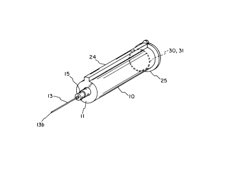
Turning now to the drawings and refernng first to FIG. 1, there is shown a
blood sample collection device having a cylindrical barrel 10 that forms an
apertured
end wall 11 at one end, while the other end 12 is open to accept a
conventional pre-
vacuumed test tube (not shown) to collect samples of blood transmitted by a
double-
ended needle 13. The needle 13 is carried by a circular needle plate 14
mounted for
reciprocating axial movement within the barrel 10. In the illustrative
embodiment of
FIGS. 1-10, the needle 13 is a conventional double-ended needle having a
plastic hub
15 rigidly attached to the needle between the two sharp ends 13a and 13b. The
hub 15
is located slightly closer to the proximal end 13a than the distal end 13b so
that the
length of needle projecting distally from the hub 15 for insertion into the
patient is
greater than the length of needle projecting proximally from the hub for
piercing the
test tube.
The mechanism for effecting axial movement of the needle plate 14 within the
barrel includes a guide tube 20 telescoped into the barrel 10 and a pin 21
projecting
radially from the plate 14 through a spiral slot 22 formed by the tube 20 and
extending
along a substantial portion of the length of the barrel 10. The outer end of
the pin 21
extends through a straight longitudinal slot 23 formed in the wall of the
barrel 10. To
close the barrel wall to prevent any leakage of blood or other liquid from the
interior
of the barrel, and to prevent undesired materials from entering the interior
of the barrel
10, the slot 23 is covered by a longitudinal channel 24 that may be formed as
an
integral part of the barrel 10 or as a separate part that is bonded to the
barrel wall
along the margins of the slot 23. The channel 24 receives the outer end of the
pin 21
and permits longitudinal movement of the pin as it passes along the slot 23.
The guide tube 20 is telescoped into the barrel 10 through the open end 12
with the outside wall of the tube 20 sliding along the inside wall of the
barrel 10. To
C: 72867( 1 K8301!. DOC)
CA 02240920 1998-06-17
permit relative rotational movement between the barrel 10 and the tube 20,
their
opposed walls are not attached to each, and do not fit so tightly as to
inhibit such
rotational movement. Relative rotational movement between the barrel 10 and
the
guide tube 20 causes the pin 21 to be cammed longitudinally along the slot 23
in the
wall of the barrel. The pin 21 cannot move in a circumferential direction
relative to
the barrel 10 because the pin is captured in the longitudinal slot 23.
Consequently,
rotational movement of the guide tube 20 relative to the barrel 10 causes the
walls of
the spiral slot 22 in the tube 20 to cam the pin longitudinally along the
straight slot 23.
Because the pin 21 is an integral part of the needle plate 14, and the needle
hub 15 is
attached to the plate 14, the needle 13 and its hub 15 and plate 14 all follow
the
longitudinal movement of the pin 21. Thus, advancing and retracting axial
movement
of the needle 13 relative to the barrel 10 can be effected by simply turning
the guide
tube 20 within the barrel 10. To facilitate such turning of the guide tube 20,
a knurled
flange 25 is formed on the proximal end of the tube 20. The user simply holds
the
barrel 10 while turning the knurled flange 25.
To retain the guide tube 20 within the barrel 10, the outer surface of the
guide
tube 20 forms a circumferential rib 30 that snaps into a corresponding groove
31
formed in the inside surface of the barrel 10 when the tube 20 is telescoped
into the
barrel 10. The fit between the tube 20 and the barrel 10 is sufficiently tight
that the
interlock between the rib 30 and groove 31 holds the two parts together in the
longitudinal direction, unless a substantial axial force is exerted on the two
parts to
separate them. Yet, because the rib 30 and groove 31 are continuous around the
circumferences of the tube 20 and barrel 10, the interlock does not inhibit
relative
rotational movement between the two parts.
In the illustrative embodiment, the proximal margin of the groove 31 forms a
large acute angle (close to 90 degrees) with the base of the groove (see FIG.
9), while
the distal margin slopes away from the base of the groove at an obtuse angle.
The
proximal side of the rib 30 is perpendicular to the axis of the tube 20, while
the distal
side is tapered with a gradual slope. These shapes facilitate the telescoping
of the
guide tube 20 into the barrel until the rib 30 snaps into the groove 31,
thereby locking
the tube 20 and barrel 10 together in the longitudinal direction, while
permitting
relative rotational movement between them.
C: 72867(1K8301!.DOC)
CA 02240920 1998-06-17
The circular needle plate 14 fits snugly inside the guide tube 20 and receives
one end of the hub 15 of the double-ended needle 13. The hub 15 and the plate
14
may be adhesively bonded to each other. The needle plate 14 is provided with
substantial margins 14a so that it does not tilt and remains steady within the
inner
tube.
In order to lock the needle 13 in either its fully advanced or fully retracted
position, a latch is provided to lock the guide tube 20 to the barrel 10 in
the
circumferential direction so that these two members cannot be rotated relative
to each
other. In the illustrative embodiment, this latch is formed as an integral
part of the
flange 25 on the proximal end of the guide tube 20. Thus, a pair of parallel
slits S 1
and 52 extend inwardly from the outer edge of the flange 25 to a living hinge
52. This
forms a small rectangular tab 53 which can be pivoted away from the end of the
barrel 10. This tab 53 is positioned directly over the end of the channel 24,
and the
distal surface of the tab 53 forms a small bead 54 which fits into the open
interior of
the channel 24 with a frictional fit. When the tab 53 is in this latched
position, as
illustrated in FIG. 9, the interlocking engagement of the bead 54 with the
interior
walls of the channel 24 prevents relative rotational movement between the
barrel 10
and the guide tube 20. When it is desired to unlatch these two members to
permit
relative rotational movement therebetween, the tab 53 is pivoted outwardly
away from
the end of the barrel 10 to remove the bead 54 from the interior of the
channel 24.
The tab 53 3is moved to this unlatched position whenever it is desired to
advance or
retract the needle holder 14 within the barrel 10.
FIG. 10 illustrates a modified needle holder 60 which eliminates the need for
a
double-ended needle. In this case, the needle holder 60 itself forms a tapered
stem 61
which is sufficiently sharp to penetrate the rubber stopper in the end of a
pre-
vacuumed sample collection tube. The rubber stopper in these pre-vacuumed
tubes is
typically pre-slit, and thus the tapered stem 61 is sufficiently sharp to
penetrate the
rubber stopper. An internal longitudinal passageway within the tapered stem 61
communicates with the hollow interior of the needle 63 which is fastened to
the distal
end of the needle holder.
The needle holder 60 in FIG. 10 also includes a modified arrangement for
mounting the needle 63 in the needle holder. In this modified arrangement, the
C: 72867( 1 K83011.DOC)
CA 02240920 1998-06-17
needle 63 is mounted on, and extends through, a hub having a threaded external
surface 64 which mates with a threaded bore 65 in the distal end of the main
body of
the needle holder 60. The hub also includes a flange 66 which seats against
the distal
end surface of the needle holder 60 when the hub is fully threaded into the
bore 65.
To use the illustrative sample collection device, the patient's skin is
cleansed
with isopropyl alcohol, the vein is made prominent by a tornique, and then the
vein is
punctured by the needle. When the needle is felt to be in the vein, the blood
sample
collection unit is stabilized and a pre-vacuumed test tube is advanced into
the barrel in
an inverted position. The end of the needle inside the barrel punctures the
rubber cap
of the test tube and fills the test tube as a result of vacuum inside the
tube. Once filled
the tube is removed and additional tubes are inserted into the barrel to
obtain
additional diagnostic samples.
After the required number of samples have been obtained, the needle is
withdrawn and the bleeding puncture area is compressed and taped. Holding the
sample collection device by one hand, the living hinge latch on the margin of
the
guide tube is released, and the guide tube is then rotated for a single turn.
The guide
tube stops automatically after one full turn, and both ends of the needle are
now
concealed within the barrel of the blood sample collection device. This is
achieved by
a specific length of the spiral slot that stops at a predetermined location.
The latch is
then closed to lock the needle device securely at this location, and the
device is then
disposed of.
The components of the sample collection device can be made by conventional
methods of machining steel tubing, and injection molding by using medical
grade
polymers such as polypropylene and others for barrel, plunger and latch
components.
The plunger seal or cap is molded from natural or synthetic elastomeric. The
spiral
sleeve is molded conventionally from polypropylene by using two slides and a
core
pin, and is press fit and locked with detentes into the wider proximal part of
the barrel.
Alternatively, molding of the spiral slot can be performed by rotating cores
with
elevated spiral. After molding the spiral around the cores, the cores are
removed while
simultaneously rotating them. The linear channel in the barrel is molded by
press
fitting a plate in the core pin and providing an identical channel in the
cavity of the
barrel. The needle plate with molded thread and arm are molded from
polypropylene.
C: 72867[ 1 K8301I. DOC)
CA 02240920 1998-06-17
Another embodiment of the present invention, illustrated in FIGS. 11-16b,
provides a blood sample collection assembly including an integral external
body 300
forming a first needle chamber 302, a second needle chamber 303, and a vacuum
tube
chamber 304. The axis of the first needle chamber 302 is orthogonal to the
axis of the
vacuum tube chamber 304. The first needle chamber 302 is a tubular or square
hollow body having a hollow tapered conical nozzle 306 integrally connected to
the
distal end thereof. The nozzle 306 forms a locking female luer taper. The
interior of
the conical nozzle 306 communicates with the interior of the first needle
chamber 302.
A cylindrical needle holder 308, having either a double-ended hypodermic
needle with a ninety degree bend or a pair of orthogonal hypodermic needles
310, 312
mounted therein, is disposed within the first needle chamber 302. The needle
holder
308 is displaceably interlocked to the first needle chamber 302 by a taper
lock
between the conically tapered portion 314 of the needle holder 308 and the
nozzle
306. The needle 310 protrudes from the distal end of the needle holder 308 and
is
coaxial with the first needle chamber 302. Prior to using the blood sample
collection
assembly, the needle 310 is covered by a conventional protective cap (not
shown) to
prevent the sharp beveled point of the needle 310 from accidently puncturing
someone. The needle 310 projects approximately one and one-quarter inches from
the
tapered portion 314 of the needle holder 308.
The other needle 312 is integrally connected to and mounted ninety degrees
away from the needle 310. During normal use, the needle 312 is positioned
along the
axis of the vacuum tube chamber 304 and the needle 312 protrudes from a side
arm
313 of the needle holder 308 into the vacuum tube chamber 304 at its base 338.
In
one embodiment, the needle 312 is a metallic hypodermic needle capable of
penetrating a rubber stopper of a conventional vacuum tube. Alternatively, the
needle
312 may be composed of plastic capable of penetrating a rubber stopper but
incapable
of penetrating skin, thereby eliminating the need for the second needle
chamber 303.
Such a plastic needle is manufactured and distributed by Baxter International
of
Deerfield, Illinois. The use of the plastic needle reduces the possibility of
accidental
needle punctures by one-half. Since both of the needles 310, 312 are hollow,
the
interior of the needle 310 communicates with the interior of the needle 312 to
form a
continuous flow path between the needles.
C: 72867(1K8301LDOC)
CA 02240920 1998-06-17
10
The needle 312 is covered by a self sealing rubber cap 330 having an open end
332 and a closed end 334 (FIG. 16a). The rubber cap 330 is retained in place
by a
plastic retaining ring 336 mounted on the circumference of the needle 312
(FIG. 16a).
As a vacuum tube 340 is inserted into the vacuum tube chamber 304, the rubber
stopper 342 of the vacuum tube 340 depresses the cap 330 so that the needle
312
pierces both the closed end 334 of the cap 330 and the rubber stopper 342
(FIG. 13).
This allows blood entering the needles 310, 312 to pass into the vacuum tube
340. As
the vacuum tube 340 is removed from the vacuum tube chamber 304, the cap 330
springs back to its position covering the needle 312 so as to check the flow
of blood
exiting from the needle 312. Thus, the rubber cap 330 acts as a valve which is
opened
by inserting a vacuum tube 340 into the vacuum tube chamber 304 and which is
closed by removing the vacuum tube 340 from the vacuum tube chamber 304. The
vacuum tube 340 recited herein may be any glass or plastic tube or tubular
stem of a
flask that is closed by a rubber stopper and contains a vacuum.
Following normal use of the blood sample collection assembly, the needle
holder 308 is retracted toward the proximal end of the first needle chamber
302,
thereby causing the needle 312 to retract into the second needle chamber 303
(FIG.
15). The second needle chamber 303 is preferably a generally rectangular
bodywhich
is narrow in one transverse direction and relatively wide in the other
transverse
direction (see FIGS. 16a-16b). The transverse dimensions are sufficiently wide
to
accommodate the width and length of the needle 312. The longitudinal dimension
of
the second needle chamber 303 is sufficiently long to accommodate the needle
312
when the needle holder 308 is fully retracted within the first needle chamber
302.
At the proximal end, the first needle chamber 302 forms a lid 316 having a
circular lid aperture 318. Furthermore, an internal tube 320 is disposed
within the first
needle chamber 302, and the tube 320 is connected to a circular knob 322 via
the lid
aperture 318. Both the tube 320 and the circular knob 322 are coaxial with the
first
needle chamber 302, and rotation of the circular knob 322 relative to the
first needle
chamber 302 causes the tube 320 to rotate relative to the first needle chamber
302.
The circular knob 322 preferably includes a textured longitudinal surface to
permit the
knob 322 to be easily gripped and rotated. The length of the internal tube 320
matches the internal longitudinal dimension of the first needle chamber 302 to
prevent
c: ~zas~axssoi~.~oc~
CA 02240920 1998-06-17
axial movement of the tube 320 relative to the first needle chamber 302.
Moreover,
the outer diameter of the internal tube 320 is slightly smaller than the inner
diameter
of the first needle chamber 302 to permit the internal tube 320 to rotate
freely, yet
stably, relative to the first needle chamber 302.
Rotation of the internal tube 320 relative to the first needle chamber 302
causes the needle holder 308 to move axially within the first needle chamber
302 from
a forward position to a retracted position. In FIG. 14c, the forward position
of the
needle holder 308 is depicted in solid lines, while the retracted position is
depicted in
dotted lines. Axial movement of the needle holder 308 relative to the first
needle
chamber 302 is effected using a helical slot 324 in the circumferential wall
of the
internal tube 320 in conjunction with a longitudinal slot 326 in the wall of
the first
needle chamber 302. The helical slot 324 and longitudinal slot 326 are
positioned
such that the side arm 313 of the needle holder 308 extends through both the
distal
end of the helical slot 324 and the distal end of the longitudinal slot 326
when the
needle holder 308 is in the forward position (FIG. 14a). Similarly, when the
needle
holder 308 is in the retracted position, the side arm 313 extends through both
the
proximal end of the helical slot 324 and the proximal end of the longitudinal
slot 326.
From the foregoing arrangement, it can be seen that the distal end of the
longitudinal
slot 326 overlies the distal end of the helical slot 324 when the needle
holder 308 is in
the forward position, and the proximal end of the longitudinal slot 326
overlies the
proximal end of the helical slot 324 when the needle holder 308 is in the
retracted
position.
During retraction of the needle holder 308, the first needle chamber 302 is
held
stationary while the internal tube 320 is rotated using the circular knob 322.
While
the internal tube 320 is being rotated, the needle holder 308 is prevented
from rotating
with the internal tube 320 by virtue of the extension of the side arm 313
through the
longitudinal slot 326 in the first needle chamber 302. Instead of rotating,
the needle
holder 308 moves axially through the first needle chamber 302. More
specifically,
axial movement of the needle holder 308 is controlled by the movement of the
side
arm 313 through the helical slot 324 in the rotating tube 320. As the side arm
313
moves through the helical slot 324, the side arm 313 cannot rotate or "swing"
with the
internal tube 320 because the side arm 313 is lodged in the longitudinal slot
326.
C: 72867(1K8301!.DOC)
CA 02240920 1998-06-17
12
Instead, the side arm 313 is forced to ascend the longitudinal slot 326 until
the side
arm 313 reaches the proximal end of the helical slot 324 (FIG. 15). At the
proximal
end of the helical slot 324, there is a detente 328 for engaging the ascending
side arm
313, thereby locking the needle holder 308 in the retracted position.
To operate the blood sample collection assembly, the protective cap is
removed, the blood sample collection site on the body of a patient is
determined, and
the skin is cleaned with an antiseptic solution. The needle 310 is then
entered into the
vein of the patient. To collect a blood sample, the vacuum tube 340 with the
rubber
stopper 342 is inserted into the vacuum tube chamber 304 (FIGS. 12-13). After
the
vacuum tube 340 is filled with the desired amount of blood, the vacuum tube
340 is
removed from the vacuum tube chamber 304. Additional blood may be collected by
inserting, filing, and removing additional vacuum tubes. The vacuum tube
chamber
304 includes tabs 344 for providing leverage while pushing a vacuum tube over
the
point of the needle 312 to puncture the rubber stopper of the vacuum tube.
While
inserting a vacuum tube, the orthogonal orientation of the vacuum tube chamber
304
relative to the first needle chamber 302 compels that the vacuum tube be
pushed into
the vacuum tube chamber 304 in a direction toward a stabilizing hand, rather
in the
direction of the sharp point of the needle 310 located in the vein. Next, the
needle 310
is withdrawn from the vein. The circular knob 322 is rotated until the needle
holder
308 is completely retracted with the side arm 313 locked in the detente 328
(FIG. 15).
With the needle holder 308 in the retracted position, the needle 310 is
concealed by
the first needle chamber 302 and the needle 312 is concealed by the second
needle
chamber 303. Finally, the blood sample collection assembly is discarded in its
entirety.
It can be seen from the foregoing description that the blood sample collection
assembly avoid the situation of advancement of one sharp point of a double -
pointed
needle towards an operator while retraction of another point of the double-
pointed
needle is attempted. In particular, since the needle 312 is orthogonal to the
needle
310, the side of the needle 312, instead of the point of the needle 312, is
advanced
toward an operator while the needle 310 is retracted into the first needle
chamber 302.
Moreover, both needles 310, 312 are retracted and concealed by operating a
single
mechanism, the circular knob 322. Due to the orthogonal orientation of the
vacuum
C: 92869( 1 K830I LDOC)
CA 02240920 1998-06-17
13
tube chamber 304 relative tot he first needle chamber 302, insertion of a
vacuum tube
is accomplished by pushing the vacuum tube in the direction of a stabilizing
hand of
an operator, rather than in the direction of the needle in the vein. This
reduces the
possibility of imparting forward thrust on the needle in the vein which, in
turn,
minimizes the possibility of double puncturing the vein. Furthermore, the
blood
sample collection assembly is compact because the needle holder 308 is
retracted
directly into the internal tube 320 itself. Because the needle holder 308
retracts into
the internal tube 320, the internal tube 320 need not extended beyond the
proximal
end of the first needle chamber 302 for needle retraction to occur. Thus, when
discarded following use, blood sample collection assembly contributes
minimally to
the bulk of refuse. The blood sample collection assembly is also compact
because,
with the needles 310, 312 mounted orthogonal to one another, the length of the
assembly is shorter than existing blood sample collection assemblies.
Refernng to FIGS. 16a-16b, the blood sample collection assembly is
constructed by injection molding the entire assembly from organic polymers,
preferably thermoplastics such as a polypropeline or ABS. To construct the
needle
chambers 302, 303 and the vacuum tube chamber 304, a polymeric sheet is
thermoformed to represent a single piece, mirror image, isometric half of
these
elements. Next, the internal tube 320 with the mounted knob 322 and the needle
holder 308 with the mounted needles 310, 312 are positioned at the proper
location on
the thermoformed sheet, and the thermoformed sheet is folded and secured shut
by
interlocking detents on the contacting surfaces (FIG. 16b). For additional
safety an
ultrasonic or solvent bond is created at the areas of contact to maintain the
integrity of
the assembly. The assembly is sterilized by conventional means.
C: 72867(1K8301l.DOC)