Note: Descriptions are shown in the official language in which they were submitted.
CA 02248260 1998-08-31
WO 97/32532 PCTIUS97/03637
1
TITLE
VASCULAR CATHETER-BASED SYSTEM FOR HEATING TISSUE
BACKGROUND OF THE INVENTION
This invention relates to a catheter-based system to position an electrode
for providing energy to shrink a vein intraluminally to change the fluid flow
dynamics
and to restore the competency of venous valves and the proper function of the
vein in
a minimally invasive procedure.
5 The human venous system of the lower limb consists essentially of the
superficial venous system and the deep venous system with perforating veins
connecting
the two systems. The superficial system includes the long or great saphenous
vein and
the short saphenous vein. The deep venous system includes the anterior and
posterior
tibial veins which unite to form the popliteal vein, which in turn becomes the
femoral
vein when joined by the short saphenous vein.
The venous systems, contain numerous one-way valves for directing blood
flow back to the heart. Venous valves are usually bicuspid valves, with each
cusp
forming a sack or reservoir for blood which, under pressure, forces the free
surfaces of
the cusps together to prevent retrograde flow of the blood and allow antegrade
flow to
the heart. When an incompetent valve is in the flow path of retrograde flow
toward the
foot, the valve is unable to close because the cusps do not form a proper seal
and
retrograde flow of blood cannot be stopped.
Incompetent valves in the venous system can occur with vein dilation.
Separation of the cusps of the venous valve at the commissure may occur as a
result.
The leaflets are stretched by the dilation of the vein and concomitant
increase in the
vein diameter which the leaflets traverse. Stretching of the leaflets of the
venous valve
allows the loose leaflets to fold on themselves and leave the valve open. This
prolapse
can allow reflux of blood in the vein. Eventually the venous valve fails,
thereby
increasing the pressure on the lower venous sections and overlying tissues.
Two venous
diseases which often involve vein dilation are varicose veins and chronic
venous
insufficiency.
CA 02248260 1998-08-31
WO 97/32532 PCT/US97/03637
2
The varicose vein condition includes dilation and tortuosity of the
superficial veins of the lower limb, resulting in unsightly discoloration,
pain and
ulceration. Varicose veins often involve incompetence of one or more venous
valves,
which allow reflux of blood from the deep venous system to the superficial
venous
system or reflux within the superficial system. Current treatments include
such invasive
open surgical procedures as vein stripping, sclerotherapy, and occasionally,
vein grafting,
venous valvuloplasty, and the implantation of various prosthetic devices. The
removal
of varicose veins from the body can be a tedious, time-consuming procedure
having a
painful and slow healing process. Complications, scarring, and the loss of the
vein for
future cardiac and other by-pass procedures may also result. Along with the
complications and risks of invasive open surgery, varicose veins may persist
or reoccur,
particularly when the valvular problem is not corrected. Due to the long,
arduous, and
tedious nature of the surgical procedure, treating multiple venous sections
can exceed the
physical stamina of the physician, and thus render complete treatment of the
varicose
vein conditions impractical.
Chronic venous insufficiency (CVT) is a problem caused by hydrodynamic
forces acting on the tissues of the body, especially the legs, ankles and
feet. As the veins
dilate due to increased pressure, the valves in the veins fail. This causes
the pressure to
increase on the next valve and vein segment down, causing those veins to
dilate, and as
this continues, the valves in the veins eventually all fail. As they fail, the
effective
height of the column of blood above the feet and ankles grows, and the weight
and
hydrostatic pressure exerted on the tissues of the ankle and foot increases.
When the
weight of that column reaches a critical point from the valve failures,
ulcerations of the
ankle begin to form, which start deep and eventually come to the surface.
These
ulcerations do not heal easily because the weight of blood which caused them
continues
to persist, and have the tendency to enlarge the ulcer.
Chronic venous insufficiency often consists of hypertension of the lower
limb in the deep, perforating and often superficial veins, and may result in
discoloration,
pain, swelling and ulceration. Existing treatments for chronic venous
insufficiency are
often less than ideal. These treatments include the elevation of the legs,
compressing the
veins externally with elastic support hose, and surgical repair by grafting
vein sections
with healthy valves from the arm into the leg. These methods have variable
effectiveness. Moreover, invasive surgery has its associated complications
with risk to
CA 02248260 1998-08-31
WO 97/32532 PCT/US97/03637
3
life and expense. Similarly, the palliative therapies require major lifestyle
changes for
the patient. For example, the ulcers will reoccur unless the patient continues
to elevate
the legs and use support hose continuously throughout the life of the patient.
Due to the time-consuming and invasive nature of the current surgical
treatments, such as vein grafting, typically only one valve is treated during
any single
procedure. This greatly limits the ability of the physician to fully treat
patients suffering
from chronic venous insufficiency. Every instance of invasive surgery,
however, has its
associated complications with risk to life and expense.
The ligation of vascular lumen by tying a suture around them,
cauterization or coagulation using electrical energy from an electrode has
been employed
as an alternative to stripping, or the surgical removal of such veins.
However, ligation
procedures close off the lumen, essentially destroying their functional
capability. For
example, it is known to introduce an electrode into the leg of a patient, and
position the
electrode adjacent to the exterior of the varicose veins to be treated.
Through a small
stab incision, a probe is forced through the subcutaneous layer between the
fascia and
the skin, and then to the various veins to be destroyed. Electrodes at the
outer end of
the probe are placed adjacent to the varicose veins. Once properly positioned,
an
alternating current of 500 kilohertz is applied to destroy the adjacent
varicose veins.
The veins lose the function of allowing blood to flow through, and are no
longer of use.
For example, ligating the saphenous vein would render that vein unavailable
for
harvesting in other surgical procedures such as coronary by-pass operations.
Ligation
techniques which functionally destroy the vein lumen would appear be
inappropriate
to a corrective procedure for restoring and maintaining the function of the
vein.
Hemorrhoids are dilated veins in and around the anus and lower rectum.
Dilation may result from an increased pressure in the hemorrhoidal vein.
Constipation,
including the frequent straining to pass hard stools increases pressure in
hemorrhoidal
veins, is a common cause of hemorrhoids. Other contributing factors include
pregnancy, a low fiber diet, and obesity. As the hemorrhoidal vein becomes
more
dilated from the increased pressure, the venous valves of the hemorrhoidal
vein may
begin to fail and become incompetent. This can exacerbate the dilation of the
hemorrhoidal vein as reflux of blood is allowed in the vein by the open
incompetent
valve. The vein may eventually form a sac-like protrusion if the condition is
allowed
to persist. Hemorrhoids are generally classified as being either internal or
external,
CA 02248260 1998-08-31
WO 97/32532 PCT/US97/03637
4
depending on their location relative to the dentate line. The dentate line is
easily
identified as the demarcation between the pink mucosa that form the anoderm.
The
dentate line separates the internal and external hemorrhoid systems. Internal
hemorrhoids are located inside the anus above the dentate line. External
hemorrhoids
are located below the dentate line. Either can extend out of the anus.
Straining or irritation caused by passing stool can injure the delicate
surface
of an internal hemorrhoid and cause bleeding. If the pressure and dilation of
the
hemorrhoidal vein continues, the internal hemorrhoids may prolapse and be
forced
through the anal opening. If a hemorrhoid remains prolapsed, considerable
discomfort,
including itching and bleeding, may result. The blood supply to these
prolapsed
hemorrhoids may become cut -off by the anal sphincter, which gives rise to a
strangulated hemorrhoid. Thrombosis may result where the blood within the
prolapsed
vein becomes clotted. This extremely painful condition can cause edema and
inflammation.
Increased pressure in the portal venous system can also cause an increase
in pressure of the superior hemorrhoidal vein (SHV) leading to an increased
diameter
of the hemorrhoid. The portal venous system allows venous drainage from the
intestinal
tissues to the liver, and can become hypertensive when the lever is cirrhotic.
The treatment methods for hemorrhoids include invasive surgery to
remove the hemorrhoid, elastic ring ligation, sclerotherapy, and the
application of
topical ointments or suppositories. The surgical removal of extensive or
severe
hemorrhoids is known as a hemorrhoidectomy. This surgical procedure can be
used on
both internal and external hemorrhoids. However, such surgery typically
involves a
long recovery period, along with the associated risks and expense of invasive
surgery.
Internal hemorrhoids may be treated by rubber band ligation, where a
legator is inserted through a scope in the anal canal. The hemorrhoid is
grasped with
forceps in the legator and held in position. The legator includes a cylinder
which is slid
upwards and releases one or more rubber bands around the base of the
hemorrhoid.
A typical diameter for the rubber band is one millimeter. The band cuts off
the
circulation of blood to the hemorrhoid, and the hemorrhoid begins to wither
away.
Provided the rubber band remains in place, the hemorrhoid typically drops off
within
seven to ten days.
CA 02248260 1998-08-31
WO 97/32532 PCT/US97/03637
Sclerotherapy, another treatment for hemorrhoids, involves injecting a
solution, such as sodium morrhuate or phenol oil, submucously into the areolar
tissue
around the hemorrhoidal vein to cause inflammation and scarring to eliminate
the
hemorrhoid. Other external treatments cause burning or coagulation to destroy
the
5 hemorrhoid. In infrared coagulation, infrared light may be applied to create
a small
tissue-destroying burn around the base of the hemorrhoid to cut off the blood
supply
to the hemorrhoid. Electrocoagulation, sometimes referred to as bipolar
diathermy, may
be utilized in a similar manner. In laser therapy, also known as vaporization,
a laser
beam causes a superficial burn to seal off the blood vessels and retain the
hemorrhoid
in a non-prolapsed position.
The prior treatments for hemorrhoids involving external ligation or
excision of the hemorrhoid may not affect the underlying causes which gave
rise to the
hemorrhoidal condition initially. Thus the condition may recur.
Varicose veins called esophageal varices can form in the venous system
along the submucosa of the lower esophagus, and bleeding can occur from the
dilated
veins. Blood returns to the heart from the portal venous system through the
veins
surrounding the esophagus. Unlike other veins, such as the saphenous vein in
the lower
leg, the veins surrounding the esophagus typically do not have valves for
bringing blood
back to the heart. The venous pressure in these esophageal veins is relatively
high, and
blood can flow back to the heart without aid of venous valves.
Esophageal varices may result from portal hypertension and other
abnormalities in the portal venous system, such as cirrhosis of the liver.
Bleeding or
hemorrhaging may result from esophageal varices, which can be difficult to
stop and, if
untreated, could develop into a life threatening condition. Such varices can
erode easily,
and lead to a massive gastrointestinal hemorrhage.
Treatments for esophageal varices include portal-caval shunts, endoscope
variceal ligation, sclerotherapy, and electrocoagulation from an electrode
within the
esophagus, such as from a tamponade device. The portal shunt involves the
surgical
joining of two veins, the portal vein and the inferior vena cava, to relieve
pressure in the
vein carrying blood into the liver. Although effective in eliminating
recurrent
hemorrhaging from varices, the attendant risks and complications of such
invasive
surgery, including encephalopathy and post-shunt hepatic failure, still exist
for the portal
shunt operation.
CA 02248260 1998-08-31
WO 97/32532 PCTIUS97/03637
6
Endoscopic variceal ligation is analogous to rubber band ligation for
treating hemorrhoids. The esophageal varices are ensnared with elastic bands
to
eradicate the varices. An endoscope is introduced into the patient and is
placed adjacent
to the esophageal varices to be treated. The varix is drawn into a drum
attached to the
tip of the endoscope. An elastic band mounted on the drum is then released
over the
varix. Endoscope variceal ligation may not achieve complete fibrosis of the
inner wall
of the esophagus, and recurrence of the varices may result. Other
complications include
bleeding from ulcers induced by the elastic bands, and esophageal obstruction
due to
occlusion of the lumen by banded esophageal varices.
In sclerotherapy, a solution, such as sodium morrhuate or ethanolamine,
is injected submucosally into the tissue around the varicose vein in the
esophagus to
cause inflammation and scarring to close off the vein and reduce the
likelihood of
bleeding. Sclerotherapy, however, may create ulcerations which can lead to
esophageal
strictures.
Electrocoagulation has also been used to treat esophageal varices. A
tamponade device having a metalized surface is introduced into the esophagus.
The
metalized surface is brought into contact with the mucous membrane of the
esophagus.
An electric current is then applied to the metalized surface to cause a
thrombosing of
the esophageal varices. This procedure may be employed to stop immediate
hemorrhaging of the esophageal varices.
The prior treatments for esophageal varices typically involve external
coagulation or obliteration of the veins, and often require multiple treatment
sessions.
Such treatments do not treat the varicosity directly, and may not affect the
underlying
causes which gave rise to the esophageal varices initially.
A need exists in the art for a system to treat dilated veins, such as those
resulting in varicose veins or from venous insufficiency, which maintains the
patency
of the veins for venous function and yet restores valvular competency. A need
also
exists in the art to treat dilated hemorrhoidal veins to reduce venous
pressure on the
hemorrhoidal region. Such treatment should maintain the functional patency of
the vein
and restore valvular competency at the origins of the hemorrhoids as well as
within the
hemorrhoid itself. A need exists in the art to treat the dilated veins which
give rise to
esophageal varices and reduce venous pressure on the esophageal region from
the portal
vein system without the attendant risks of invasive surgery. Further need
exists to
CA 02248260 2005-11-25
7
provide a less invasive procedure which can treat multiple venous sites
quickly and easily. The
need exists to restore and normalize flow patterns, dynamics, and pressure,
and shrink sections of
dilated veins to a normal or reduced diameter. Where bleeding occurs, there is
a need to achieve
hemostatis in bleeding varices and minimize recurrence of bleeding.
SUMMARY OF THE INVENTION
In one embodiment of the invention there is provided an apparatus for applying
energy to
cause shrinkage of a vein, the apparatus comprising: a catheter having a
working end and an outer
diameter, the outer diameter of the a catheter is less than the inner diameter
of the vein; means for
heating a venous treatment area to cause a reduction in the diameter of the
vein, the means for
heating is located at the working end of the catheter; and a plurality of
bowable members
connected to the working end of the catheter, the bowable members having
sufficient strength to
limit the reaction in the diameter of the vein so that the vein remains patent
for continued venous
function.
In another embodiment of the invention there is provided use of the
aforementioned
apparatus for applying energy to cause shrinkage of a vein.
In another embodiment of the invention there is provided the use of an
apparatus for
applying energy to cause shrinkage of a vein, the apparatus comprising: a
catheter having a
working end and an outer diameter, the outer diameter of the catheter being
less than the inner
diameter of the vein; a device for heating a venous treatment area to cause
shrinkage of the vein,
the heating device being located at the working end of the catheter; and means
for controlling
shrinkage of the vein so that the vein remains patent for venous function.
CA 02248260 2005-11-25
7a
Briefly, and in general terms, the present invention provides a less invasive
and faster method for solving the underlying problems of varicose veins and
venous
insufficiency, and uses a novel repair system, including a catheter for
placement of an
electrode for delivering radio frequency energy. The present invention
includes a
method of applying energy to cause shrinkage of a vein, the method comprising
the
steps of introducing a catheter having a working end and means for heating
located at
the working end, to a treatment site in a vein; positioning the means for
heating at the
treatment site in the vein; applying energy from the means for heating to
controllably
heat the treatment site and cause shrinkage of the vein; and terminating the
emission of
energy from the means for heating after sufficient shrinkage of the vein has
occurred so
as to restore valvular competency or so that the vein remains patent so as to
continue
to function as a blood conduit. The method of the present invention is a
minimally
invasive procedure which eliminates the need for open surgical procedures for
venous
repair, including venous valvuloplasty, and the transplantation of an arm vein
into the
leg.
An apparatus for performing the method of applying radiant energy to
cause shrinkage of a vein, comprises a catheter having a working end, means
for heating
a venous treatment area to cause shrinkage of the vein, wherein the means for
heating
is located at the working end of the catheter, and means for preventing
further shrinkage
after sufficient shrinkage of the vein, so that the vein continues to
function. The
heating means may include RF electrodes to heat and shrink the vein. Balloons,
or
other mechanisms for controlling the outer diameter of the heating means, may
be used
to limit the amount of shrinkage. Feedback control systems may be applied to
these
mechanisms, or may be used to control the application of energy to heat the
venous
tissue, in order to control the amount of shrinkage.
CA 02248260 1998-08-31
WO 97/32532 PCTNS97/03637
8
Features of the present invention include restoring the competence of
venous valves, normalizing flow patterns, dynamics, and pressure, and reducing
sections
of dilated varicose veins to a normal diameter for cosmetic purposes. The
treated veins
remain patent and can continue to function and return blood to the heart.
One feature of the present invention is to provide a procedure for restoring
venous valvular competency by controllably shrinking the otherwise dilated
lumen of
the vein to a desired diameter.
Another feature of the present invention is to control or adjust the
effective diameter of the catheter or electrode configuration in order to
control the
amount of circumferential shrinking experienced by the vein wall. An
extendable
member located adjacent to the working end of the catheter can increase the
effective
diameter of the catheter and limit the shrinkage of the vein.
Another feature of the present invention is to provide a catheter electrode
which generates a radio frequency field around the circumference of the
catheter in
order to shrink the vein wall circumferentially and omnidirectionally while
minimizing
lengthwise contraction when the catheter electrode is positioned
intraluminally within
the vein.
Yet another feature of the present invention is to generate a field at a
specific frequency around the catheter in order to minimize coagulation within
the vein,
and to control the spread of heating within the venous tissue.
An additional feature of the present invention is to protect the venous
valve leaflets by minimizing the heating effect on the venous valves by the
selective
positioning of the electrodes within the vein.
Another feature of the present invention is to deliver cooling fluid to the
bloodstream in order reduce the likelihood of heating the blood to a point of
coagulation.
An additional feature of the present invention is to prevent shrinkage of
the vein past the end of the catheter.
Another feature of the present invention is to maintain the electrodes in
apposition to the venous tissue to ensure that the heat is delivered towards
the venous
tissue, and not the blood moving through the vein.
Another feature of the present invention is to use electrodes which are
bowable members that can be deflected radially outward for maintaining contact
with
CA 02248260 1998-08-31
WO 97/32532 PCTIUS97/03637
9
the venous tissue. The bowable members are conductive longitudinal electrodes
substantially covered by an insulating film, except for the portion which is
to come into
apposition with the venous tissue.
Another feature of the present invention is a balloon located on one side
of the catheter having electrodes on the opposite side. Inflation of the
balloon will
move the electrodes into apposition with the vein wall on the opposite side.
Yet another feature of the present invention is to provide a procedure
which can treat multiple venous sites quickly and easily.
An additional feature of the present invention is that no foreign object or
prothesis remain in the vasculature after treatment.
These and other aspects and advantages of the present invention will
become apparent from the following more detailed description, when taken in
conjunction with the accompanying drawings which illustrate, by way of
example, the
principles of the invention.
BRIEF DESCRIPTION OF THE DRAWINGS
FIGURE 1 shows a cross-sectional view of a dilated vein having
incompetent venous valves in a lower limb which are to be treated in
accordance with
the present invention;
FIGURE 2 shows a representative view of a venous section from FIG. 1
taken along lines 2-2 which is to be treated in accordance with the present
invention;
FIGURE 3 shows a partial cross-sectional view of a catheter having
electrodes being delivered antegrade to a venous treatment site in accordance
with the
present invention;
FIGURE 4 shows the partial cross-sectional view of the venous section of
FIG. 2 after being treated in accordance with the present invention;
FIGURE 5 shows a partial cross-sectional view of the catheter and vein
shown in FIGURE 3 being delivered to another venous treatment site in
accordance
with the present invention;
CA 02248260 1998-08-31
WO 97/32532 PCT/US97/03637
FIGURE 6 shows a partial cross-sectional view of a catheter being
delivered retrograde and deflected -laterally to a venous treatment site in
accordance with
the present invention;
FIGURE 7 shows a partial cross-sectional view of an embodiment of the
5 catheter having a bulbous tip and ring electrodes for treating a dilated
vein in accordance
with the present invention;
FIGURE 8 shows a partial cross-sectional view of an embodiment of the
catheter having a flush tip at the working end and ring electrodes for
treating a dilated
vein in accordance with the present invention;
10 FIGURE 9 shows a partial cross-sectional view of an embodiment of the
catheter having a cap electrode for treating a dilated vein in accordance with
the present
invention;
FIGURE 10 shows a partial cross-sectional view of another embodiment
of the catheter having a cap electrode and a balloon to center the placement
of the
electrode within the vein to be treated;
FIGURES 11a, 11b, and 11c show partial cross-sectional views of another
embodiment of the catheter having a bendable tip which deflects laterally for
causing
apposition between the electrodes of the catheter and the vein wall in
accordance with
the invention;
FIGURES 12a and 12b show partial cross-sectional side and top views,
respectively, of another embodiment of the catheter having a balloon on one
side of the
catheter and longitudinal electrodes on the other side at the working end of
the catheter
for moving the electrodes into appositional contact with the vein wall in
accordance
with the invention;
FIGURE 13 shows another embodiment of the catheter having bendable
electrodes which deflect outwardly for increasing the effective diameter at
the working
end of the catheter in accordance with the invention;
FIGURE 14 shows another embodiment of the catheter having a balloon
and bendable members with electrodes which deflect outwardly for increasing
the
effective diameter at the working end of the catheter in accordance with the
invention;
FIGURE 15a shows a cross-sectional view of an embodiment of the
catheter shown in FIGURE 14 having four equidistantly spaced electrodes in
accordance
with the present invention;
CA 02248260 1998-08-31
WO 97/32532 PCT/US97/03637
11
FIGURE 15b shows a cross-sectional view of an embodiment of the
catheter shown in FIGURE 14 having four electrodes preferentially spaced to
form two
pairs of electrodes in accordance with the present invention;
FIGURE 16 shows a partial cross-sectional view of another embodiment
of the catheter having four equidistantly spaced electrodes, and being
delivered
retrograde to a venous treatment site in accordance with the present
invention;
FIGURE 17 shows a partial cross-sectional view of an embodiment of an
over-the-wire balloon catheter having four equidistantly spaced apart
electrodes on the
surface of the balloon in accordance with the present invention;
FIGURE 18 shows a cross-sectional view taken along the lines 18-18 of the
over-the-wire balloon catheter of FIG. 17 in accordance with the present
invention;
FIGURE 19 shows a partial cross-sectional view of another embodiment
of the catheter having electrodes located within the balloon portion in
accordance with
the present invention;
FIG. 20 is a side view of an embodiment of a catheter having bowable
electrodes in accordance with the invention coupled with a block diagram of a
heat
treatment system;
FIG. 21 is a partial side view of the working end of the catheter illustrated
in FIG. 20, and having electrodes which deflect outwardly for increasing the
effective
diameter at the working end of the catheter in accordance with the present
invention;
FIG. 22 is a cross-sectional view along lines 22-22 of the electrode for the
catheter depicted in FIG. 21;
FIG. 23 is a cross-sectional view along lines 23-23 of FIG. 20, and depicts
a catheter having four equidistantly spaced electrodes in accordance with the
present
invention;
FIG. 24 is a cross-sectional view of another embodiment of the catheter
depicted in FIG. 23, this embodiment having four electrodes preferentially
spaced to
form two pairs of electrodes in accordance with the present invention;
FIG. 25 is a cross-sectional view of another embodiment of the catheter
depicted in FIG. 23, this embodiment having two pairs of opposing electrodes
in
accordance with the present invention;
FIG. 26 is a cross-sectional view of the catheter along lines 26-26 of FIG.
20;
CA 02248260 1998-08-31
WO 97/32532 PCT/US97/03637
12
FIG. 27 is a partial side view of the working end of another embodiment
of a catheter having a balloon and bendable members with electrodes in
accordance with
the present invention;
FIG. 28 is a cross-sectional view along lines 28-28 of FIG. 26;
FIG. 29 shows a partial cross-sectional view of the venous system of the
hemorrhoid region which is to be treated in accordance with the present
invention;
FIGS. 30a, 30b, 30c, and 30d are side views of an embodiment of a catheter
treating a venous treatment site within a dilated vein in accordance with the
present
invention.
FIG. 31 is a partial profile view of the anatomical region of the esophageal
region, including a vein to be treated in accordance with the present
invention;
FIGS. 32a, 32b and 32c are side views of an embodiment of the catheter
constructed and delivered to a venous treatment site within a dilated vein for
treatment
in accordance with the present invention.
DETAILED DESCRIPTION OF THE EMBODIMENTS
As shown in the exemplary drawings, the invention is directed toward the
intravenous treatment of veins using a catheter to deliver at least one
electrode to a
venous treatment site. As used herein, like reference numerals will designate
similar
elements in the various embodiments of the present invention to be discussed.
In
addition, unless otherwise noted, the term working end will refer to the
direction
toward the treatment site in the patient, and the term connecting end will
refer to the
direction away from the treatment site in the patient. The invention will be
described
in relation to the treatment of the venous system of the lower limbs. It is to
be
understood, however, that the invention is not limited thereto and may be
employed
intraluminally to treat veins in other areas of the body such as hemorrhoids,
esophageal
varices, and venous-drainage-impotence of the penis. Furthermore, although the
invention will be described as using RF energy from the electrode, it is to be
understood
that other forms of energy such as microwaves, ultrasound, direct current,
circulating
heated fluid, radiant light, and lasers can be used, and that the thermal
energy generated
from a resistive coil or curie point element may be used as well.
A partial cross-sectional view of a dilated vein from a lower limb having
incompetent valves is shown in FIG. 1. These veins are often disposed within
muscle
CA 02248260 1998-08-31
WO 97/32532 PCTIUS97/03637
13
tissue. Veins have bicuspid valves, and in a normal and competent valve, each
cusp
forms a sack or reservoir for blood which, under pressure, forces the free
edges of the
cusps together to prevent retrograde flow of the blood and allow only
antegrade flow
to the heart. The arrow leading out the top of the vein represents the
antegrade flow
of blood back to the heart. The venous valves prevent retrograde flow as blood
is
pushed forward through the vein lumen and back to the heart.
When an incompetent valve encounters retrograde flow, the valve is unable
to close, the cusps do not seal properly and retrograde flow of blood may
occur.
Incompetent valves may result from the stretching of dilated veins. As the
valves fail,
increased pressure is imposed on the lower veins and the lower valves of the
vein, which
in turn exacerbates the failure of these lower valves. A cross-sectional
perspective view
of a dilated vein taken along lines 2-2 of FIG. 1 is illustrated in FIG. 2.
The valve cusps
can experience some separation at the commissure due to the thinning and
stretching of
the vein wall at the cusps.
The method of the present invention for the minimally invasive treatment
of venous insufficiency can be performed using a catheter 10 to deliver
electrodes 12 to
a venous treatment site in order to restore the competency of a vein. One
embodiment
of the catheter 10 for delivering the electrodes 12 to the venous treatment
site is shown
in FIG. 3. The electrodes 12 may be two RF ring electrodes 14 and 16 located
at the
working end 11 of the catheter 10. This and other embodiments of the catheter
10 will
be described in greater detail later. Further, the method is contemplated to
be used with
any suitable appliance for applying radiant energy, thermal energy, or other
forms of
energy to heat and shrink the venous tissue in the repair or reconfiguration
of
incompetent veins in order to restore venous function or valvular competency.
Particular discussion will be directed to the treatment of incompetent and
varicose veins
in the legs, although the method of the present invention is well suited to
treating veins
in other areas of the body.
When treating the veins of the lower limbs, the patient is typically placed
onto a procedure table with the feet dependent in order to fill the veins of
the leg. The
leg of the patient is prepped with antiseptic solution. A percutaneous
introducer is
inserted into the vein using the well-known Seldinger technique to access the
saphenous
or deep vein system. Alternatively, a venous cut-down can be used to access
the vein
to be treated. The procedure for the repair of incompetent veins can be
accomplished
CA 02248260 1998-08-31
WO 97/32532 PCT/1JS97/03637
14
by a qualified physician with or without fluoroscopic or ultrasonic
observation, or under
direct visualization. Further, the physician could palpate the treatment area
to
determine the location of the catheter, and the treatment site, during the
procedure
when treating the superficial venous system.
The catheter 10 could be passed within the vein after insertion through the
introducer, and advanced through to the venous treatment site. Alternatively,
a guide
wire for the catheter may be inserted into the vein. The wire is advanced
antegrade to
the venous treatment site, such as the level of the most proximal incompetent
vein site
which is to be repaired. The catheter is then inserted upon the wire and is
fed up the
leg through the vein to the level of the venous section where retrograde flow
exists. In
either case, the catheter 10 delivers the electrodes 12 to the venous
treatment site.
Fluoroscopy, x-ray, ultrasound, or a similar imaging technique could then be
used to
direct the specific placement of the catheter and confirmation of position
within the
vein. X-ray contrast material can be injected through or around the catheter
to identify
the incompetent venous sections to be repaired.
From the antegrade approach, the catheter can be pushed through the
venous valve so that the electrodes are positioned across the valve of the
incompetent
venous section to be treated. The catheter 10 travels antegrade through the
venous
valves, as shown in FIG. 3, and is positioned so that the electrodes 12 are
near a dilated
section of the vein to be treated. The electrodes may be positioned so as to
extend past
the incompetent venous valve. When the electrodes 12 of the catheter 10 are
positioned
at the venous treatment site, the RF generator is activated to provide
suitable RF energy,
preferably at a selected frequency from a range of 250 kHz to 350 MHZ. One
suitable
frequency is 40 Mhz. One criteria for the selection of the applied frequency
is the
minimization of coagulation in the vein. Another criteria is to control the
spread and
depth of the thermal effect in the tissue. The extent of heating or depth of
penetration
into the tissue generally increases with lower frequencies, and decreases as
the frequency
increases. A microprocessor can be used to select a frequency for treating
different veins
according to the above criteria. For example, the microprocessor can include a
table
stored in memory for associating specific frequencies for treating various
veins and vein
diameters according to the criteria of minimizing coagulation and controlling
the spread
or depth of the heating effect. The energy emitted from the electrodes is
converted
within the venous tissue into heat. As the temperature of the venous tissue
increases,
CA 02248260 1998-08-31
WO 97/32532 PCT/US97/03637
the venous tissue begins to shrink. The shrinkage is due in part to
dehydration and the
structural transfiguration of the collagen fibers in the vein. Although the
collagen
becomes compacted during this process, the collagen still retains some
elasticity. When
RF energy is applied near the locus of the dilated vein and venous valve,
shrinkage of
5 the vein can restore valvular competency by reducing the dilation which is
preventing
the proper functioning of the venous valve.
The working end 11 of the catheter 10 near the electrodes 12 physically
limits the amount of shrinkage. The working end 11 is preferably sufficiently
sized or
enlarged to prevent the complete ligation of the vein. Other schemes, such as
an
10 inflatable balloon, may be used to mechanically limit or control the amount
of shrinkage
in the vein.
Vein dilation is reduced after RF energy applied from the electrodes 12
heats the surrounding venous tissue to cause shrinkage. RF energy is no longer
applied
after there has been sufficient shrinkage of the vein to alleviate the
dilation of the vein
15 near the valves, so as to restore venous function or valvular competency.
Sufficient
shrinkage may be detected by fluoroscopy, external ultrasound scanning,
intravascular
ultrasound scanning, impedance monitoring, temperature monitoring, direct
visualization
using an angioscope, or any other suitable method. For example, the catheter
10 can
be configured to deliver x-ray contrast medium to allow visualization by
fluoroscopy for
assessing the condition of the vein and the relationship of the catheter to
the treatment
area of the vein during the shrinkage process. As an alternative to
fluoroscopy, external
ultrasound techniques such as B-scanning using distinct ultrasound signals
from different
angles, or intravascular ultrasound can be used to acquire a more
multidimensional view
of the vein shrinkage at the treatment site, which improves the detection of
uneven
shrinkage in the vein. An angioscope may also be used to directly visualize
and
determine the extent and degree of vein shrinkage.
After treatment, the commissure and the cusps of the venous valves should
be closer together with little separation or prolapse, which indicates a
restoration of the
competency of the valve. A cross-sectional view of the venous valve after
being treated
with RF energy is shown in FIG. 4. Valvular competence may be determined by
contrast injection or Doppler probe measurement.
Substantial shrinkage may be achieved very rapidly, depending upon the
specific treatment conditions. Because the shrinkage can proceed at a rather
rapid rate,
CA 02248260 1998-08-31
WO 97/32532 PCTIUS97/03637
16
the RF energy is preferably applied at low power levels. As previously
discussed, the
frequency of the RF energy is selected to minimize coagulation and to control
the spread
of the heating effect at the treatment site. The properties of the treatment
site, such as
temperature, can be monitored to provide feedback control for the RF energy in
order
to minimize coagulation. Other techniques such as impedance monitoring, and
ultrasonic pulse echoing, can be utilized in an automated system which shuts
down the
application of RF energy from the electrodes to the venous section when
sufficient
shrinkage of the vein is detected and to avoid overheating or cauterization of
the vein.
Monitoring these values in an automatic feedback control system for the RF
energy can
also be used to control the spread, including the depth, of the heating
effect. In all
instances, the application of RF energy is controlled so as to shrink the
venous tissue
sufficiently to restore and maintain the competency of the venous valve.
After treating the venous section shown in FIG. 3, the catheter 10 is
moved to the next lower venous valve suffering from insufficiency as shown in
FIG. 5.
The electrode 12 may be placed across the venous valve as discussed previously
in
connection with FIG. 3. However, an alternative placement of the electrode 12
may be
used. For example, as shown in FIG. 5, the electrode 12 is positioned just
below or
retrograde to the cusps of the venous valve. Placement of the electrode below
the valve
when applying RF energy can be advantageous in minimizing the effect of
localized RF
heating on the thin cusps of the venous valve while still achieving shrinkage
of the vein
to restore venous function or valve competency.
Where the catheter is designed with a fluid delivery lumen, a cooling fluid
can be delivered through the delivery lumen to the bloodstream during RF
heating of
the vein being treated. The delivered cooling fluid minimizes any heating
effect on the
blood, and reduces the risk of heating the blood to the point of coagulation.
The fluid
may be delivered through ports formed along the side of the catheter near the
working
end and the electrodes.
While the method has thus far been described as restoring valvular
competency, the invention is not so limited. A contiguous axial section of
dilated vein
can be treated by applying RF energy along the dilated venous section, even if
the
section is extensive. The dilated vein is shrunk and reduced to a normal
diameter under
the controlled application of RF energy in accordance with the present
invention. Such
treatment can be used in the cosmetic treatment of varicose veins. Further,
thickening
CA 02248260 1998-08-31
WO 97/32532 PCT/US97/03637
17
of the vein may occur during treatment, which can reduce the likelihood of the
recurrence of varicose veins and venous insufficiency.
The catheter 10 can be repositioned to treat as many venous sections and
valves as necessary. RF energy is applied to each venous section to be
repaired, until all
of the desired venous sections are repaired and the valves are rendered
competent.
Multiple incompetent valves and insufficient or dilated venous sections may be
treated
and repaired in a single minimally invasive procedure. If desired, a second
introducer
can be inserted into the limb of a patient in order to access either the deep
or the
superficial vein system, whichever has yet to be treated. The catheter can
then be used
to treat incompetent venous sections in the other vein system.
Instead of the antegrade approach, as shown in FIGS. 3 and 5, the catheter
can deliver the electrodes to the venous treatment site from a retrograde
direction. The
catheter 10 is introduced through the skin and into the vein in a retrograde
direction.
The catheter 10 can penetrate the vein above and adjacent to the incompetent
venous
section to be treated. The electrodes are advanced until contact with the cusp
of the
venous valve is observed by fluoroscopy, ultrasound, or other detection
method. The
catheter is then pulled back slightly to allow treatment of the dilated
section of vein.
The electrodes are activated to deliver RF energy to the venous tissue and
shrink the
vein. The shrinkage of the vein can be limited to prevent ligation and allow
the
continued function of the vein. The outer diameter of the catheter or an
extendable
member can be controlled to limit the magnitude of the vein shrinkage.
More specific application of the RF energy to the separating commissures
of venous valves can be effective in restoring venous function and valvular
competency.
The catheter 10 can be configured to position the electrodes within the vein
and to
appose the electrodes with the venous section to be repaired. The catheter is
capable
of being deflected, torqued, or otherwise moved to allow for proper placement
of the
electrode. Alternatively, a permanent bend may be formed near the working end
of the
catheter, which can then be turned and twisted in order to achieve the desired
apposition. Manipulating the working end of the catheter enables preferential
heating
along the vein wall being treated, if desired, where the electrodes are placed
closer to one
side of the vein wall.
The electrodes 12 on a deflected catheter, as shown in FIG. 6, can be
placed in close apposition to the vein walls near the commissure from a
retrograde
CA 02248260 1998-08-31
WO 97/32532 PCT/US97/03637
18
approach. The catheter may also be manipulated to place the electrodes in
close
apposition to the commissures of the venous valve to cause local shrinkage
near the
commissures to remedy any separation of the commissures from vein dilation and
to
restore venous function and valvular competency. After treating one end of the
valvular
commissure, the catheter may then be moved to place the electrodes near the
commissure at the opposite end of the valve. Thus, after selectively applying
RF energy
to one side of the vein wall, the catheter can be turned 180 degrees around to
apply
energy to the other side of the vein wall, so as to promote the restoration of
the
function of the vein. Alternatively, an asymmetrical balloon as shown in FIG.
12, or
another such positioning device, can be used to appose the electrodes against
the venous
section to be treated. The balloon may be deflated and then inflated to allow
easier
movement and repositioning of the catheter.
After treating one section of the vein, the catheter can be moved to the
level of the next section of vein to be repaired. The same procedure would
then be
repeated for each subsequent instance of vein repair. The treatment may be
repeated
several times until sufficient shrinkage is achieved to restore venous
function and
valvular competence, while the vein retains patency. After completing the
treatment for
the incompetent venous sections, the electrode containing catheter is removed
from the
vein.
An embodiment of the catheter 10 having electrodes 12 on the working
end 11 which causes localized heating of the surrounding venous tissue and
shrinkage
of the vein described, as shown on FIGS. 3 and 5, is shown in more detail in
FIG. 7.
The electrodes 12 include two ring electrodes 14 and 16. The end ring
electrode 14 can
act as the active electrode, and the ring electrode 16 can act as the return
electrode, or
vice versa. The end ring electrode 14 is preferably spaced away from the tip
of the
working end of the catheter which may be formed from plastic or some other non-
conductive material. The RF field created by the ring electrodes 14 and 16
should not
extend past the end of the catheter. The inert non-conductive tip of the
working end
of the catheter helps prevent shrinkage past the end of the catheter by
limiting the
extent and formation of the RF field. This non-conductive tip acts as a shrink-
limiting
mandrel to prevent the veins from shrinkage to a diameter less than the
catheter tip and
can extend 2 to 25 mm past the electrode 14. Both electrodes 14 and 16 are
preferably
made from stainless steel. An insulator material 18 is located between the end
electrode
CA 02248260 1998-08-31
WO 97/32532 PCTIUS97/03637
19
and the ring electrode. The catheter 10 and electrodes 12 should be
constructed from
materials which would allow visualization under fluoroscopy, x-ray,
ultrasound, or other
imaging techniques. For example, the catheter 10 can be configured to deliver
x-ray
contrast medium to allow visualization by fluoroscopy. Contrast media injected
into
the vein can be used to assess the condition of the vein and the relationship
of the
catheter to the treatment area of the vein by phlebography during the
shrinkage process.
The catheter 10 includes a stranded, twisted center conductor 20
surrounded by a layer of insulation 22 which is preferably formed from TFE
Teflon .
A silver-coated copper braid 24 surrounds the insulated center conductor, and
provides
flexible and torqueable characteristics to the catheter shaft. A sheath 26
covers the
copper braid 24. The sheath 26 is preferably made of an electrically
resistive,
biocompatible material with a low coefficient of friction such as Teflon . The
center
conductor 20 is connected to a power source 64 such as an RF generator, to
provide RF
energy to the electrodes 12.
While the electrodes 12 have been described as ring electrodes, other
electrode configurations and arrangements can be used. For example,
equidistantly
spaced longitudinal electrodes ' can be used to provide omnidirectional and
circumferential shrinkage and to minimize lengthwise contraction of the vein.
The
electrodes form an RF field circumferentially around the electrode.
It is to be understood that although a bipolar arrangement is described, a
monopolar arrangement may also be used. In a monopolar arrangement, an inside
electrode, such as a mesh or wire electrode, is inserted into a cavity in a
patient's body.
An outer electrode having a much larger surface area than the inside electrode
is placed
on the outer surface of the patient's body near the treatment site. For
example, an
external metal plate is placed on the skin over the region to be treated by
the inside
electrode. The electrodes are connected to a RF generator which produces an
electric
field within the patient's body. Because the surface area of the inner
electrode is much
smaller than that of the outer electrode, the density of the electric field is
much higher
around the inside electrode. The electric field reaches its highest density
between the
two electrodes in the region near the inside electrode. The increased density
of the field
around the inside electrode allows localized heating of the tissues
surrounding the inside
electrode. The degree of heating may be dependent on such factors as the
impedance
and dielectric constant of the tissue being heated.
CA 02248260 1998-08-31
WO 97/32532 PCTIUS97/03637
The end ring electrode 14 and the ring electrode 16 are preferably located
between the sensors 60 for measuring values such as impedance. In measuring
impedance, as will be described in further detail later, the area between the
electrodes
often provides the most relevant data. It is to be understood that the sensors
60 may
5 be used to measure other values including temperature and ultrasound
signals. Further,
the positioning of the sensors 60 on the catheter 10 can vary depending on the
value
being measured. For example, when measuring temperature, it may be desirable
to place
the sensor on or immediately adjacent to the electrode. The temperature sensor
can
sense the temperature of the tissue around the electrodes. When measuring echo
signals
10 of pulsed ultrasound, the sensors may be placed between the electrodes, or
at the tip of
the catheter. When measuring pulse echo ultrasound signals, the catheter is
preferably
rotated to resolve an image of the environment surrounding the catheter and
the sensors.
The sensors 60 measure parameters which can be used to determine the
extent of vein shrinkage. For example, the sensors 60 can be sensing
electrodes which
15 measure the impedance of the venous tissue in contact between the end
electrode 14 and
the ring electrode 16. A constant RF current is emitted from the active end
electrode
14 to the return ring electrode 16. Also, the impedance may be measured
between the
electrodes 14 and 16 directly. The voltage across the electrodes is measured
by the
sensing electrodes to detect the impedance of the volume between the
electrodes. The
20 voltage measured is proportional to the impedance Z between the electrodes,
where Z
= V/I and the current, I, is constant. The impedance changes as a function of
the
diameter of the vein because there is less blood and less conductance as the
venous
diameter decreases. As the volume decreases due to shrinkage, the amount of
conductive
volume between the electrodes decreases, and the increased impedance causes a
corresponding increase in the measured voltage. This technique allows for the
measurement of vein shrinkage in relative terms. The signals from the sensing
electrodes
can be input to a monitor, or microprocessor 62 which could send control
signals to the
RF generator 64 in order to control the application of RF energy to the
electrodes in
accordance with the relative impedance measured. Alternatively, the signals
from the
sensing electrodes can be displayed visually on a monitor in order to allow
for manual
control by the physician.
In an alternate embodiment, the sensors 60 can instead be temperature
sensors such as thermistors. The temperature sensors may be included on the
catheter
CA 02248260 1998-08-31
WO 97/32532 PCTIUS97/03637
21
near the electrodes on the working end to monitor the temperature surrounding
the
electrodes and the venous section being treated. Application of RF energy from
the
electrodes may be halted when the monitored temperature reaches or exceeds the
specific
temperature at which venous tissue begins to shrink. The signals from the
temperature
sensors can be input to the microprocessor 62 for controlling the application
of RF
energy to the electrodes in accordance with the monitored temperature.
Instead of sensing electrodes or thermistors, another embodiment includes
ultrasonic piezoelectric elements which emit pulsed ultrasound waves as the
sensors 30.
The piezoelectric elements are operated in a pulse-echo manner to measure the
distance
to the vein wall from the catheter shaft. Again, the signals representative of
the pulse-
echo would be input to the microprocessor 62, or to a monitor to allow for
manual
control, and the application of RF energy would be controlled in accordance
with the
distance computed between the catheter and the vein wall.
The working end 11 of the catheter 10, as shown in FIG. 7, is rounded to
provide an atraumatic tip which minimizes any incidental damage as the
catheter is
manipulated within the vein. The working end 11 of the catheter 10 can have an
enlarged dimension which limits the amount of local vein shrinkage. An
enlarged
atraumatic tip may be achieved using a bulbous shape for the working end 11.
Different
sized working ends 11 and electrodes 12 can be manufactured separately from
the
catheter 10 for later assembly with the shaft of the catheter 10 so that a
single catheter
shaft may be used with working ends having a variety of diameters. A working
end
having a specific size or shape could then be used with the catheter 10
depending on the
type of vein being treated. For example, certain larger veins have a diameter
of seven
to eight millimeters (mm), while other veins only have a diameter of 2 to 3.5
mm.
Alternatively, the working end 11 and the ring electrodes 14 and 16 can be
flush with
the shaft of the catheter as shown in FIG. 8. Other methods, such as
monitoring the
amount of shrinkage by fluoroscopy, may be used to determine and control the
amount
of shrinkage. In other respects, the construction of the catheter in FIG. 8 is
similar to
that of FIG. 7, as previously discussed.
Another embodiment of the catheter 10 includes an end electrode 14 which
is a cap electrode formed on the tip of the working end 11 of the catheter 10.
As shown
in FIG. 9, the end electrode 14 is preferably fabricated from stainless steel.
The end
electrode 14 acts as the active electrode, and the ring electrode 16 acts as
the return
CA 02248260 1998-08-31
WO 97/32532 PCT/US97/03637
22
electrode. The cap electrode 14 of the catheter 10 is rounded to provide an
atraumatic
tip so as to minimize any damage to the surrounding venous tissue as the
catheter is
manipulated through the vein. The outer diameters (O.D.) of the electrodes 14
and 16
in one example size is 7 French or about 2.3 mm. Alternatively, the cap
electrode and
the working end 11 of the catheter 10 may have an enlarged dimension from the
remainder of the catheter. The electrodes and the working end, as shown in the
exemplary FIG. 9, are substantially flush with the remainder of the catheter.
The braid
sheath 26 covers the silver-coated, copper braid 24 of the catheter, and the
sheath is flush
with the outer diameter of the ring electrode 16. An insulator tube 18 is
located
between the end electrode and the ring electrode. At the working end of the
catheter,
a solder fill 28 is formed between the center conductor 20 and the end
electrode 14. The
center conductor 20 is isolated from the ring electrode 16 by insulation 22.
The end cap
electrode of FIG. 9 does not limit shrinkage of the vein adjacent to the tip
of the
catheter and therefore can allow the vein to shrink completely if desired.
In another embodiment, an inflatable balloon 40 coaxially placed over the
braided shaft can center the catheter 10 and the electrodes 14 and 16 within
the vein
lumen in order to avoid unintended electrode contact with the vein lumen which
could
otherwise result in uneven heating of portions of the vein lumen. As shown in
FIG. 10,
the balloon 40 is located adjacent to the electrode 16 which is closer to the
connecting
end of the catheter. The balloon 40 is preferably expandable and compliant,
and
fabricated from an elastic material such as latex, which can provide
intermediate
diameters. The balloon can be inflated with saline or other conductive
solutions.
As discussed in connection with FIG. 6, it can be desirable to maintain
selective apposition between the electrodes and the venous tissue at the
treatment site.
An embodiment of the catheter 10, shown in FIGS. 11a, 11b and 11c, is capable
of being
deflected by a shaft deflection wire 29. The catheter includes a silver-coated
copper
shield 24 and an outer layer of insulation 26. The electrodes 12 can be four
circumferentially spaced longitudinal electrodes, as previously discussed.
FIGS. 11a and
11c only show two of four longitudinal electrodes. The catheter 10 further
includes a
stiffening jacket 25 formed around the catheter shaft, except for the working
end of the
catheter. A central hollow wire lumen 27 extends through the length of the
catheter.
The shaft deflection wire 29 has a'stiff bend formed near its working end, and
is pushed
through the wire lumen 27 of the catheter. The end of the wire 29 after the
stiff bend
CA 02248260 1998-08-31
WO 97/32532 PCT/US97/03637
23
which advances through to the tip of the working end of the catheter is
preferably
flexible and pliant. The stiffening jacket 25 prevents the catheter shaft from
being
deflected by the shaft deflection wire 29 until the deflection wire reaches
the working
end of the catheter. The bend in the deflection wire 29 moves the working end
11 of
the catheter to one side. The electrodes 12 can then be selectively placed in
apposition
with the specific venous tissue to be treated. A contrast medium can also be
administered to the treatment site through the lumen 27. Further, a cooling
solution
or fluid may be delivered to the treatment site through the lumen 27. Side
ports 30 for
the lumen can be formed at the working end near the electrodes 12 for
delivering the
contrast medium and the cooling.fluid. Alternatively, the lumen 27 could be
closed at
the tip of the working end of the catheter in order to allow an injection of
contrast
media or cooling solution to be forced out the side ports 30. Closing the
lumen 27 at
the tip further allows the deflection wire 29 to be made more stiff without
concern for
the stiffer wire extending past the catheter.
Another embodiment uses an asymmetrical balloon 40 to deflect the
electrodes 12 at the working end 11 of the catheter to one side. The
electrodes 12 are
a pair of longitudinal electrodes located on one side of the catheter. As
shown in FIGS.
12a and 12b, the balloon 40 is located on the opposite side of the catheter.
When the
balloon 40 is inflated, the opposite side of the working end 11 accommodating
the
longitudinal electrodes is moved into apposition with the venous tissue to be
treated.
After treating the dilated venous section, the balloon 40 can be deflated, and
the catheter
removed from the vasculature. It should be noted that the other mechanisms for
deflecting the working end of the catheter may be used. For example, a
bendable
actuation wire may be used on one side of the catheter in order to perform a
function
similar to that of the asymmetrical balloon. The catheter further includes the
jacket 26,
the braid 24, and the TFE insulation 22, and is similar in construction to the
previously
discussed embodiments.
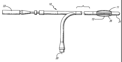
In another embodiment, as shown in FIG. 13, the catheter 10 includes
bowable electrodes 12 in the form of four conductive elongate members. The
bowable
electrodes 12 are similar to longitudinal electrodes formed along the
circumference of the
catheter, but are not fixed to the catheter. The catheter itself can fit
through a suitably
sized sheath for the procedure. For example, a 9 French sheath, which has
about a 3
mm diameter, may be used. The working end 11 of the catheter includes a
movable tip
CA 02248260 1998-08-31
WO 97/32532 PCTIUS97/03637
24
31 manually controlled by a diameter actuator 33 located at the connecting end
of the
catheter. The movable tip 31 is connected to the diameter actuator 33 by a
central wire
(not shown) running through the catheter. The diameter actuator 33 may be
threaded
onto the connecting end of the catheter. Maneuvering the actuator 33 into and
out of
the connecting end of the catheter causes a corresponding movement in the
movable tip
31 at the working end of the catheter. If the movable tip 31 is pulled toward
the
connecting end by the diameter actuator 33, then the electrodes 12 are bowed
outwardly.
The bowable electrodes 12 preferably expand out to treat veins up to 8 mm. If
the
movable tip 31 is pushed out by the diameter actuator 33, the bowable
electrodes 12 are
then retracted towards the shaft of the catheter. Consistent contact of the
electrode can
be maintained with the vein wall.
The extent of shrinkage can be controlled by the effective diameter of the
catheter and the electrode combination. The electrodes 12 may be bowed
radially
outwards as part of the effective diameter of the catheter so as to come into
apposition
with the vein wall. As RF energy is applied, the vein begins to shrink down to
the
effective diameter of the catheter. The effective diameter of the catheter is
reduced
under the control of the physician to control the amount of shrinkage. As the
effective
diameter is decreased, the electrodes continue to maintain apposition with the
venous
tissue. As before, the extent of vein shrinkage can be monitored by
fluoroscopy, or any
other suitable method. After shrinking the vein to the desired diameter, the
application
of RF energy from the electrodes 12 is ceased. The desired diameter can be the
final
effective diameter of the catheter, as defined by the deflected electrodes 12.
The electrodes 12 may be fabricated from spring steel or nitinol so that the
electrodes 12 would be biased to return to a reduced diameter profile. Where
the entire
length of the bowable longitudinal electrode is conductive, insulation 35 may
be
provided over the majority of the electrode surface in order to prevent any
unintended
heating effects. The ends of the electrodes are insulated from each other to
prevent
creating variable field densities at the ends, especially as the effective
diameter increases
which would create even greater field disparities between the ends and the
bowed
midsection. The insulation 35 can be polyimide or another type of insulating
film.
Insulation 35 provided along the back of the electrodes away from the vein
wall further
prevents heating of the blood flowing in the vein, which should also reduce
the
likelihood of coagulation. The remaining exposed area of the electrode is
preferably the
CA 02248260 1998-08-31
WO 97/32532 PCT/US97/03637
area which contacts the vein wall during apposition. The heating effect is
then focused
along the vein wall. The exposed surface area of the electrode should be as
great as
allowable while maintaining a consistent distance between the exposed sections
of the
electrode along the circumference of the effective diameter. The larger the
exposed
5 surface of the electrodes apposed against the vein wall during shrinkage,
the greater the
surface area of the vein wall affected by the electric field generated by the
electrodes.
Another embodiment of the catheter 10, as shown in FIG. 14, includes
bowable elongate members 32 having one end anchored to the working end 11 of
the
catheter, and the other end slidably connected to the catheter towards the
connecting
10 end. The catheter shown in FIG. 14 is similar to that shown in FIG. 13,
except that
instead of having the elongate members act as the electrodes themselves, the
electrodes
12 are located on the elongate members 32. The elongate members 32 preferably
include
a flat central area 34 for the electrodes 12. The central area 34 remains
substantially flat
as the elongate members 32 are deflected and bowed outwardly. The
substantially flat
15 central area allows for a more uniform contact with the vein wall. The flat
area
establishes a larger surface area to assure contact between the electrode 12
on the
elongate member and the vein wall. It is to be understood that the flat area
34 need not
be centrally located on the elongate member 32. The flat area should be
located so as
to be the first area that contacts the vein wall. The elongate members 32
shown in FIG.
20 14 are connected to a sliding sleeve 36 formed along the exterior of the
catheter shaft.
As the electrodes 12 are moved radially outwards and inwards, the slidable
sleeve 36 is
moved towards and away from the working end.
The balloon 40 can be furnished between the catheter shaft, and the
elongate members 32. Manual manipulation of the sliding sleeve is not required
in this
25 embodiment, and the sleeve need not travel any substantial length of the
catheter. The
balloon 40 is inflated and comes into contact with the elongate members 32. As
the
balloon 40 is further inflated, the electrodes 12 are moved outwardly in a
radial direction
as the elongate members are deflected and bowed by the expanding balloon 40.
The
balloon is preferably inflated using a non-conductive fluid, especially where
the elongate
members contain the electrodes, or where the elongate member itself is
conductive so
as to act as the electrode. When the proper diameter for the electrodes is
reached, the
inflation of the balloon ceases, and the application of the RF energy begins.
The
balloon 40 covers a greater surface area over the venous treatment site, and
ensures
CA 02248260 1998-08-31
WO 97/32532 PCTIUS97/03637
26
proper electrode placement relative to the vein wall while controlling the
amount of
venous shrinkage. More precise control over the shape and diameter of the
balloon can
also be possible using the bowable members. As RF energy is applied, the vein
begins
to shrink down. The effective diameter of the catheter is reduced under the
control of
the physician to control the amount of shrinkage. As the effective diameter is
decreased,
the electrodes continue to maintain apposition with the venous tissue. The
application
of RF energy from the electrodes 12 is terminated after shrinking the vein to
the desired
diameter, which is the final effective diameter as defined by the diameter of
the balloon
40 and the deflected elongate members 32. The balloon 40 is then is deflated
to a
minimal profile. The elongate members 32 are preferably fabricated from spring
steel
or nitinol so that the elongate members 32 would be biased to return to a
reduced
diameter profile when the balloon is deflated.
A cross-sectional view of the electrodes 12 of FIG. 14 along lines 15-15 is
shown in FIG. 15a. In the four-electrode configuration, a preferred embodiment
is to
have the electrodes 12 spaced equidistantly apart along the circumference of
the catheter.
The polarity of each electrode is preferably opposite to the polarity of the
immediately
adjacent electrodes. Thus, a uniform RF field would be created along the
circumference
of the catheter by the alternating electrodes. In another embodiment, as shown
in FIG.
15b, if adjacent electrodes were to be moved closer together, two effective
pairs of active
electrodes of opposite polarity would be formed along the circumference of the
catheter.
While an RF field would still be formed along the entire circumference of the
catheter,
the RF field would be strongest between the closest adjacent electrodes of
opposite
polarity. Shrinkage of the vein would be concentrated where the RF field was
strongest.
In an alternative embodiment of that discussed in connection with FIG.
14, the outer sleeve 36 can extend down the length of the catheter to allow
the operator
or physician to mechanically control the effective electrode diameter during
the
application of RF energy, so that a separate balloon 40 is not required.
Moving the
slidable sleeve toward the working end 11 of the catheter causes the
electrodes to deflect
and radially bow outward to an increased diameter. The outer sleeve 36 can be
moved
a preset distance to cause the electrodes to bow outwardly to a known
diameter.
Bowing the electrodes outwardly also places the electrodes in apposition with
the venous
tissue to be treated. Moving the sleeve 36 toward the connecting end of the
catheter
pulls back and flattens the electrodes against the catheter before insertion
or withdrawal
CA 02248260 1998-08-31
WO 97/32532 PCT/US97/03637
27
from the vein. Moving the sleeve controls the diameter of the electrode
deployment for
proper treatment of vein lumen having different diameters, and for providing
varying
degrees of vein shrinkage. For example, the electrodes could be placed in
contact with
the venous tissue, and the effective diameter could be mechanically reduced to
control
shrinkage while RF energy was being applied.
In another embodiment, instead of an outer sleeve, the ends of the elongate
members that would otherwise be attached to the outer sleeve are instead
slidably
located within longitudinal slots or channels disposed along the circumference
of the
catheter. The ends of the bowable members would slide towards the working end
within these channels as the members are deflected or bowed outwardly, and
retreat
back towards the connecting end in order to return to their original
configuration.
In another alternate embodiment, the electrodes and the elongate members
could be replaced by a single wire mesh or braided electrode, preferably when
applying
RF energy in a monopolar configuration. As before, the balloon could radially
extend
the mesh electrode outward into apposition with the vein wall. The balloon can
also
control the amount of vein shrinkage.
An alternative method for changing the effective diameter of the catheters
in FIGS. 13 and 14 is to move the electrodes 12 into direct contact with the
vein wall.
As the electrodes emit RF energy, the vein wall shrinks and pushes the
electrodes
inwardly towards the catheter. The vein shrinkage reduces the effective
diameter
directly, rather than by the active control of the physician, thereby
eliminating the need
for constant fine mechanical adjustments to the effective diameter. A
mechanism such
as a push rod or fixed-diameter balloon can be included to prevent further
radial
contraction of the electrodes at a specific effective diameter, thereby
controlling and
limiting the amount of vein shrinkage. This has the advantage of maintaining
the
electrodes in apposition with the venous tissue so that the tissue is heated
more than the
surrounding blood, without requiring the physician to constantly adjust the
effective
diameter of the catheter while applying the RF energy.
Other devices which are controllably expandable or extendable can be used
to limit the shrinkage of the vein to a desired size. For example, mandrels
can be
advanced out through the sides of the catheter to define a diameter limit for
shrinking
the venous section. As another example, a bowable conductive deflection wire
can be
located on one side of the catheter for achieving apposition with the vein
wall.
CA 02248260 1998-08-31
WO 97/32532 PCTIUS97/03637
28
Furthermore, even the non-expandable catheter shaft and electrode shown in
FIG. 7 can
be used to limit the amount of vein shrinkage during the procedure. The vein
would
merely shrink down to the fixed diameter of the catheter.
Other methods may be used with the catheter for maintaining apposition.
For example, a pressure cuff may be used to apply external pressure to the leg
to
compress the treatment area so that the vein wall comes into contact with the
electrodes.
Apposition of the electrodes with the venous tissue would be maintained by the
applied
external pressure. Such external compression may be used when treating the
superficial
veins. Methods other than ' the aforementioned mechanical methods may also be
used
to control the magnitude of vein shrinkage. Such non-mechanical methods
include
controlling the time and temperature of the venous RF treatment.
The working end of the catheter 10 could be constructed to have a bend
near the working end as shown in FIG. 11 so that the catheter can be rotated
to create
a stirring effect within the vein in order to achieve more uniform heating of
the venous
tissue for more even shrinkage. Rather than a permanent bend, the catheter can
be
manufactured to provide a controllable bend near the working end. For example,
the
bend may be formed from a shape-memory metal, manipulatable by a system of
wires,
a torquable braid, or a permanent bend in the catheter.
Another method for controlling the heat transfer to achieve more uniform
heating is by using an external tourniquet to reduce blood flow or compress
the vein
around the catheter at the venous treatment site. By reducing blood flow
either by
external compression or an intravenously inflated occlusive balloon, the
influence of
blood flow through the vein, which can carry heat away from the treatment
site, is
minimized. The heat transfer to the venous tissue during the procedure is less
impacted
by the blood flow, and the shrinkage rate of the vein would therefore be more
predictable. Sufficient pressure may also be established by the external
tourniquet to
cause the vein to come into apposition with the electrodes.
In another embodiment, as shown in FIG. 16, an occlusive centering
balloon 40 is used to retain a static pool of blood near the venous treatment
site. A
single occlusive balloon 40 may be used in conjunction with the venous valve
to retain
a pool of blood to be heated, wherein the electrodes 12 are located between
the venous
valve and the occlusive balloon 40. Two occlusive balloons (not shown) may be
formed
on either end of the electrodes to create a static pool of blood at a venous
treatment site
CA 02248260 1998-08-31
WO 97/32532 PCT/US97/03637
29
away from the venous valve. Such an arrangement isolates and protects the
venous valve
when treatment of the valve is not desired. The occlusive balloons may also be
used to
center the electrode within the vein lumen.
Although not limited to the occlusive balloon embodiment shown in FIG.
16, the catheter 10 further includes the electrodes 12 arranged in
longitudinal fashion
around the circumference of the catheter. This embodiment is similar to the
embodiments disclosed and described in connection with the FIGS. 13 and 14,
however,
the electrodes in this instance are fixed on the catheter and do not bow
outwards. This
fixed diameter arrangement allows a RF field to be formed along the
circumference of
the catheter. Such an arrangement can provide omnidirectional shrinkage and
avoid
lengthwise contraction of the vein. The particular positioning and orientation
of the
longitudinal electrodes is preferably as shown in FIG. 15a.
A balloon expandable embodiment, as shown in FIG. 17, includes the four
longitudinal electrodes 12 arranged in longitudinal fashion around the
circumference of
the balloon 40 of the catheter 10. This embodiment is similar to the
embodiments
disclosed and described in connection with FIGS. 13 and 14, so as to provide
omnidirectional shrinkage and minimize lengthwise contraction of the vein. The
particular positioning and orientation of the longitudinal electrodes is
preferably
equidistant as shown in FIG. 15a. The catheter 10 as shown in FIG. 17 is an
over-the-
wire type in which the catheter travels over a guide wire 42 through a
guidewire lumen
52. The catheter 10 further includes the braided shield 24 surrounding the
guidewire
lumen 52. A braid tube 54 is formed around the braid 24. The lumen 56 for the
balloon 40, and the balloon tube 55, encircle the braid tube 54. The braid
tube forms
a sealing barrier against the inflation fluid leaking into the guidewire lumen
52 from the
balloon lumen. The exterior of the catheter includes a retainer tube 57
holding the
conductor leads 20, which connect the electrodes 12 to an RF generator. A
cross-section
of the shaft of the catheter 10 along lines 18-18 of FIG. 17 is shown in FIG.
18.
In another embodiment, the electrodes 12 are located under the balloon
40 of the catheter 10. This embodiment, which is shown in FIG. 19 and which is
similar to that shown in FIGS. 17 and 18, allows for conductive heating of the
venous
tissue. The catheter 10 shown in FIG. 19 is an over-the-wire type in which the
catheter
travels over the previously introduced guide wire 42. The balloon is inflated
and
expands to come into contact with the venous tissue. As discussed previously,
the
CA 02248260 1998-08-31
WO 97/32532 PCTIUS97/03637
inflated balloon 40 can be used to control or limit the magnitude of shrinkage
of the
vein to the outer diameter of the inflated balloon 40. The effective diameter
can be
controlled by the selective inflation and deflation of the balloon 40. The
inflation
medium of the balloon 40 is preferably a conductive fluid, such as saline
solution, so
5 that a significant amount of the RF energy will still be transferred to the
surrounding
venous tissue. However, the inflation medium may absorb a certain amount of
the RF
energy, which will then be converted to heat. This diffusion of the RF energy
could
provide greater control over the shrinkage of the vein. Alternatively, a
conventional
heater coil or curie point element could be used in place of the electrodes 12
in order
10 to directly heat the inflation medium, which in turn would conductively
transfer the
heat to the venous tissue.
Another embodiment of the catheter 10 having electrodes 12 on the
working end 11 which causes localized heating of the surrounding venous tissue
and
shrinkage of the vein is shown in FIG. 20. The catheter 10 includes electrodes
12 in the
15 form of four conductive elongate members which can be bowed outward. The
bowable
electrodes are formed along the circumference of the catheter, but are not
fixed to the
catheter. The catheter itself is fit through a suitably sized sheath for the
procedure. For
example, a 7 French sheath, which has about a 2.3 millimeter (mm) diameter,
may be
used. The sheath is composed of a biocompatible material with a low
coefficient of
20 friction. The working end 11 of the catheter includes a tip 15 which is
attached to one
end of each electrode, and the other end of each electrode is connected to a
sliding sleeve
36 formed along the exterior of the catheter shaft. The outer sleeve extends
down the
length of the catheter to allow the physician to directly and mechanically
control the
effective electrode diameter during the application of RF energy. As the
slidable sleeve
25 36 is moved towards and away from the working end in response to a control
actuator
33, the electrodes 12 are urged radially outwards and inwards, respectively.
The tip 15
essentially remains stationary while the slidable sleeve is moved. Moving the
sleeve 36
back toward the connecting end of the catheter pulls back and flattens the
electrodes
against the catheter before insertion or withdrawal from the vein. Moving the
sleeve 36
30 forward toward the working end of the catheter causes the electrodes to
deflect and
radially bow outward to an increased diameter. The contact area of the
electrodes is
bowed outwardly as the opposite ends of the longitudinal electrode are moved
closer
together. The outer sleeve may be moved a preset distance to cause the
electrodes to
CA 02248260 1998-08-31
WO 97/32532 PCTIUS97/03637
31
bow outwardly to a known diameter. Bowing the electrodes outwardly also places
the
electrodes in apposition with the venous tissue to be treated. By manipulating
the
slidable sleeve to adjust the effective diameter of the catheter defined by
the radial
bowing of the electrodes, contact between the electrodes and the vein wall can
be
maintained as the vein shrinks. The control actuator 33 is a switch, lever,
threaded
control knob, or any other suitable mechanism, preferably one which can
provide fine
control over the movement of the slidable sleeve. By using the control
actuator to move
the slidable sleeve, the effective diameter of the electrode can be controlled
for treating
vein lumen having different diameters, and for providing varying degrees of
vein
shrinkage.
The tip 15 has a nosecone shape, or can have any shape which allows
tracking of the catheter over the guide wire and through bends in the venous
vascular
system. The nosecone tip can be fabricated from a polymer having a soft
durometer,
such as 70 Shore A. Alternatively, the nosecone can be constructed from a
spring
covered with a thin layer of polyethylene shrink tubing.
The extent of shrinkage is controlled by the effective diameter of the
catheter and the electrode combination. The electrodes 12 are bowed radially
outward
as part of the effective diameter of the catheter so as to come into
apposition with the
vein wall. After being placed in contact with the venous tissue, and the
effective
diameter could be mechanically reduced to control shrinkage while RF energy
was being
applied. The electrodes 12 are preferably operated as bipolar electrodes. As
RF energy
is applied to the electrodes, an RF field is created around the effective
diameter of the
catheter as defined by the bowed electrodes, and the vein becomes heated and
begins to
shrink. The effective diameter of the catheter is reduced under the control of
the
physician to control the amount of shrinkage. As the effective diameter is
decreased,
the electrodes continue to maintain apposition with the venous tissue. The
extent of
vein shrinkage is monitored by fluoroscopy, or any other suitable method.
After
shrinking the vein to the desired diameter, the application of RF energy from
the
electrodes 12 is ceased. The desired diameter of the vein is the final
effective diameter
of the catheter, as defined by the deflected electrodes 12.
The electrodes 12 have an elongated shape and may be fabricated from
stainless steel, spring steel, or nitinol, so that the electrodes 12 would be
biased to return
to a reduced diameter profile. The electrodes are rounded wires to facilitate
flexing of
CA 02248260 1998-08-31
WO 97/32532 PCTIUS97/03637
32
the catheter at the working end while being delivered through the bands of
tenuous
venous vasculature. The diameter of the electrodes are preferably between
about 0.005
to 0.015 inches (about 0.12 to 0.35 mm), but can be up to about 0.03 inches
(about 0.7
mm). Other shapes including rectangular wires having relatively large flat
surfaces for
contacting the vein wall can be used. Such rectangular wires can have widths
ranging
from 0.005 to 0.05 inches (0.12 mm to 1.2 mm), and preferably between 0.015
and 0.030
inches (0.35 mm to 0.7 mm), to allow four to eight electrodes around the
catheter shaft.
The entire length of the bowable longitudinal electrode is conductive, and
insulation 35 is provided over the majority of the electrode surface, as shown
in FIGS.
21 and 22, in order to prevent any unintended heating effects. Only a modest
portion
of the conductive surface is exposed to act as the electrode. The heating
effect is greatest
when the electrodes are close together since the electrical field density
(power density)
is greatest at this point. The ends of the electrodes are insulated from each
other to
prevent creating electrical field densities that are larger at the ends
compared to that
around the middle of the electrode. As the effective diameter increases,
greater field
disparities between the ends and the outwardly bowed midsections could be
created if
no insulation were provided. The insulation 35 can be polyimide, parylene, or
another
type of insulating material. The insulation 35 provided along the sides and
the back of
the electrodes opposite from the vein wall further prevents heating of the
blood flowing
in the vein, which should also reduce the likelihood of coagulation. Where the
wire has
a rectangular shape, then the exposed area which functionally acts as the
electrode would
then occupy only one face of that wire. As shown in FIG. 22, the insulation 35
surrounding the electrode can further cover the peripheral edges of the
exposed face of
the electrode to further isolate the blood flow from unintended heating
effects.
The exposed area of the electrode is preferably the area which directly
contacts the vein wall during apposition. The heating effect is then focused
into the
vein wall. The exposed surface area of the electrode should be as great as
allowable
while maintaining a consistent distance between the exposed sections of the
electrode
along the circumference of the effective diameter. The larger the exposed
surface of the
electrodes apposed against the vein wall during shrinkage, the greater the
surface area of
the vein wall affected by the electric field generated by the electrodes. The
exposed area
for the electrode can be substantially flat to enhance uniform contact with
the vein wall
and for controlling the diameter of the vein.
CA 02248260 1998-08-31
WO 97/32532 PCTIUS97/03637
33
A sensor 60 such as a small thermocouple for measuring temperature is
attached to the electrode 12. As shown in the cross-sectional view of FIG. 22,
the
temperature sensor 60 is soldered in place through a hole in the electrode so
that the
sensor is nearly or substantially flush with the exposed surface of the
electrode. The
sensor can accurately sense the temperature of the vein wall in apposition
with the
exposed electrode surface. The leads to the sensor are situated on the
opposite side of
the electrode which is insulated.
A cross-sectional view of the electrodes 12 of FIG. 20 along lines 23-23 is
shown in FIG. 23. In the four-electrode configuration, a preferred embodiment
is to
have the electrodes 12 spaced equidistantly apart along the circumference of
the catheter.
Although the catheter has been described as having a four electrode
configuration, it is
to be understood that the catheter may include a different number of
electrodes, for
example, six, eight, or more bowable electrodes, in order to lessen the inter-
electrode gap
and reduce the amount of power required to heat the venous tissue. The
polarity of
each electrode is preferably opposite to the polarity of the immediately
adjacent
electrodes to provide for omnidirectional and circumferential shrinkage of the
vein.
Thus, a relatively uniform RF field would be created along the circumference
of the
catheter by the alternating electrodes. In another embodiment, as shown in
FIG. 24, if
adjacent electrodes were to be moved closer together, two effective pairs of
active
electrodes of opposite polarity would be formed along the circumference of the
catheter.
While an RF field would still be formed along the entire circumference of the
catheter,
the RF field would be strongest between the closest adjacent electrodes of
opposite
polarity. Shrinkage of the vein would be concentrated where the RF field was
strongest.
In another embodiment, the RF field may be further focused directionally
using two pairs of electrodes arranged so as to be isolated from one another.
For
example, as shown in FIG. 25, the positive electrodes of each electrode pair
would be
adjacent to one another, and no field is formed along the circumference of the
effective
diameter between the two pairs of electrodes. Opposing RF fields are
established by the
two pairs of electrodes to create two discrete heating zones along the
circumference.
These heating zones may be directed to cause heating at isolated areas within
the vein
(i.e., not circumferentially) so as to direct treatment to the specific area
of variceal
CA 02248260 1998-08-31
WO 97/32532 PCT/US97/03637
34
bleeding from the vein. Specific or isolated instances of variceal bleeding
may be treated
by such directional application of RF energy to the vein.
The working end of the catheter further includes a guide wire lumen 39
for accepting the guide wire 13. The tip of the guide wire 13 is preferably
rounded.
The guide wire lumen 39 is preferably insulated so as to prevent or minimize
any
coupling effect the electrodes 12 may have on the guide wire 13. The guide
wire can
be removed before the application of RF energy to the electrodes. A cross-
sectional
view of the catheter 10 taken along lines 26-26 of FIG. 20 is shown in FIG.
26. The
guide wire 13 is shown centrally located within a guide wire lumen 38. The
guide wire
lumen 38 is surrounded by a layer of insulation material 22, which in turn is
surrounded
by a copper braid 24 for stability and stiffness, as well as for providing
flexible
torqueability to the catheter. An insulation sheath 26 covers the copper braid
24, and
contains the conductive leads 20 to the electrodes as well. In a bipolar
arrangement, the
conductive leads 20 have opposing polarity. In an over-the-rail type catheter,
the guide
wire is outside the catheter until arriving at the working end of the
catheter, upon
which, the guide wire enters the guide wire lumen. The guide wire lumen 39 is
preferably located within the insulation material 22 in order to electrically
isolate the
guide wire 13 from the electrodes 12. The guide wire lumen can also allow for
the
delivery or perfusion of medicant and cooling solution to the treatment area
during
application of the RF energy.
Another embodiment of the catheter 10, as shown in FIG. 27, includes
bowable elongate members 32 having one end anchored to the working end 11 of
the
catheter, and the other end slidably connected to the catheter towards the
connecting
end. The catheter shown in FIG. 27 is similar to that shown in FIG. 20, except
that
instead of having the elongate members act as the electrodes themselves, the
electrodes
12 are located on the elongate members 32. The elongate members 32 preferably
include
a flat central area for the electrodes 12. The central area remains
substantially flat as the
elongate members 32 are deflected and bowed outwardly. The substantially flat
central
area allows for a more uniform contact with the vein wall. The flat area
establishes a
larger surface area to assure contact between the electrode 12 on the elongate
member
and the vein wall. It is to be understood that the flat area need not be
centrally located
on the elongate member 32. The flat area should be located so as to be the
first area
that contacts the vein wall. The elongate members 32 at the working end of the
CA 02248260 1998-08-31
WO 97/32532 PCTIUS97/03637
catheter are connected to a movable tip manually controlled by a diameter
actuator
located at the connecting end of the catheter. The movable tip 17 is connected
to the
diameter actuator by an actuation wire 37 running centrally through the
catheter, as
shown in FIG. 28. The diameter actuator may be threaded onto the connecting
end of
5 the catheter. Maneuvering the diameter actuator into and out of the
connecting end of
the catheter causes a corresponding movement in the movable tip at the working
end
of the catheter via the actuation wire. If the movable tip 17 is pulled toward
the
connecting end by the diameter actuator, then the electrodes 12 are bowed
outwardly.
The bowed electrodes 12 preferably expand out to treat veins having diameters
of up to
10 ten mm or more. If the movable tip 17 is pushed forward by the actuator
wire 37, the
electrodes 12 are then retracted towards the shaft of the catheter. Contact
between the
electrode and the vein wall can be maintained with the vein wall as the vein
shrinks.
In one embodiment, the balloon 40 is located between the catheter shaft
and the elongate members 32. Manual manipulation of a sliding sleeve or a
movable tip
15 is not required in this embodiment, and the sliding sleeve, if used, need
not travel any
substantial length of the catheter. The balloon 40 may be either an elastic
material, such
as latex, or a noncompliant material. The balloon 40 is inflated and comes
into contact
with the elongate members 32. As the balloon 40 is further inflated, the
electrodes 12
are moved outwardly in a radial direction as the elongate members are
deflected and
20 bowed by the expanding balloon 40. The balloon is preferably inflated using
a non-
conductive fluid, especially where the elongate members contain the
electrodes, or where
the elongate member itself is conductive so as to act as the electrode. When
the proper
diameter for the electrodes is reached, the inflation of the balloon ceases,
and the
application of the RF energy begins.
25 The balloon 40 covers a greater surface area over the venous treatment
site,
and ensures proper electrode placement relative to the vein wall while
controlling the
amount of venous shrinkage. More precise control over the shape and diameter
of the
balloon is possible using the bowable members. The balloon can also be used to
control
the effective diameter of the catheter at the working end. As RF energy is
applied, the
30 vein begins to shrink down to the effective diameter of the catheter. The
effective
diameter of the catheter is reduced under the control of the physician to
control the
amount of shrinkage. As the effective diameter is decreased, the electrodes
continue to
maintain apposition with the venous tissue. The application of RF energy from
the
CA 02248260 1998-08-31
WO 97/32532 PCT/US97/03637
36
electrodes 12 is terminated after shrinking the vein to the desired diameter,
which is the
final effective diameter as defined by the diameter of the balloon 40 and the
deflected
elongate members 32. The balloon 40 is then is deflated to a minimal profile.
The
elongate members 32 can be fabricated from stainless steel, spring steel, or
nitinol so that
the elongate members 32 would be biased to return to a reduced diameter
profile when
the balloon is deflated.
In another embodiment, the ends of the elongate members are instead
slidably located within longitudinal slots or channels disposed along the
circumference
of the catheter. The ends of the bowable members would slide towards the
working end
within these channels as the members are deflected or bowed outwardly, and
retract
back towards the connecting end in order to return to their original
configuration.
In another alternate embodiment, single wire mesh or braided electrode,
preferably when applying RF energy in a monopolar configuration. As before,
the
balloon could radially extend the mesh electrode outward into apposition with
the vein
wall. The balloon further controls the amount of vein shrinkage.
An alternative method for changing the effective diameter of the catheter
is to move or deflect the electrodes into direct contact with the vein wall
and then allow
the vein wall to alter the effective -diameter. As the electrodes emit RF
energy, the vein
wall shrinks and pushes the electrodes inwardly towards the catheter. The vein
shrinkage reduces the effective diameter directly, rather than by the active
control of the
physician, thereby eliminating the need for constant fine mechanical
adjustments to the
effective diameter. A mechanism such as a push rod or fixed-diameter balloon
may be
included to prevent further radial contraction of the electrodes at a specific
effective
diameter, thereby controlling and limiting the amount of vein shrinkage. This
has the
advantage of maintaining the electrodes in apposition with the venous tissue
so that the
tissue is heated more than the surrounding blood, without requiring the
physician to
adjust the effective diameter of the catheter while applying the RF energy.
The method of the present invention for the minimally invasive treatment
of venous insufficiency can be performed using a catheter to deliver at least
one
electrode at the working end of the catheter to a venous treatment site in
order to
restore the proper function of a vein leading to the hemorrhoidal region. An
over-the-
wire or rail wire guided catheter can be used to deliver the one or more
electrodes
through the tortuous bends in the venous system to the hemorrhoidal treatment
site.
CA 02248260 1998-08-31
WO 97/32532 PCT/US97/03637
37
The electrode applies RF energy at a suitable frequency to minimized
coagulation for a sufficient amount of time to shrink, stiffen, and fixate the
vein, yet
maintain venous function or valvular competency. This intraluminal approach
avoids
the risks and morbidity associated with more invasive surgical techniques such
as
hemorrhoidectomy, while significantly reducing reflux of blood in the area
without
necrosing or removing the venous tissue.
When treating the veins of the lower hemorrhoidal region, the access site
is prepped and a percutaneous introducer is inserted into the vein. The
procedure for
the repair of incompetent veins can be accomplished by a qualified physician
with
fluoroscopic guidance, ultrasonic observation, or direct visualization. A
guide wire is
passed into the vein through the introducer, and advanced through to the
venous
treatment site. Alternatively, the catheter may be inserted into the vein
directly and
manipulated without a guide wire. The guide wire preferably has a spring wound
tip.
The guide wire is advanced retrograde to the venous treatment site, such as
the most
distal incompetent vein site which is to be repaired. Several intravenous
paths may be
taken to the hemorrhoidal treatment site.
A partial cross-sectional view of the venous system leading to the
hemorrhoidal region is shown in FIG. 29. Hemorrhoids are generally defined as
internal
or external depending on whether they are formed above or below the dentate
line DL,
respectively. Internal hemorrhoids IH are commonly formed when the smaller
veins
draining to the superior hemorrhoidal vein SHV or the middle hemorrhoidal vein
MHV
become dilated. External hemorrhoids are commonly formed when the smaller
veins
draining to the inferior hemorrhoidal vein IHV become dilated.
One method of delivering the catheter 10 and guide wire 13 is to introduce
the guide wire 13 into the external iliac vein EI on the side opposite to the
dilated veins
of the hemorrhoid. The guide wire is steered across the bifurcated branch of
the inferior
vena cava IVC to the inferior iliac vein II. The guide wire is then maneuvered
into
either the middle hemorrhoidal vein MHV to treat internal hemorrhoids, or the
pudendal vein PV and then the inferior hemorrhoidal vein IHV to treat external
hemorrhoids. The guide wire 13 is deployed and maneuvered into the middle
hemorrhoidal vein MHV to treat an internal hemorrhoid. The guide wire 13 is
maneuvered through the venous system until it reaches the dilated veins of the
hemorrhoid. The catheter 10 is then delivered to the venous treatment site
over the
CA 02248260 1998-08-31
WO 97/32532 PCTIUS97/03637
38
guide wire 13, as shown in FIG. 29. The working end 11 of the catheter 10
includes one
or more electrodes for applying RF energy once properly positioned at the
venous
treatment site to cause shrinkage of the vein. The working end of the catheter
further
includes a flexible nose cone tip to allow tracking of the catheter over the
guide wire
and through bends in the venous vascular system. Fluoroscopy, x-ray,
ultrasound, or
a similar imaging technique could be used to direct the specific placement of
the catheter
and to confirm position within the vein. X-ray contrast material can be
injected
through or around the catheter to identify the incompetent venous sections to
be
repaired. This approach advantageously allows the guide wire or catheter to
avoid sharp
bends or turns while being steered to the venous treatment site. It is to be
understood
that other access sites can be used to treat either internal or external
hemorrhoids.
Another method of delivering the catheter and guide wire is to introduce
the guide wire into the superior hemorrhoidal vein and maneuver the guide wire
through the superior hemorrhoidal vein SHV to the hemorrhoidal region. The
guide
wire is maneuvered into position, and the catheter is then delivered over the
guide wire
to the venous treatment site for the internal hemorrhoid. The venous treatment
site is
within the lumen of a dilated vein.
When the electrodes 12 of the catheter 10 are positioned at the venous
treatment site, an RF generator is activated to provide suitable RF energy,
preferably at
a low power level, and, preferably at a selected frequency from a range of 250
kHz to
350 MHZ. For example, one suitable frequency is 510 kHz. Another suitable
frequency
is 460 kHz. One criterion for the selection of the applied frequency is to
control the
spread, including the depth, of the thermal effect in the tissue. Another
criteria for the
selection of the applied frequency is the capability of filtering circuits to
eliminate RF
noise from thermocouple signals.
The energy emitted from the electrodes is converted within the venous
tissue into heat. As the temperature of the venous tissue increases, the
venous tissue
begins to shrink. The shrinkage is due in part to dehydration and the
structural
transfiguration of the collagen fibers in the vein. Although the collagen
becomes
compacted during this process, the vessel wall collagen still retains
elasticity.
RF energy can be applied to heat the dilated venous section of a
hemorrhoid. The dilated vein is shrunk to a normal or reduced diameter under
the
controlled application of RF energy which heats the venous tissue. Venous
pressure on
CA 02248260 1998-08-31
WO 97/32532 PCT/US97/03637
39
the lower venous sections of the hemorrhoid may be lessened, due to the
decrease in the
cross-sectional area of the vein. Valve competency in the lower venous
sections may
also be restored indirectly by the lessening of the venous pressure.
Thickening of the
vein will also occur during treatment, which can reduce the likelihood of the
recurrence
of vein dilation. The temperature and power of the RF energy may also be
controlled
to both shrink the hemorrhoid and cause the wall of the hemorrhoidal vein to
become
affixed to adjacent tissue.
Although applying RF energy can shrink the vein dilation near the
formation of the hemorrhoid, extending the shrinkage to include higher venous
sections
can be advantageous in further lessening the effect of higher and increased
venous
pressure on the hemorrhoidal system. A contiguous axial section of dilated
vein can be
treated by applying RF energy along the dilated venous section, even if the
section is
extensive. For example, hemorrhoids are sensitive to pressures from the portal
system,
which can be transferred to the hemorrhoids through the superior hemorrhoidal
vein
SHV. Treatment of the superior hemorrhoidal vein by general shrinkage along an
extensive section of the vein above the hemorrhoid can offset the dilating
forces that
arise from any increased pressures from the portal system. Such treatment may
be
desirable even if there is no significant dilation in the superior
hemorrhoidal vein SHV.
The catheter 10, as shown in Fig. 30a, is introduced over the guide wire
13 through the venous system. The tip 15 of the working end 11 of the catheter
10 is
in the form of a nosecone which is flexible in order to travel over the guide
wire and
through bends in the venous system. As shown in FIG. 30b, the catheter 10 is
delivered
into the dilated venous section which may include an incompetent valve. The
electrodes
are then placed in apposition with the vein wall, preferably by mechanically
bowing the
electrodes 12 outwardly from the catheter 10 as shown in FIG. 30c. The
application of
RF energy from the electrodes causes the vein to shrink, and the effective
diameter of
the catheter, as defined by the bowed out electrodes, is mechanically
decreased to
control the amount of vein shrinkage. The bowed electrodes are held in
position to
define a specific effective diameter, as shown in FIG. 30d, to avoid occluding
the vein.
The catheter may be moved along the length of the dilated venous section to
cause
general shrinkage where the dilation is extensive.
RF energy is no longer applied from the electrodes after there has been
sufficient shrinkage of the vein' to alleviate the dilation of the vein.
Substantial
CA 02248260 1998-08-31
WO 97/32532 PCT/US97/03637
shrinkage may be achieved very rapidly, depending upon the specific treatment
conditions, including the power level of the applied RF energy. The properties
of the
treatment site, such as temperature, can be monitored to provide feedback
control for
the RF energy. Other techniques such as impedance monitoring, and ultrasonic
pulse
5 echoing, can be utilized in an automated system which shuts down the
application of
RF energy from the electrodes to the venous section when sufficient shrinkage
of the
vein is detected and to avoid overheating or cauterization of the vein.
Monitoring these
values in an automatic feedback control system for the RF energy can also be
used to
control the power level and heating effect.
10 Sufficient shrinkage of the vein may be detected by fluoroscopy,
venography, external ultrasound scanning, intravascular ultrasound scanning,
impedance
monitoring, temperature monitoring, direct visualization using an angioscope,
or any
other suitable method. For example, the catheter 10 can be configured to
deliver an x-
ray contrast medium to allow visualization by fluoroscopy for assessing the
condition
15 of the vein and the relationship of the catheter to the treatment area of
the vein during
the shrinkage process. As an alternative to fluoroscopy, external ultrasound
techniques
such as B-scanning using distinct ultrasound signals from d+ferent angles, or
intravascular
ultrasound can be used to acquire a more multidimensional view of the vein
shrinkage
at the treatment site, which improves the detection of uneven shrinkage in the
vein. An
20 angioscope may also be used to directly visualize and determine the extent
and degree
of vein shrinkage.
Where the catheter is designed with a fluid delivery lumen, a cooling fluid
can be delivered through the delivery lumen to the bloodstream during RF
heating of
the vein being treated. The fluid may include radiodense contrast material.
The
25 delivered cooling fluid minimizes any heating effect on the blood, and
reduces the risk
of heating the blood to the point, of coagulation. The fluid may be delivered
through
ports formed along the side of the catheter ne. he working end and the
electrodes.
The working end 11 of the catheter 10 near the electrodes 12 can be used
to physically limit the amount of shrinkage. The working end 11 is preferably
30 sufficiently sized or enlarged to prevent the complete occlusion of the
vein. Other
schemes, such as an inflatable balloon, may be used to mechanically limit or
control the
amount of shrinkage in the vein or to displace blood from the treatment site.
Such
CA 02248260 1998-08-31
WO 97/32532 PCTIUS97/03637
41
mechanical schemes can also be used to assure apposition between the
electrodes and the
venous tissue during treatment.
While providing for generalized shrinkage of the vein, the catheter may
also be used to more directly treat the venous valves. The hemorrhoidal veins
have
bicuspid valves, and in a normal and competent valve, each cusp forms a sack
or
reservoir for blood which, under pressure, forces the surfaces of the cusps
together to
prevent retrograde flow of the blood and allow only antegrade flow to the
heart. The
arrows leading out the top of the inferior vena cava IVC and the superior
hemorrhoidal
vein SHV, as shown in FIG. 29, represent the antegrade flow of blood back to
the heart.
The venous valves prevent retrograde flow as blood is pushed forward through
the vein
lumen and back to the heart. In an incompetent valve, the cusps do not seal
properly
and retrograde flow of blood may occur. Incompetent valves may result from the
stretching of dilated veins. As the valves fail, increased pressure is imposed
on the lower
veins and the lower valves of the vein, which in turn exacerbates the failure
of these
lower valves. Hemorrhoids may occur or, become aggravated as a result. The
valve
cusps can experience some separation at the commissure due to the thinning and
stretching of the vein wall at the cusps. When RF energy is applied within the
dilated
vein near the incompetent venous valve, shrinkage of the vein can restore
valvular
competency by reducing the dilation which is preventing the proper functioning
of the
venous valve.
In treating venous valves, the electrodes on the catheter are advanced until
contact with the cusp of the venous valve is observed by fluoroscopy,
ultrasound, or
another detection method. The catheter is then pulled back slightly to allow
treatment
of the dilated section of vein. The electrodes are activated to deliver RF
energy to the
venous tissue and shrink the vein. The application of RF energy should be
controlled
to avoid unintentionally heating the valvular cusps. The shrinkage of the vein
can be
limited to prevent occlusion and allow the continued function of the vein. The
outer
diameter of the catheter or an extendable member can be controlled to limit
the
magnitude of the vein shrinkage.
After treatment, the commissure and the cusps of the venous valves should
be closer together with little separation or prolapse, which indicates a
restoration of the
competency of the valve. Valvular competence may be determined by contrast
injection
or Doppler probe measurement. For example, a radiopaque contrast solution can
be
CA 02248260 1998-08-31
WO 97/32532 PCT/US97/03637
42
infused through the catheter lumen to assess valve competence via descending
venography. It should be noted that reducing vein dilation by general
shrinkage in a
section above the section containing the incompetent venous valves could
restore
valvular competency by reducing the venous pressure on the valve and the
dilation of
the vein, which reduces the necessary span of the cusps. Also, direct
placement of the
electrodes across a vein valve can result in shrinkage of the loose, floppy
leaflets, thereby
preventing prolapse and reflux of blood through the valve.
The catheter 10 can be repositioned within the vein so as to treat as many
venous sections and valves as necessary. RF energy is applied to each venous
section to
be repaired, until all of the desired venous sections are repaired and the
valves are
rendered competent. Multiple incompetent valves and dilated venous sections
may be
treated and repaired in a single minimally invasive procedure. If desired, a
second
introducer can be inserted into the patient in order to treat incompetent
venous sections
in the other vein systems, such as the superior hemorrhoidal vein.
Another area of venous insufficiency suitable for treatment in accordance
with the present invention involves esophageal varices. Varicose veins called
esophageal
varices can form in the venous system along the submucosa of the lower
esophagus, and
bleeding can occur from the swollen veins. Properly sized catheters can be
used in
accordance with the present invention to deliver the electrodes to the site of
venous
insufficiency along the esophageal varices. Endovascular access for the
catheter is
preferably provided through the superior mesenteric vein or portal vein to
shrink the
portal vein branches leading to the lower esophagus. Proper positioning of the
electrode
within the vein can be confirmed using fluoroscopic or ultrasound techniques.
The
electrodes apply RE energy or other radiant energy at a suitable frequency to
shrink the
vein and reduce the swelling and transmission of high portal venous pressure
to the
veins surrounding the esophagus while maintaining the function of the vein.
The
amount of shrinkage of the vein can be limited by the diameter of the catheter
or
electrodes themselves can be expanded to a predetermined diameter which limits
shrinkage of the vein to that diameter.
Varicose veins called esophageal varices can form in the venous system
along the submucosa of the lower esophagus, and bleeding can occur from the
swollen
veins. A properly sized catheter 10 is used to deliver the electrodes 12 to
the site of
venous dysfunction along the esophageal varices. Endovascular access for the
catheter
CA 02248260 1998-08-31
WO 97/32532 PCTIUS97/03637
43
is preferably provided through the superior mesenteric vein or portal vein to
shrink the
portal vein branches leading to the lower esophagus. Proper positioning of the
electrode
within the vein may be confirmed using fluoroscopic or ultrasound techniques.
The
electrodes apply RF energy or other forms of energy at a suitable power or
frequency
to shrink the vein and reduce the swelling and transmission of high portal
venous
pressure to the veins surrounding the esophagus while maintaining the function
of the
vein. The amount of shrinkage of the vein is limited by the diameter of the
catheter
itself, and the catheter or electrodes themselves may be expanded to a
predetermined
diameter which limits shrinkage of the vein to that diameter.
When treating the veins of the lower esophageal region, the access site is
prepped and a percutaneous introducer is inserted into the vein. The procedure
for the
repair of incompetent veins may be accomplished by a qualified physician with
fluoroscopic guidance or ultrasonic observation, or direct visualization. A
guide wire
13 is passed into the vein through the introducer, and advanced through to the
venous
treatment site. The wire is advanced to the venous treatment site, such as the
level of
the most proximal incompetent vein site which is to be repaired. Preferably,
the guide
wire and catheter are advanced antegrade to the esophageal treatment site.
Alternatively,
the catheter may be inserted into the vein directly and manipulated without a
guide
wire.
As shown in FIG. 31, in a partial view of the venous system leading to the
esophageal region, the catheter 10 is advanced over the guide wire 13 to a
dilated section
of the vein. One method of delivering the catheter and guide wire is to
introduce the
guide wire through the superior mesenteric vein SMV to the portal vein PV and
coronary vein CV which branches and leads to the lower esophagus E to form the
esophageal veins EV. As an alternate route, the guide wire could be introduced
into the
inferior mesenteric vein, and routed through the splenic vein SV, the portal
vein PV,
and the coronary vein CV to arrive at the esophageal varix to be treated.
The guide wire is deployed and manipulated so as to reach the treatment
site for treating the esophageal varices. The venous treatment site is
preferably within
the lumen of a dilated vein. The catheter 10 is then delivered to the venous
treatment
site over the guide wire 13 as shown in FIG. 31. Fluoroscopy, x-ray,
ultrasound, or a
similar imaging technique could be used to direct the specific placement of
the catheter
and to confirm position within the vein. X-ray contrast material may be
injected
CA 02248260 1998-08-31
WO 97/32532 PCT/US97/03637
44
through or around the catheter to identify the dilated venous sections to be
treated.
Hemorrhaging or bleeding of the esophageal varices may also be identified in
this
manner.
Once the dilated venous section is reached, the one or more electrodes 12
are activated to apply RF energy to the dilated venous section. While the
electrodes
may be maintained in the center of the vein, the electrodes are preferably
placed in
apposition with the vein wall. The electrodes in apposition with the venous
tissue
ensures that the heating effect is delivered towards the venous tissue, and
not the blood
moving through the vein, and allow control over the shrinkage of the vein. One
method of achieving apposition is by bowing the electrodes out away from the
body of
the catheter. This is illustrated in FIGS. 32a, 32b, and 32c. The electrodes
have an
elongate longitudinal structure having opposite ends attached to a stationary
and a
moveable portion, respectively, at the working end of the catheter. The
bowable
electrodes are actuated by moving the outer sleeve of the catheter while
maintaining the
tip of the catheter stationary. Alternately a central wire could be used to
move the tip
while keeping the opposite end of the bowable electrode in place.
The one or more electrodes 12 at the working end 11 of the catheter 10
apply RF energy once properly positioned and apposed at the venous treatment
site to
cause shrinkage of the vein. An RF generator is activated to provide suitable
RF energy
to the electrodes, preferably at a low power level, and preferably at a
selected frequency
from a range of 250 kHz to 350 MHZ. For example, one suitable frequency is 510
kHz.
One criteria for the selection of the applied frequency is to control the
spread, including
the depth, of the thermal effect in the tissue. Another criteria is
compatibility with
filter circuits which can be used to eliminate RF noise from thermocouple
signals.
The energy emitted from the electrodes is converted within the venous
tissue into heat. As the temperature of the venous tissue increases, the
venous tissue
begins to shrink. The shrinkage is due in part to dehydration and the
structural
transfiguration of the collagen fibers in the vein. Although the collagen
becomes
compacted during this process, the vessel with collagen still retains
elasticity.
Substantial shrinkage may be achieved very rapidly, depending upon the
specific treatment conditions, including the diameter of the vein being
treated and power
level of the applied RF energy. The properties of the treatment site, such as
temperature, may be monitored to provide feedback control for the RF energy.
Other
CA 02248260 1998-08-31
WO 97/32532 PCTIUS97/03637
techniques, such as impedance monitoring and ultrasonic pulse echoing, may
also be
utilized in an automated system which shuts down the application of RF energy
from
the electrodes to the venous section when sufficient shrinkage of the vein is
detected and
to avoid overheating or cauterization of the vein. Monitoring these values in
an
5 automatic feedback control system for the RF energy may also be used to
control the
heating effect.
Sufficient shrinkage of the vein may be detected by fluoroscopy, external
ultrasound scanning, intravascular ultrasound scanning, impedance monitoring,
temperature monitoring, direct visualization using an angioscope, or any other
suitable
10 method. For example, the catheter 10 may be configured to deliver x-ray
contrast
medium to allow visualization by fluoroscopy for assessing the condition of
the vein and
the relationship of the catheter to the treatment area of the vein during the
shrinkage
process. As an alternative to fluoroscopy, external ultrasound techniques such
as B-
scanning using distinct ultrasound signals from different angles, or
intravascular
15 ultrasound may be used to acquire a more multidimensional view of the vein
shrinkage
at the treatment site, which improves the detection of uneven shrinkage in the
vein. An
angioscope may also be used to directly visualize and determine the extent and
degree
of vein shrinkage.
The working end 11 of the catheter 10 near the electrodes 12 physically
20 limits the amount of shrinkage. The electrodes 12 at the working end 11 are
bowed
outwards into apposition with the vein wall, and then gradually reduced
inwardly
towards the catheter during the application of RF energy. The final effective
diameter
of the electrodes 12 at the working end 11 is preferably sufficient to prevent
the
complete occlusion of the vein. Other schemes, such as an inflatable balloon,
may be
25 used to mechanically limit or control the amount of shrinkage in the vein
to a desired
diameter. RF energy is no longer applied from the electrodes after there has
been
sufficient shrinkage of the vein to alleviate the dilation of the vein.
Methods other than
the aforementioned mechanical methods may also be used to control the
magnitude of
vein shrinkage. Such non-mechanical methods include controlling the time and
30 temperature of the venous RF treatment.
The dilated venous section is heated and shrunk to a normal or reduced
diameter under the controlled application of RF energy in accordance with the
present
invention. A contiguous axial section of dilated vein may be treated by
applying RF
CA 02248260 1998-08-31
WO 97/32532 PCTIUS97/03637
46
energy along the dilated venous section, even if the section is extensive. To
treat an
extensive venous section, the catheter is moved in intervals to progressively
shrink the
venous section, or moved back and forth along the extensive section during the
application of RF energy. Further, thickening of the vein may occur during
treatment,
and reduce the likelihood of the recurrence of vein dilation and bleeding.
Applying RF energy shrinks the dilation of esophageal varices, and
extending the shrinkage to include other sections in the portal venous system
can be
advantageous in further lessening the effect of increased venous pressure on
the
esophageal varices. Esophageal veins can be sensitive to pressures from the
portal
system. Treatment of the branches of the portal vein before the esophagus by
general
shrinkage along an extensive section of the vein before the esophagus can
reduce the
dilating effect on the esophageal veins caused by increased pressures from the
portal
venous system.
It is to be understood that other mechanisms may be employed to position
or appose the electrodes with the venous section to be repaired without bowing
or
expanding the electrodes away from the catheter itself. The catheter may be
made
capable of being deflected, torqued, or otherwise moved to allow for the
proper
placement of the electrode. The catheter may be manufactured to provide a
controllable
bend near the working end. For example, the bend may be formed from a shape-
memory metal, manipulatable by a system of wires, a torquable braid, or a
permanent
bend in the catheter. Manipulating the working end of the catheter enables
preferential
heating along the vein wall being treated, if desired, where the electrodes
are placed
closer to one side of the vein wall. Preferential heating of the vein can also
be used to
effect hemostasis.
It is to be understood that although a bipolar arrangement is described, a
monopolar arrangement may also be used. In a monopolar arrangement, an inside
electrode, such as a mesh or wire electrode, is inserted into a patient's
body. An outer
electrode having a much larger surface area than the inside electrode is
placed on the
outer surface of the patient's body near the treatment site. For example, an
external
metal plate is placed on the skin over the region to be treated by the inside
electrode.
Alternatively, a metalized balloon is introduced into the esophagus and
inflated to come
into contact with the mucosal lining of the esophagus to act as the inactive
return
electrode. The electrodes are connected to a RF generator which produces an
electric
CA 02248260 1998-08-31
WO 97/32532 PCT/US97/03637
47
field within the patient's body. Because the surface area of the inner
electrode is much
smaller than that of the outer electrode, the density of the electric field is
much higher
around the inside electrodes. The electric field reaches its highest density
between the
two electrodes in the region near the inside electrode. The increased density
of the field
around the inside electrode allows localized heating of the tissues
surrounding the inside
electrode. The degree of heating may be dependent on such factors as the
impedance
and dielectric constant of the tissue being heated. It is to be understood
that different
numbers and configurations of electrodes can be used to produce the desired
discretionary heating effect.
As can be readily ascertained from the disclosure herein, the procedure of
the present invention is accomplished without the need for prolonged
hospitalization
or postoperative recovery. The curative restoration of venous function is
possible
without the need for continued lifestyle changes, such as frequent leg
elevation, the
wearing of relatively uncomfortable elastic support stockings or prolonged
treatment of
recurrent venous stasis ulcers. Moreover, the need for surgical
transplantation of veins
would not be necessary.
Early treatment of venous disease could prevent more serious
complications such as ulceration, thrombophlebitis and thromboembolism. The
cost of
treatment and complications due to venous disease would be significantly
reduced. There
would be no need for extensive 'hospitalization for this procedure, and the
need for
subsequent treatment and hospitalization would also be reduced from what is
currently
needed. Furthermore, the minimally invasive nature of the disclosed methods
would
allow the medical practitioner to repair or treat several venous sections in a
single
procedure in a relatively short period of time.
It is to be understood that the type and dimensions of the catheter and
electrodes may be selected according to the size of the vein to be treated.
Although the
present invention has been described as treating venous insufficiency of the
lower limb
such as varicose veins in the leg, the present invention may be used to
intraluminally
treat venous insufficiency in other areas of the body.
Another area of venous insufficiency relates to erectile impotency of the
penis. A significant number of all physically-induced cases of impotence
result from
excessive drainage of blood from the penile venous system. Venous-drainage-
impotence
can be treated using the present invention. Catheters having a sufficiently
small
CA 02248260 1998-08-31
WO 97/32532 PCT/US97/03637
48
diameter can be used to deliver the electrodes through the dorsal vein of the
penile
venous system to shrink this venous outflow path. Fluoroscopic or ultrasound
techniques can be used to properly position the electrode within the
incompetent vein.
RF energy or other radiant energy is applied from the electrodes at a suitable
frequency
to shrink the surrounding venous tissue in order to reduce the excessive
amount of
drainage from the penis while maintaining venous function or valvular
competency.
The amount of shrinkage of the vein can be limited by the diameter of the
catheter
itself, or the catheter or electrodes themselves can be expanded to the
appropriate size.
Ligation of these veins should be avoided so as to allow for the proper
drainage of blood
from an engorged penis which is necessary for proper penile function.
While several particular forms of the invention have been illustrated and
described, it will be apparent that various modifications can be made without
departing
from the spirit and scope of the, invention. Accordingly, it is not intended
that the
invention be limited, except as by the appended claims.