Note: Descriptions are shown in the official language in which they were submitted.
CA 02250777 1998-10-O1
WO 98/33443 PCT/US98/01894-
VASCULAR FILTER
FIELD OF THE INVENTION
The present invention relates to the treatment of vascular
disease of either surgery or percutaneous angioplasty and
stenting. More particularly, the invention relates to a device
that reduces macro- and micro-embolism during the treatment of
vascular stenosis.
BACKGROUND OF THE INVENTION
A variety of surgical and non-surgical angioplasty
l0 procedures have been developed for removing obstructions from
blood vessels. Balloon angioplasty utilizes a balloon-tipped
catheter which may be inserted within a stenosed region of the
blood vessel. By inflation of the balloon, the stenosed region
is dilated. Surgery involves either removing the plaque from
the artery or attaching a graft to the artery so as to bypass
the obstructing plaque. Other techniques, such as atherectomy,
have also been proposed. In atherectomy, a rotating blade is
used to shave plaque from an arterial wall.
One problem common with all of these techniques is the
accidental release of portions of the plaque or thrombus,
resulting in emboli which can lodge elsewhere in the vascular
system. Such emboli are, of course, extremely dangerous to the
patient, frequently causing severe impairment of the distal
circulatory bed. Depending upon the vessel being treated, this
may result in a stroke or myocardial infarction or limb
ischemia.
Vascular filters or embolism traps for implantation into
the vena cava of a patient are well known, being illustrated
by, for example, U.S. Patents. Nos. 4,727,873 and 4,688,553-._
Additionally, there is a substantial amount of medical
literature describing various designs of vascular filters and
reporting the results of the clinical and experimented use
thereof. See, for example, the article by Eichelter & Schenk
entitled "Prophylaxis of Pulmonary Embolism," Archives of
CA 02250777 1998-10-O1
WO 98/33443 PCT/L1S98/01894-
-2
Surgery, Vol. 97, August 1968, pp. 348 et seq. See, also, the
article by Greenfield, et al., entitled "A New Intracaval _
Filter Permitting Continued Flow and Resolution of Emboli",
Surgery, Vol. 73, No. 4, pp. 599-606 (1973).
Vascular filters are used, often during a postoperative
period, when there is a perceived risk of a patient
encountering a pulmonary embolus resulting from clots generated
at the surgical site or the like. As a typical use of vascular
filters, the filter is mounted in the vena cava to catch large
emboli passing from the surgical site to the lungs.
The vascular filters of the prior art are usually
permanently implanted in the venous system of the patient, so
that even after the need for the filter has abated, the filter
remains in place for the lifetime of the patient, absent
surgical removal. U.S. Pat. No. 3,952,747 describes a
stainless steel filtering device which is permanently implanted
transvenously within the inferior vena cava. The filtering
device is intended to treat recurrent pulmonary embolism. U.S.
Pat. No. 4,873,978 describes a catheter device comprising a
catheter body having a strainer mounted at it distal end. The
strainer is shiftable between an opened configuration where it
extends substantially across the blood vessel to entrap passing
emboli, and a closed configuration where it retains the
captured emboli during removal of the catheter. A mechanism
actuable at the proximate end of the catheter body allows
selective opening and closing of the strainer. Typically, the
strainer is a collapsible cone having an apex attached to a
wire running from the distal end to the proximate end of the
catheter body.
Permanent implantation is often deemed medically
undesirable, but it has been done because vascular filters are
implanted in patients primarily in response to potentially life
threatening situations. Accordingly, the disadvantages of
permanent implantations of a vascular filter are often
accepted.
To avoid permanent implantation, it would be highly
desirable to provide an-apparatus and method for preventing
CA 02250777 1998-10-O1
WO 98/33443 PCT/US98/01894 -
-3-
embolisms associated with conventional surgery and angioplasty
procedures. In particular, it would be desirable to provide a
device which could be located within the vascular system to
collect and retrieve portions of plaque and thrombus which have
dislodged during the surgery or angioplasty procedure.
OBJECT OF THE INVENTION
It is an object of this invention to provide a vascular
filter for reducing macro- and micro-embolism.
It is also an object of the invention to provide a
vascular filter which is readily removable from the vascular
system, or elsewhere, of a patient when the filter is no longer
needed.
It is a further object of the invention to provide a
3.5 vascular filter having a configuration which does not require
hooks to penetrate and grip the blood vessel walls, so that the
implantation results in less blood vessel injury.
It is a yet further object of the invention to provide a
vascular filter of very low profile which is part of a
guidewire and can be used in small vessels
These and other objects of the invention will become more
apparent from the description below.
SUMMARY OF THE INVENTION
The present invention generally relates to the surgical
and interventional treatment of vascular disease. For example,
during angioplasty and stenting of carotid stenosis, there is
occurrence of macro- and micro-embolism which increases the
risk of a minor or major stroke. The device of the present
invention for reducing macro- and micro-embolism is very useful
in helping to prevent the risk of stroke. However, this device
would also be useful in any angioplasty or surgical procedure
where embolization is a risk.
The filters of the present invention will decrease
embolism while allowing brain, or other distal tissue,
perfusion. The filters-are incorporated into a guidewire which
CA 02250777 1998-10-O1
WO 98/33443 PCT/US98/01894-
-4
is used for the entire procedure from crossing a lesion to
deploying a stmt. The filter consists of a thin membrane -
attached to the guidewire_and supported by fine metal spines.
The filter membrane has a pore size such that blood flow is not
impeded when the filter membrane is expanded but micro- and
macro-emboli are blocked. The attachments of the filter
membrane to the guidewire allow expansion of the filter
membrane with a firm fit inside the artery. Expansion of the
filter membrane is aided by the forward flow of blood against
the filter. The attachments also allow for collapse of the
filter membrane at the end of the procedure so it fits tightly
against the guidewire and can be withdrawn through the guide
catheter. The filter design results in a very low profile so
that the initial crossing of the lesion is minimally traumatic.
Also, the small diameter and small profile facilitate use of
the device in small or larger arteries with minimal or no
obstruction of blood flow.
BRIEF DESCRIPTION OF THE DRAWINGS
The above and other objects and advantages of the
invention will be apparent upon consideration of the following
detailed description, taken in conjunction with the
accompanying drawings, in which the reference characters refer
to like parts throughout and in which:
Fig. 1 is a lateral, partly cross-sectional view of the
distal end of a guidewire of one embodiment of the invention
with the filter membrane in a collapsed position;
Fig. 2 is a lateral, partly cross-sectional view of the
distal end of a guidewire of Fig. 1 with the filter membrane in
an expanded, deployed position;
Fig. 3 is a proximal end-on view of the filter membrane
shown in Fig. 2; ~-
Fig. 4 is a lateral, partly cross-sectional view of
another embodiment of the invention;
Fig. SA~is a lateral, partly cross-sectional view of a
further embodiment of the invention;
CA 02250777 1998-10-O1
WO 98/33443 PCT/US98/01894-
-5
Fig. 5B is a lateral, partly cross-sectional view of the
embodiment of the invention shown in Fig. 5A with the filter
membrane in an expanded, deployed position;
Fig. 6 is a partly cross-sectional view of a control
handle for the invention;
Fig. 7 is a partly cross-sectional view of another
embodiment of the invention;
Fig. 8 is a partial cross-sectional view of an embodiment
of the invention wherein the filter membrane has curved
supports;
Fig. 9 is a partial cross-sectional view of yet another
embodiment of the invention wherein the filter membrane has a
spiral wire;
Fig. 10 is a top, cross-sectional view of the embodiment
of the invention shown in Fig. 9;
Fig. 11 is a partial cross-sectional view of another
embodiment of the invention having inflatable support spines;
and
Figs. 12 and 13 represent partial cross-sectional views of
another embodiment of the invention in collapsed and deployed
positions, respectively.
DETAILED DESCRIPTION OF THE INVENTION
The present invention relates to a vascular filter for use
in percutaneous angioplasty and stenting and provides for the
prevention of distal embolism during endovascular procedures.
Further, the filter device of the invention allows for distal
perfusion while preventing embolism.
The device consists of a thin, perforated filter membrane
which is capable of blocking emboli and which is attached to
. the distal end of a guidewire. The device preferably uses thin
fibers which are moveable and are attached to the filter
membrane to deploy and collapse the filter membrane. The
invention also contemplates the use of metal spines or
inflatable spines attached to the filter membrane to deploy the
CA 02250777 1998-10-O1
WO 98/33443 PCT/US98/01894-
-6
filter membrane. The fibers or spines can also be attached to
a moveable core which is slidable within the guidewire and is-
used to deploy and collapse the filter membrane.
The filter membrane deploys in an umbrella-like fashion
with the unattached edge of the membrane moving upward, i.e.,
distally, and outward until it is in firm contact with an
artery wall. When the filter membrane is deployed, it spans
the cross-sectional area of the vessel lumen being treated for
a stenosis such as carotid stenosis, or another condition
likely to produce emboli.
The invention can perhaps be appreciated better by
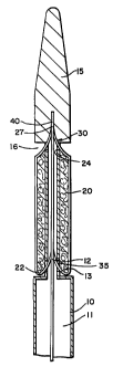
referring to the drawings. Fig. 1 illustrates a lateral,
cross-sectional view of a distal end of a guidewire 10 with a
filter membrane 20 attached thereto. Fig. 1 shows guidewire 10
with a shapeable, tapered soft tip 15 at its extreme distal end
which provides flexibility and maneuverability to guidewire 10.
The filter membrane in Fig. 1 is in a collapsed position.
Filter membrane 20 has a fixed portion 24 which is movably
attached to guidewire 10, and filter membrane 20 lies adjacent
guidewire 10 proximal to fixed portion 24 when filter membrane
20 is in the collapsed state. A moveable core 40 runs through
a center lumen 11 of guidewire 10 and preferably extends
distally a short distance beyond fixed portion 24 of filter
membrane 20. Deploying wires or fibers 30 are each firmly
attached at one end 27 to moveable core 40 distal to fixed
portion 21 of filter membrane 20. The deploying fibers 30 are
attached at their other ends to filter membrane 20 at
attachment points 22.
Collapsing fibers 35 are each firmly attached at one end
12 to the portion of moveable core wire 40 which is interior to
filter membrane 20 when it is in the collapsed state.
Collapsing fibers 35 are each attached at their other end 13 to
filter membrane 20 at attachment points 22. Accordingly,
collapsing fibers 35 lie interior to filter membrane 20 when
filter membrane 20 is in the ccsllapsed state.
Filter membrane 20 is deployed when the operator pulls
moveable core 40 proximally through the interior of guidewire
CA 02250777 1998-10-O1
WO 98/33443 PCT/US98/01894- -
10. Prior to retraction of moveable core 40, deploying fibers
30 are sufficiently relaxed so as not to create any tension at
filter membrane attachment points 22. Upon retraction of
moveable core 40, tension is created in deploying fibers 30.
There will preferably be from 2 to 6 each of evenly-spaced
deploying fibers 30 and collapsing fibers 35, 3 or 4 being most
preferred. The deploying fibers 30 and collapsing fibers 35
can be made of any flexible, medically acceptable material,
including stainless steel, nitinol, or another metal or
metallic alloy or a non-metallic substance such as graphite or
a suitable polymer. In addition, guidewire 10 and moveable
core 40 can be made from similar materials, as would be
appreciated by those skilled in the art. Typically, guidewire
10 could have an external diameter of from about 0.014 mm to
about 0.035 mm, a wall thickness of from about 0.002 mm to
about 0.010 mm, and a length of from about 25 cm to about 300
cm. Also, moveable core 40 could have a diameter of from about
0.003 mm to about 0.010 mm and a length of from about 30 cm to
about 350 cm.
Fig. 2 illustrates the filter device of the invention in a
deployed position on the inside of an artery wall 60. Moveable
core 40 is in a retracted state, i.e., pulled proximally
through the interior of guidewire 10. Tension is created in
deploying fibers 30, and filter membrane 20 extends to a
deployed position where the outer edge 14 of filter membrane 20
contacts artery wall 60. In this deployed position, collapsing
fibers 35 are in a relaxed state and extend from filter mem-
brane attachment points 22 to fixed attachment points 28 on
moveable core 40.
The flow of blood in Fig. 2 is toward the distal end of
guidewire 10. As such, the force of the flow of blood pushes
on deployed filter membrane 20 and helps to maintain filter
membrane 20 in the deployed position.
For withdrawal of guidewire 10 and the filter device,
filter membrane 20 is collapsed so that it sits tightly against
guidewire 10. This is accomplished by extending moveable core
distally through guidewire 10, thus relaxing deploying
CA 02250777 1998-10-O1
WO 98/33443 PCT/US98/01894
_g_
fibers 30 and creating tension in collapsing fibers 35: The
tension in collapsing fibers 35 collapses the filter membrane-
20, allowing it to fit tightly against guidewire 10 in recess
16 as depicted in FIG. 1.
Fig. 3 illustrates the filter device of the invention from
a distal end view in Fig. 2 with filter membrane 20 deployed.
Guidewire 10 is centrally located, and structural wires 50 are
seen extending from guidewire 10 to the outer edge 14 of filter
membrane 20. These wires 50 provide structural integrity and
rigidity to filter membrane 20. Fig. 3 depicts four, evenly-
spaced structural wires 50, but there can be more or less
structural wires 50. Preferably there are from two to six
structural wires 50, which may be spaced regularly or
irregularly. The wires 50 may preferably be comprised of
stainless steel or another medically acceptable metal or alloy.
Filter membrane 20 of the invention is preferably a mesh
such as that depicted in Fig. 3. The mesh should have pores of
a size sufficient to block and capture any micro- and macro-
emboli which may flow downstream from the site where the
stenosis is being treated, but large enough such that blood
flow is not impeded. The mesh used in the filter device of the
invention can have a pore size of from about 20 to about 300
microns, preferably from about 30 to about 100 microns, more
preferably from about 40 to 60 microns. Moreover, the size of
filter membrane 20, i.e., the distance from guidewire 10 to
free ends 22, is such as to allow a firm fit between filter
membrane 20 and artery wall 60. The diameter of filter
membrane 20 will be directly related to the artery being
treated, with typical diameters ranging from about 2 mm to
about 40 mm, most preferably from about 2 mm to about 20 mm.
The membrane can be comprised of fabric or non-fabric
meshes, such as those used in known hemodialysis filters or
heart-lung bypass machine filters. Suitable materials include
polymers or physiologically acceptable metals or alloys.
In alternative embodiments of the invention shown in Figs.
4, 5A and SB., filter membrane 20 will be suspended between from
two to six, preferably three or four, thin metal wires 51 which
CA 02250777 1998-10-O1
WO 98/33443 PCT/US98/01894-
-9-
serve as spines for filter membrane 20. Wires 51 may be
comprised of stainless steel or another metallic alloy, _
nitinol, or another shape-memory material. Wires 51 will be
constructed so that they assume a 90° angle with guidewire 10
when they are in an unconstrained state. This will result in
expansion of the filter membrane 20 to a position normal to
guidewire 10. A set of thin fibers 17 are attached at
attachment points 18 to filter membrane outer edge 14 and are
used to collapse filter membrane 20.
Fig. 4 shows an embodiment of this invention in which
metal wires 51 are allowed to regain their 90° angle
unconstrained state by use of a moveable core 40 that runs
through guidewire 10. Prior to retraction of moveable core 40,
fibers 17 are sufficiently tensed so as to restrain wires 51.
Upon retraction of moveable core 40, tension in fibers 17 is
released and wires 51 are allowed to revert to their relaxed
shape, which will result in expansion of filter membrane 20 to
a position normal to guidewire 10.
Figs. 5A and 5B show an embodiment of the invention
wherein wires 51 are restrained by fibers 17 that run through
guidewire 10 and that are controlled at a remote location. In
Fig. 5A, there is sufficient tension in fibers 17 to maintain
wires 51 in a constrained position. In Fig. 5B, tension in
fibers 17 has been relaxed such that wires 51 are allowed to
revert to their relaxed shape, which will result in expansion
of filter membrane 20 to a position normal to guidewire 10.
Fig. 6 depicts a control handle especially suitable for
the embodiment of the invention shown in Figs. 5A and 5B. The
proximal end 32 of guidewire 10 is rotatably attached to handle
33, such that rotation of handle 33 causes handle 33 to move
distally or proximally relative to proximal guidewire end 32.
For example, handle 33 may have threads 34 which engage threads
on guidewire proximal end 32. Fibers 17 attached to filter
membrane 20 are secured in a base 36 of handle 33. Then, as
35 handle 33 is turned, the fibers 17 move distally or proximally
to open or close filter membrane 20.
CA 02250777 1998-10-O1
WO 98/33443 PCT/US98/01894-
-10
As handle 33 is turned clockwise in the direction of arrow
A and fibers 17 are allowed to move distally in the direction-
of arrow C, the tension on the filter membrane fibers 17
decreases and wires 51 are allowed to assume their natural 90°
angle with respect to the guidewire, resulting in opening of
filter membrane 20. Similarly, when handle 33 is turned
counter-clockwise in the direction of arrow B and fibers 17 are
pulled proximally in the direction of arrow D, the tension on
filter fibers 17 increases, causing filter membrane 20 to
collapse tightly against guidewire 10. Of course, the
direction of turn of handle 33 as discussed above can be
reversed, as long as threads 34,35 are properly formed to allow
appropriate movement of handle 33 relative to guidewire
proximal end 32.
In yet another embodiment of the invention, shown in Fig.
11, filter membrane 20 can be supported by inflatable spines
135 supporting the filter membrane 20. Spines 135 supporting
the filter membrane 20 are from two to six hollow plastic tubes
which are inflatable using, for example, a standard balloon
angioplasty inflation device or endoflator in fluid connection
through channel 137 with spines 135. Inflation of spines 135
causes them to become rigid and deploys filter membrane 20. "
The underside of the filter membrane is attached to very thin
fibers 17 which are attached to moveable core 40 inside hollow
guidewire 10. Filter membrane 20 is collapsed by deflating the
spines 135 and withdrawing the moveable core 40 in the
direction of arrow E until the membrane 20 fits tightly against
guidewire 10.
A catheter-based configuration is also possible, as shown
in FIG. 7. In this design, the guidewire is not part of the
filter catheter; the guidewire and filter catheter are two
separate components. The filter catheter has an entry hole for
the guidewire below the attachment of the filter membrane and
the guidewire exits out the end of the filter catheter. The
filter catheter could be designed to accommodate a variety of
guidewire sizes, most commonly a 0.014 inch guidewire. The
advantages of this design are that a variety of guidewires
CA 02250777 1998-10-O1
WO 98133443 PCT/US98/01894 -
-11
could be used; the lesion could be crossed with the guidewire
prior to crossing with the filter catheter; the filter catheter
could be removed from the artery without removing the
guidewire; and the filter catheter could be made smaller.
In the embodiment of the invention shown in Fig. 7 a
catheter 101 comprises a longitudinally extending lumen 103,
which has an annular recess 105 adjacent the distal end of
catheter 101. Positioned within recess 105 is a filter 107
comprised of structural wires 109 and a filter membrane 111.
The distal end of each of wires 109 is attached at point 113 in
recess 105. Fibers 117 extend from the proximal ends 119 of
wires 109 proximally to a control means such as described in
Fig. 6.
Catheter 101 contains guidewire port 125 located proximal
to recess 105. It is intended that in use the distal portion
128 of a guidewire 127 will be threaded into the distal end 129
of catheter 101 and out through port 125.
Alternatively, and not shown here, a catheter 101 could
comprise a longitudinally extending lumen and a shorter
tracking lumen that extends from distal end 129 to a point
proximal to recess 105. The distal~end of guidewire 127 would
then be threaded into the distal opening of the tracking lumen
and out the proximal end of the tracking lumen.
Spiral or curved structural wires may be used to deploy
the filter membrane instead of straight wires. Fig. 8
illustrates the use of four curved wires 120. The angulation
of the filter attachment point of wires 120 relative to their
guidewire attachment has the effect of wrapping the filter
fabric around the guidewire in the undeployed state. This
leads to a lower profile for the undeployed filter.
Figs. 9 and 10 illustrate the use of a single spiral
structural wire 130 which is attached to the filter 107. As
tension fiber 131 is released, wire 130 unwinds and deploys
filter 107 in a conical configuration. This configuration has
the simplicity of using a single wire and, when the tension on
fiber 131 is increased, allows filter 107 to be wrapped very
CA 02250777 1998-10-O1
WO 98/33443 PCT/US98/01894
-12
tightly around the guidewire shaft 131, resulting in filter 107
having a low profile in its undeployed state.
Another modification shown in Figs. 12 and 13 comprises a
retractable sheath 140 at the distal end of guidewire 142 which
covers filter membrane 144 in the collapsed state. Sheath 140,
the distal portion of which is affixed to guidewire tip 146,
which is affixed to the distal end of moveable core 148, would
prevent an edge 150 of filter membrane 144 from becoming
entangled in an artery or guide catheter as it was being
withdrawn from a patient.
More specifically, when guidewire 142 with tapered tip 146
is inserted percutaneously into a patient, sheath 140 covers
collapsed filter membrane 144. After the filter membrane is
determined by fluoroscopy to be in proper position, moveable
core 148 is pushed distally to cause sheath 140 to "release"
filter membrane 144, which has spines 152, to cause filter
membrane 144 to deploy, as shown in Fig. 13.
It will be seen that the objects set forth above, among
those made apparent from the preceding description, are
efficiently attained and, since certain changes may be made in
carrying out the method and in the apparatus set forth without
departing from the spirit and scope of the invention, it is
intended that all matter contained in the above description and
shown in the accompanying drawings shall be interpreted as
illustrative and not in a limiting sense.
It is also to be understood that the following claims are
intended to cover all of the generic and specific features
herein and described and all statements of the scope of the
invention which, as a matter of language, might be said to fall
therebetween.
CA 02250777 2001-11-30
-13-
ITEM LISTING
No. Item
Guidewire
5
11 Guidewire lumen
12 End of collapsing fiber
13 End of collapsing fiber
14 Filter membrane outer edge
Guidewire soft tip
10
16 Recess
17 Collapsing fiber
18 Attachment point
Filter membrane
22 Filter membrane attachment point
15
24 Filter membrane fixed portion
27 Fiber attachment point
28 Fiber attachment point
Deploying fibers
20 32 Guidewire proximal end
33 Handle
34 Handle threads
Guidewire proximal end threads
36 Handle base
25 40 Moveable core wire
50 Structural wires
51 Deploying wires
60 Artery wall
101 Catheter
30 103 Lumen
105 . Recess
107 Filter mesh structure
109 Filter wire
111 Mesh
35 113 Attachment point
117 Deployment collapse wire
120 Curved filter structural wires
125 Guidewire port
127 Guidewire
CA 02250777 1998-10-O1
WO 98/33443 PCT/IJS98/01894- -
-14-
128 Guidewire distal end
129 Spiral wire
131 Fiber
132 Guidewire shaft
135 Inflatable spines
137 Inflation channel
140 Sheath
142 Guidewire
144 Filter member
146 Tapered guidewire tip
148 Moveable core
150 Filter membrane edge
152 Filter membrane spine