Note: Descriptions are shown in the official language in which they were submitted.
CA 02261488 1999-O1-21
FIELD OF THE INVENTION
The present invention relates to the field of cardiac surgery apparatus, and
more
specifically to the apparatus used in cardiac surgery performed directly on
the beating
heart.
In the present invention, the term "cardiac surgery" comprises the following
types of
surgery: coronary artery bypass graft surgery (CABG) performed directly on a
beating
heart (beating heart bypass surgery), CABG performed on an arrested heart
(traditional
l0 CABG), heart valve repair surgery or valve replacement surgery, and surgery
to correct
either an atrial septal wall or ventricular septal wall defect.
In the present invention the terms "device" , "cardiac devices", and
"apparatus"
comprise the surgical apparatus, instrumentation, and devices utilized during
cardiac
surgery.
In the present invention the term "cardiac organs" comprises the heart, the
heart's
arteries and veins, the surrounding tissue and vessels, in particular the
mediastinum,
the pericardium, the thymus, the pleura, and the space between the two lungs.
In the present invention, the terms "closed chest" signifies a surgical
intervention
whereby the patient's thoracic structure TS (ribcage), and bone defining said
thoracic
structure, are not cut, broken, spread apart or substantially not displaced
from their
normal anatomical position and orientation.
BACKGROUND OF THE INVENTION
Cardiac surgery, and more specifically traditional CABG, has been performed
since the
1970's on a regular basis with the support of the cardio-pulmonary machine,
whereby
the patient's blood is oxygenated outside the body, through extracorporeal
circulation
(ECC). The development of the cardio-pulmonary machine for ECC enables
surgical
interventions to take place on the arrested heart. This allows the surgeon to
manipulate and operate on a perfectly still heart. The arrested heart can be
positioned
to expose and provide best access to the target artery requiring bypass
grafting.
1
CA 02261488 1999-O1-21
Traditional CABG is still referred to as the gold standard in coronary artery
revascularization since it enables complete revascularization to be achieved,
that is the
treatment of all diseased arteries requiring bypass grafts. It thereby also
reduces the
likelihood of future surgical re-interventions, which reduces costs to the
healthcare
system and alleviates anxiety for the cardiac patient.
However, there are two main invasive aspects associated to traditional CABG -
the
sternotomy incision and the ECC.
Even with the constant technological improvements achieved during the last
twenty-five
years, the advantages offered with ECC have been offset by the morbidity
(complications) and mortality related to the ECC itself. ECC represents the
most
invasive clinical aspect of traditional cardiac surgery, particularly in CABG
surgery.
The inflammatory response, as well as the systemic microembolisms generated by
ECC, induce to some extent a dysfunctional state of the brain, lungs, and
kidneys,
which tends to increase with the aging of the patient. Furthermore, evidence
suggests
that when ECC can be avoided, the left ventricular function (pumping
efficiency) of the
heart is better preserved, thereby also reducing the risks of post-operative
complications and the need for ventricular assist devices to wean the arrested
heart
back to normal function.
In addition to being the most invasive aspect of traditional CABG, ECC is also
the most
costly device to operate during this procedure.
Median sternotomy is less clinically invasive than ECC, but has the perception
of being
more invasive due to the surgical scaring that results from the surgery. Full
median
sternotomy can result in: temporary disturbance in the respiratory mechanism,
increased risk of operative shock, dehiscence, and re-operation from bleeding
complications. Moreover, long exposure of the mediastinum to air can lead to
hypothermia, infection and compromise of the neuro-endocrine response.
Patients with
severe chronic obstructive pulmonary disease (COPD) or severe emphezema or
with
severe pulmonary insufficiency are therefore at higher risk of complications
when
exposed to sternotomy incisions.
As a result, alternative CABG procedures that do not rely on the very invasive
and
costly use of ECC offer distinct advantages to both the patient and the
progressively
discriminating cost sensitive health care system. Furthermore, if the
sternotomy
2
CA 02261488 1999-O1-21
incision can also be eliminated this would offer distinct advantages in
minimizing
surgical scaring. An even further advantage can be realized, if complete
revascularization can be achieved on the beating heart through closed chest
approach
since it not only manages healthcare costs incurred with future surgical
interventions
but also from the patient's perspective, the anxiety and inconvenience
associated with
future re-interventions.
In recent years, the drive for less invasive surgical apparatus and cost-
effective
medical approaches has placed emphasis on cardiac surgery as well. However,
unlike
l0 other organ surgeries, gall bladder for instance, the beating motion of the
heart
complicates the surgical intervention.
Port access surgery (HeartportTM) consists of replacing the full median
sternotomy by
a series of port incisions in the chest, through which coronary artery
revascularization
is performed. However, the most invasive aspect, ECC, is retained in this
surgery.
Femoral cannulation and aortic cross-clamping must be performed to place
patient on
ECC. This approach also requires lung deflation to provide working volume and
to
access remote territories of the heart. Unlike traditional CABG, the heart
cannot be
"verticalized" with respect to the chest cavity in order to access the
posterior territory.
2o Performing the surgery remotely through small ports is difficult, often
leading to
unwanted tissue dissection that requires the conversion to traditional CABG
through full
sternotomy in order to complete the surgical procedure.
It would be advantageous to have a surgical apparatus and medical approach
which
maintains, as much as possible, the normal anatomical position and orientation
of the
heart during the surgical intervention. This invention replaces the unnatural
verticalization required to access the posterior territory with the full
sternotomy
approach.
3o In minimally invasive direct coronary artery bypass graft surgery (MIDCAB),
ECC is
avoided and coronary artery revascularization is performed directly on the
beating
heart with the help of a mechanical stabilizer, through a mini-sternotomy or
mini-
thoracotomy incision. This surgical approach allows access to only one or two
of the
anterior arteries of the heart, most commonly the left anterior descending
artery (LAD).
Demographically only 5-15% of the population is afflicted with single vessel
disease;
the majority of cardiac patients (70%) suffer from triple vessel disease,
whereby at
least one artery on each of the anterior, inferior and posterior territories
of the heart
3
CA 02261488 1999-O1-21
requires a bypass graft. As a result, this approach is also referred to as
"limited access
bypass surgery".
The beating heart approach employed with mechanical stabilization has also
been
developed to enable grafting of the difficult to access posterior arteries,
such that
complete revascularization can be achieved on the beating heart. One such
surgical
device that immobilizes a portion of the beating heart around the target
artery, and
helps "verticalize" the beating heart is described in Canadian Patent
Application
2,216,893 filed by Cartier and Paolitto, entitled "Sternum Retractor for
Performing
Bypass Surgery on a Beating Heart". A median sternotomy is required in order
for the
apex of the "verticalized" beating heart to clear the ribcage while exposing
the posterior
territory. Although less invasive than ECC, the sternotomy incision with its
associated
complications is retained in this approach.
Percutaneous transluminal angioplasty (PCTA) or Coronary Stenting are
intraluminal
surgical procedures which achieve coronary artery revascularization through
the
enlarging of restricted vessels by balloon angioplasty (PTCA) and in some
cases also
supplemented by the scaffolding effect of the tubular mesh stent. Sternotomy
incisions
and ECC are avoided since the entire procedure takes place through the
patient's
artery. However, the high incidence off restenosis (repeat restriction of the
artery), and
its inapplicability to triple vessel disease does not make this procedure
suitable to the
majority of cardiac patients that require complete revascularization.
Other emerging technologies, such as Transmyocardial Revascularization (TMR)
or
Percutaneous Myocardial Revascularization (PMR) are reserved for non-
reconstructible
disease.
For the great majority of patients, those with triple vessel disease, the aim
of any
coronary revascularization cardiac surgery is to achieve complete
revascularization.
That is, the revascularization of all diseased arteries in the least invasive
manner. The
aim is to overcome the limitations in current approaches, which at the expense
of a
less invasive intervention compromise the thoroughness or completeness of the
surgical procedure. This limits the likelihood of re-intervention in
approaches where
the benefits are short-lived (restenosis associated with PTCA and Stenting) or
the
disease progresses in areas of the heart that were inaccessible at the first
intervention
(limitations associated with surgical apparatus and technique).
4
CA 02261488 1999-O1-21
It would therefore be advantageous to provide a surgical approach and
associated
apparatus that can cater to the entire demographically representative group of
patients
without the invasive aspects of ECC and median sternotomy, that achieves
complete
revascularization.
It would be a further advantage if this surgical approach and associated
surgical
apparatus is cost effective in lowering the initial healthcare costs of the
procedure and
minimizing future costs by reducing likelihood of re-intervention.
This invention describes a surgical apparatus that allows the manipulation and
positioning of the beating heart, along with the deployment of coronary
stabilizers that
serve to immobilize a portion of the beating heart around the target artery,
through a
transabdominal tunnel, thereby allowing complete revascularization without the
invasiveness of ECC and sternotomy incision. The grafting is either performed
through additional ports through the patient's chest or through the same
transabdominal
tunnel. Stereoscopic camera lenses, that transmit images to the surgeon so
that closed
chest interventions can be performed remotely, are placed at the distal
surgical
worksite either through the transabdominal tunnel or through additional port
incisions in
the patient's chest. Carbon dioxide is used to displace abdominal organs in
deployment of the transabdominal tunnel or to prevent air embolisms in the
chest
cavity during the revascularization procedure. Passages in the transabdominal
tunnel
are provided for the channeling of carbon dioxide gas.
SUMMARY OF THE INVENTION
It is therefore the object of the present invention to improve the efficacy
and safety of
cardiac surgery, more specifically CABG, by providing a surgical apparatus
that
eliminates ECC, and achieves complete revascularization directly on the
beating heart
through a closed chest approach, more specifically without sternotomy
incision.
It is a further object of the present invention to improve safety of the
cardiac surgery,
more specifically CABG, by providing a surgical apparatus that improves the
surgical
outcome for the patient.
It is therefore a further object of the present invention to provide a
surgical apparatus
which allows cardiac surgery, more specifically CABG, while eliminating the
likelihood
5
CA 02261488 1999-O1-21
of bone breakage or bone displacement associated with traditional sternotomy
or
thoracotomy heart exposure.
It is therefore a further object of the present invention to provide a
surgical apparatus
that expands the patient base for traditional CABG, more specifically by
including
patients with severe COPD, severe emphezema, severe pulmonary insufficiency.
It is therefore a further object of the present invention to provide a
surgical apparatus
that allows cardiac surgery, more specifically CABG, while decreasing the
risks of
l0 operative shock associated with traditional CABG.
It is a further object of the present invention to provide a surgical
apparatus that
decreases the initial cost of cardiac surgery, more specifically CABG, and
futute costs
of surgical re-intervention associated with the limitations of alternative
coronary artery
revascularization surgeries.
It is a further object of the present invention to position and orient the
beating heart
through a device acting on a distal remote location away from the target work-
site on
said beating heart where the surgical intervention is to be performed.
It is a further object of the present invention to improve the invasiveness of
beating
heart CABG, by providing a means of positioning and orienting the beating
heart
without impeding or restricting the natural beating function of the heart.
It is an additional object of the present invention to apply the concepts and
principles of
this invention as they relate to beating heart CABG to other types of cardiac
surgeries.
BRIEF DESCRIPTION OF THE DRAWINGS
3o The invention will further be described, by way of example only, with
reference to the
accompanying drawings wherein:
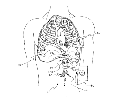
Figure 1 - is a frontal view of the patient with sectioned thoracic cavity
illustrating preferred embodiment according to the present invention;
Figure 2 is a partial sectional view illustrating the insertion of a
laparascopic
cannula to create a sagittal tunnel towards the diaphragm;
6
CA 02261488 1999-O1-21
Figure 3A is a sectional view of the patient's thorax, illustrating the multi-
lumen
channel 10 and heart manipulator 20 prior to C02 insufflation into
the pleural space;
Figure 3B is a sectional view of the patient's thorax, illustrating the
transabdominal device 1 after C02 insufflation into the pleural
space;
l0 Figure 4 is a sectional view of the diaphragm tissue retractor 40 in closed
position within the extraperitoneal space;
Figure 5A is a sectional view of the diaphragm tissue retractor in deployed ,
position with multi-lumen channel 10 inserted within;
Figure 5B is a partial sectional view of the multi-lumen channel 10 engaged
with the diaphragm accessing the pleural space, and of the channel
clamp 510;
Figure 6 is a partial sectional view through a portion of the securing
platform
50;
Figure 7 is longitudinal sectional view illustrating the transabdominal device
1
engaged with the apex of the beating heart during CABG surgery;
Figure 8A is a traverse sectional view through the multi-lumen channel 10
illustrating the HML and PAL and a open ended clamp variant for the
articulation mechanism 170;
Figure 8B is a traverse sectional view through the multi-lumen channel 10
illustrating a multi lumen variant of said channel and a closed clamp
variant for the articulation mechanism 170;
Figure 9 is longitudinal sectional view of the heart manipulator 20;
Figure 10 is a perspective view of the coronary stabilizer 30;
7
CA 02261488 1999-O1-21
Figure 11A is a perspective view of the transabdominal device 1 deployed to
provide surgical intervention on the posterior territory of the beating
heart;
Figure 11 B is a perspective view of the transabdominal device 1 deployed to
provide surgical intervention on the anterior territory of the beating
heart;
Figure 12 is a partial sectional view exposing the pleural space and
illustrating
l0 a pericardium retraction device 69 inserted through the PAL of multi-
lumen channel 10 to assist in positioning the beating heart during
posterior artery CABG surgery.
DESCRIPTION OF THE PREFERRED EMBODIMENTS
is
The present invention reduces the invasiveness of cardiac surgery, more
specifically
CABG surgery, while also reducing associated initial and future healthcare
costs, by
providing a device which enables closed chest, beating heart coronary artery
revascularization.
The patient's beating heart is positioned and oriented through a multi-luminal
transabdominal device (MLTAD). Subsequent surgical interventions can then be
performed through at least a section of said transabdominal tunnel or
alternatively
through additional port incisions through the patients closed chest.
The features and principles of this invention can be applied, in whole or in
part, to other
types of cardiac surgery requiring the strategic positioning and orientation
of the heart
and transabdominal introduction of surgical apparatus within the closed chest
pleural
space, but the description of the preferred embodiments will focus on beating
heart
CABG surgery.
In broad terms, the surgical procedure for the set-up and deployment of the
surgical
apparatus relating to this invention consists of:
1. Stereoscopic camera lens inserted into the pleural space via a port
incision
between the patients ribs;
2. Single lung deflation, preferably the left lung, is performed to augment
the
closed-chest pleural space (PLS) -- surgical work space;
8
CA 02261488 1999-O1-21
3. Abdominal incision (AI) is performed in the left upper quadrant of the
patient;
4. Insertion of a laparoscopic cannula into the abdominal
incision to reach the
extra-peritoneal space (EPS);
5. Carbon dioxide insufflation via the laparoscopic cannula
to assist the
dissection of the extra-peritoneal space, and laterally
displace the viceral
organs (VO) contained within the peritoneum (PER);
6. From the site of the abdominal incision, creation
of an upward sagittal tunnel
to access the diaphragm (DG), preferably at the left
leaflet location;
7. Insertion of a guide wire through the center of laparoscopic
cannula, up the
sagittal tunnel, through the diaphragm, to attain
the pleural space;
8. Over the guide wire, slide an enlarging cannula with
conical tip that
progressively enlarges the diaphragm (Seldinger Approach);
9. Over the enlarging cannula, slide a diaphragm tissue
retractor 40 that
pierces through the diaphragm and creates an opening
through its
subsequent radial deployment;
10. Once the desired opening is achieved in the diaphragm,
slide through the
center of the diaphragm retractor 40, the multi-lumen
channel 10 in a
manner that its distal end is now in communication
with the pleural space;
11. Retrieve the diaphragm retractor leaving the perimeter
of the retracted
diaphragm tissue engaged with the multi-lumen channel
10;
12. In order to further augment the surgical work space,
carbon dioxide is
channeled into the closed chest pleural space thereby
forcing downward the
dome of the diaphragm, along with the engaged multi-lumen
channel 10;
13. Secure channel 10 to the surgical table via securing
platform 50;
14. If the internal mammary artery (IMA) is required for a bypass graft,
proceed
to surgical harvesting of the IMA by inserting a cauterizing scalpel or
ultrasonic cutter through one of the pleural access lumens (PAL) in the
multi-lumen channel 10;
15. With the surgical harvesting of the IMA completed, also incise the
pericardium tissue of the beating heart to expose the myocardium, and
retrieve the ultrasonic cutter;
16. Through the heart manipulation lumen (HML) of channel 10, engage a
portion of the heart, preferably the apex, with the heart manipulator 20;
17. Rotate multi-lumen channel 10 with respect to the centerline of its HML,
to
obtain the best exposure and access to the desired coronary artery territory
via the eccentric PAL;
9
CA 02261488 1999-O1-21
18. Distally position and orient the beating heart attached to the heart
manipulator, through extracorporeal proximal movement of manipulation
handle;
19. Deploy the coronary stabilizer 30 through the multi-lumen channel 10, into
the pleural space, to attain the desired configuration for the specific artery
requiring grafting;
20. Place the coronary stabilizer on the myocardium thereby immobilizing the
portion of the beating heart around the target artery to be grafted;
21. Once immobilization is achieved, secure the coronary stabilizer with
respect
to the multi-lumen channel 10;
22. With help of stereoscopic vision, perform closed-chest anastomosis either
through trans-thoracic ports between the patient's ribs, or transabdominally
through the access lumen in channel 10;
23. Once the anastomosis is completed, retrieve coronary stabilizer and repeat
procedure (steps 16 - 21) for the other arteries, if multi vessel CABG is
performed;
24. Once all diseased arteries are revascularized, retrieve all components of
the
transabdominal device 1, and proceed to closing all incisions via standard
medical practice.
In the preferred embodiment according to this invention, the transabdominal
device
TAD 1, is comprised of a multi-lumen channel 10, a heart manipulator 20, a
coronary
stabilizer 30, diaphragm tissue retractor 40, a securing platform 50, and
thoracoscopic
surgical instruments 60 (Figure 1).
The diaphragm tissue retractor 40 is comprised of a hollow inner body 460,
tissue-
retracting petals 410, a translating sleeve 440, and a deployment lever 430
activated
outside the patient's body (Figure 4). The diaphragm is pierced firstly by the
guide wire
400, and subsequently distended by enlarging cannula 402 configured with a
tissue
piercing tip 401. The distal end of the inner body 460 is configured with a
plurality of
tissue retracting petals 410 which, in their closed position, form a conical
leading end
profile with a hollow tip well suited to being insertable and slidable over
the enlarging
cannula 402. The cylindrical tip 411, formed by the tissue retracting petals
in their
closed position, engages between the perimeter of pierced diaphragm and the
outer
diameter of the enlarging cannula 402. Each portion of the petals forming said
cylindrical tip 411 is deployed radially outward to enlarge the starting
orifice in the
diaphragm to the desired opening, thereby capable of receiving the multi-lumen
l0
CA 02261488 1999-O1-21
10
channel 10 (Figure 5A). Deployment is achieved through lever 430 which
displaces a
translating sleeve 440 with a sliding fit 441 over the exterior of inner body
460, thereby
engaging the cam like interface 445 and 415 between the retracting petals and
the said
translating sleeve. The radially inward force from the cam interface rotates
each of the
petals about hinge 420, thereby retracting the diaphragm tissue. Unlike the
Seldinger
method of gradually increasing the opening in tissue by progressive insertion
of a
conical tip cannula, the present embodiment allows the significant enlargement
of the
diaphragm orifice without risk of injury to the above-lying thoracic organs,
that would
likely result if an enlarging cannula would be used exclusively.
Once the diaphragm tissue has been retracted, the multi-lumen channel 10 is
inserted
through the hollow inner body 460. The permanent weir 130 extends past the end
of
the retracting petals 410. Subsequently, the deployment lever 430 is released,
the
petals and diaphragm tissue contract slightly and the tissue retractor 40 is
retrieved
from the body. This results in the diaphragm engaged around the distal end of
multi
lumen channel 10, upstream from the permanent weir. This configuration is
beneficial
since it allows the said channel 10 to mechanically pull down on the engaged
diaphragm, or if C02 will be inserted within the pleural space, the pressure
loads on
said channel 10 will maintain it engaged with the diaphragm through the
permanent
weir 130.
C02 can be introduced into the pleural space either through a lumen in channel
10, or
alternatively through a trans-thoracic port. The pressurized C02 serves to
augment the
pleural space by pushing down on the dome of the diaphragm, and consequently
through the permanent weir which serves as a axial buttress, the said channel
10 also
moves down and out of the body, leaving a shorter length of engaged channel
within
the body.
Channel 10 may be configured with a fastened proximal end 110, that can at
this point
be removed to yield a more ergonomic extracorporeal work space. The resulting
shorter length channel 10 allows more angular range in articulation of
instruments that
may be inserted through said channel during surgery.
Fastened interface 111 may be threaded, bayoneted, detented, wedged or of any
other
quick assembly interface.
11
CA 02261488 1999-O1-21
In order for the pleural space to remain pressurized with C02, all lumens
within said
channel 10 are provided with a seal, preferably but not limited to a diaphragm
type
seal. In the preferred embodiment, the seal 160 is a membrane with a plurality
of
nipples 161, through which a variety of surgical instruments may be easily
inserted
either before or during surgery, more specifically the heart manipulator 20
and coronary
stabilizer 30 (Figure 7).
Once engaged with the diaphragm, and C02 pressurization of the pleural space
has
been introduced (if desired), said channel 10 is positioned and oriented with
respect to
to the patient's body, and more specifically the cardiac organs that will be
subject to the
surgical intervention. This level of adjustment is referred to as "coarse
adjustment".
Multi-lumen channel 10 is secured in any substantially stable position and
orientation
relative to the surgical table 3, via securing platform 50. Said platform 50
is comprised
of a channel clamp 510, an articulation rod assembly 540, and a table clamp
570
(Figure 5A and 5B)
The preferred embodiment of the channel clamp 510 comprises a set of three
discs
511, 512, 513 whose inner diameters match the outer diameter 101 of the multi-
lumen
channel 10, such that the said discs can be slidingly rotated over said outer
diameter
101. Disc 512 is also rotatably engaged to discs 511 and 513 through eccentric
shoulders 514 and 515 protruding from both faces of disc 512. A rotation of
disc 512,
relative to discs 511 and 513, will radially offset disc 512 relative to said
discs 511 and
513, to the extent that the three discs will place the multi-lumen channel 10
in shear,
thereby achieving the desired clamping. Clamping techniques such as just
described
are commonly used in shafting design. The outer discs 511 and 513 are
permanently
attached to a 'U' shaped block 516 such that the inner portion of the 'U' does
not come
in contact with disc 512. Block 516 is permanently attached to a support rod
517 that
has a sphere 518 at the end opposite to block 516. The sphere is pivotingly
engaged in
socket 550 such that the channel clamp 510 is free to rotate and pivot about
the center
point of said sphere within the conical limits defined by the surface 542 of
nut 541.
when nut 541 is loose.
The end of articulation rod 543, closest to table clamp 570, has another
socket 560 that
rotatably engages sphere 571 of table clamp 570. The location of hole 561 in
nut 560
is strategically placed to give optimum positioning of the articulation rod
assembly 540
and channel clamp 510 with respect to the patient. The sphere 571 is
permanently
12
CA 02261488 1999-O1-21
attached to the clamp block 572 via rod 573. Articulation rod assembly 540 is
free to
rotate and pivot about the center point of sphere 571 within the conical
limits defined
by the surface 563 of nut 562, when said nut is loose. Clamp block 572 is
secured to
the surgical table 3 by tightening at least one screw 574 with the aid of
pivoted handle
575.
In addition to the present degrees of freedom allowed by the preferred
embodiment, an
additional degree of freedom can be obtained by making articulation rod 543 of
variable length.
l0
Alternatively, the clamping method at joints 550 and 580 can be pneumatic,
hydraulic,
electromechanical, or magnetic.
Alternatively, the channel clamp 510 with any other portion of the securing
platform 50
can be attached to a surgical robot instead of the surgical table.
In the preferred embodiment, the inside of channel 10 is configured with at
least one
hollow lumen that is substantially sealed to prevent pressure communication
between
the patient's pleural space and extracorporeal atmosphere. The heart
manipulator
occupies at least a portion of the hollow lumen, and the coronary stabilizer
30 at least
another portion of said lumen.
Alternatively, said channel 10 can be configured with two designated lumens
(Figure
8A); the HML lumen reserved for the heart manipulator, and the PAL lumen
providing
access to the pleural space and primarily occupied by the coronary stabilizer
during
beating heart CABG surgery. The PAL lumen, preferably when the coronary
stabilizer
is not occupying lumen, can be used provide access to the pleural space for
the
instruments used in the following surgical interventions: (i) IMA harvesting,
(ii) incision
of the pericardium sac, (iii) transabdominal port anastomosis, (iv) insertion
of vascular
conduit in bypass surgery, (v) doppler patency verification of newly-grafted
vascular
conduit, and (vi) assist in heart positioning and orientation through
pericardium
retraction sutures.
Alternatively, a plurality of lumens 125 (Figure 8B), each for a designated
purpose can
be incorporated for any combination of the above outlined (i) to (vi) surgical
interventions. Designated lumens 120 are also possible for surgical services
such as
C02 pressurization of pleural space , illumination of the closed chest cavity
through
13
CA 02261488 1999-O1-21
fiber optic bundle, and visioning of closed cavity through stereoscopic camera
lenses
(Figure 8A).
Figure 12 illustrates a pericardium retraction device 69 inserted through the
PAL of
channel 10. In order to assist in positioning and orienting the beating heart
during
posterior revascularizations, a suture 67 can be placed through the incised
pericardium
tissue 68, and pericardium traction applied through the said device 69. This
helps to
lift the heart within the thoracic cavity. The amount of protrusion of device
69 along
with its fine adjustment position and orientation with respect to the channel
10 will
determine the vector direction of the pericardium retraction load applied
through suture
67.
Figure 7 illustrates a sectional view through multi-lumen channel 10, with the
heart
manipulator 20 and coronary stabilizer 30 assembled.
Once the multi-lumen channel is secured with the channel clamp 510, the heart
manipulator 20 is preferably deployed before the coronary stabilizer. Heart
manipulator
is comprised of heart contacting member 200, conduit member 220, and
detachable
handle 240 (Figure 9).
Heart tissue engaging member 200 is comprised of flexible polymer
substantially-
conical sheath 204, detachable from hollow conduit member 220 through a barb
fitting
interface formed by mating members 202 and 222.
Member 200 engages with the beating heart, preferably the apex, through
negative
pressure.
Sheath 204 may be embodied with structural ribs 201 to bias the stiffness of
said
sheath in certain directions, thereby serving to facilitate the interface with
the beating
heart when negative pressure is applied through hollow passage 223 in conduit
means
220.
Alternatively, sheath 204 can be designed to have variable elastic properties
either by
function of its thickness or by its variable composition in fabrication.
Reinforcement
fibers can also be used in the fabrication of the polymeric sheath to bias its
elasticity
along certain axes. This is especially beneficial in the embodiment where the
conduit
means 220 is rigid, whereby 204 acts as a buffer in elastic gradient and
encourages the
14
CA 02261488 1999-O1-21
deforming beating heart to remain in compliant contact with perimeter 205 of
said
sheath.
The contact perimeter 205 is configured with a tapered beveled edge,
deformable skirt
203. This deformable skirt achieves a compliant seal perimeter 205, regardless
of the
beating heart's spatial orientation.
The deformable skirt 203 provides local readjustment of the plane formed by
the
perimeter 205 depending on how loads are applied to and reacted by the beating
heart.
Any manipulation force applied in a direction substantially parallel to the
axis of 220,
the beveled edge distorts equally around the perimeter, in a direction toward
the
opening of said perimeter. If the force is applied in a skewed direction
relative to the
axis of conduit means 220, the beveled edge will distort unevenly around the
perimeter
in a fashion to replicate a plane substantially perpendicular to direction of
application of
said manipulation force or heart reaction force to imposed negative pressure
loads.
Alternatively, can have plurality of conical sheaths 204 fed by a common
negative
pressure conduit 220.
Alternatively, the heart contacting member 200 can be comprised of a
mechanical
tissue clamping means, of a hydrogel or tissue adhesive-like coating or layer
disengaged by positive pressure through 220, of a hemi-cylindrical cradle with
perforations to allow anchoring of a suture to the apex tissue, of a non
flowing static
suction cup.
The outer diameter of conduit member 220, when detachable handle 240 is
removed,
allows its insertion into the articulation joint 170 of multi lumen channel
10.
The proximal end of 220 has barb fitting suction interface 221, that mates
with the
negative pressure source available in operating rooms.
The heart manipulator 20 can be positioned and oriented with respect to the
multi-
lumen channel 10. This position and orientation will be referred to as "fine
adjustment".
The motion degrees of freedom that yield this fine adjustment are required to
first
enable engagement of the heart contacting member 200 with the desired portion
of the
heart, and subsequently are required to allow re-positioning and re-
orientation of
engaged heart during surgery with respect to the patient's thoracic cavity. In
this
CA 02261488 1999-O1-21
manner, all coronary territories are accessible by the coronary stabilizer 30,
with heart
being located strategically within pleural space
More specifically these motion degrees of freedom allow conduit 220 to be
slidingly and
pivotingly engaged through articulation mechanism 170.
The articulation mechanism 170 is insertable transversally through channel 10,
thereby
facilitating cleaning and sterilization if re-usable components are used. Said
articulation mechanism is comprised of knob 190, two mating jaws 191 that when
l0 engaged together form a longitudinal cylindrical surface that can rotate
within bushing
192. Each jaw is provided with a hemi-cylindrical surface 193, such that when
mating
jaws engage, said hemi-cylindrical surfaces can apply a substantially
diametrical
clamping load to the outer diameter of the therewithin contained articulation
cylinder
194. A cylindrical passage 195, perpendicular to the centerline of the
articulation
cylinder 194, is provided to receive the conduit 220. The surface of the
cylindrical
passage 195 is interrupted by at least one substantially longitudinal split
196, such that
the clamping load imposed by the jaws on the puck will be transmitted to the
outer
diameter of conduit member 220.
Articulation mechanism 170 allows all the required degrees of freedom, at
least 4., that
is: the translation through articulation cylinder 194 of member 220 along the
axis of
said member 220, the rotation within cylinder 194 of member 220 about its
centerline,
the articulation of member 220 about the centerline of articulation cylinder
194, and the
pivoting of member 220 about the cylinder of thread 197. Once the desired
position
and orientation of manipulator 20 is achieved, the fine adjustment is secured
via knob
190 external to the multi-lumen channel 10.
The same articulation mechanism 170 can be employed for the coronary
stabilizer 30,
but it acts on the outer surface of proximal shaft means 360.
Figure 8A illustrates a variant to the articulation mechanism 170, that is, an
open-
ended clamp design that allows the transverse insertion of a shaft member on
surgical
instrument to be inserted through lumen of channel 10. This is advantageous if
want to
substitute surgical devices inside the lumen of channel 10 without wanting to
disrupt
the bulk of the surgical set-up.
16
CA 02261488 1999-O1-21
Once the heart has been positioned and oriented by the heart manipulator, the
multi-
lumen channel 10 is rotated such that the eccentric access lumen, or the
portion of
lumen not obstructed by the manipulator, is aligned with the target coronary
territory.
Figure 11 B shows the device deployed for anterior artery revascularization;
the access
lumen in the top half of the channel 10, the beating heart oriented downward.
Figure
11A shows the device deployed for the posterior artery revascularization; the
access
lumen on the bottom half of the channel, the beating heart oriented upward.
The same
applies for any coronary artery regardless of its location on the heart; the
channel is
rotated in such a manner to always offer optimum access and surgical approach
of the
l0 coronary stabilizer to the target artery. The present invention, therefore
allows the
synergistic deployment of the surgical apparatus -- the channel 10 is always
positioned
with respect to the heart manipulator, and more specifically its heart
manipulation
lumen (HML) as a function of the desired pleural access lumen (PAL).
The fine adjustment of the coronary stabilizer 30, that is of the proximal
shaft member
360 with respect to multi-lumen channel 10, is achieved in the same manner as
the
heart manipulator 20, and secured through knob 190. Rotation C about the
center line
of proximal rod 360 is through the rotation of 360 within passage 195 of
articulation
cylinder 194.
The coronary stabilizer 30 is comprised of three main subassemblies (Figure
10): (i)
extracorporeal control section, proximal to the surgeon (371, 331, 387, 386,
380, 385);
(ii) the heart contacting section, within the closed chest cavity, distal to
the surgeon
(300, 310, 320, 321, 322, 330, 341); and (iii) the center adjustment assembly
(340,350,
351, 360, 361, 362, 370) for transmitting the surgeon's desired manipulation
from the
control section to the heart-contacting section.
The control section comprises a securing bolt 385, and a multi-socket cradle
380. The
cradle is machined with three smaller diameter spherical sockets to interface
with the
3o proximal sphere ends (not shown) of the articulation transmission cables
340. These
interfaces with the cables can be permanently engaged by flaring the perimeter
of the
concave spherical surface in cradle around the sphere end of the cable, or
easily
disassembled if cradle is made from a resilient material or of a snap-in"
design.
The cradle 380 is also machined with a larger central spherical socket to
interface with
the substantially spherical end (not shown) of the inner rod 386. The
perimeter of this
concave spherical surface is flared only locally in three locations. The
substantially
17
CA 02261488 1999-O1-21
spherical end of inner rod 170 has three flats that allow it to be insertable
past the
flared edge of the cradle. The cradle is then rotated approximately 60 degrees
with
respect to the centerline of rod 386, thereby achieving its fully assembled
position.
This allows all the movements of a spherical joint with the two components
slidingly
linked in one assembly. The inner rod 386 has three longitudinal grooves,
machined
along most of its length, to serve as channels for the transmission cables
340.
The center socket in cradle 380 is pierced by a small threaded hole, at its
topmost
point, to receive securing bolt 385. This bolt exerts a force on the spherical
end of rod
170, thereby clamping the spherical end against the flared edges of the cradle
380.
This results in a locked assembly through an action / reaction mechanism.
Loosening
the bolt 385 permits sliding at the spherical interface, and repositioning of
articulation
transmission cables 340.
An annular brace 387 is inserted over the inner rod 386, to retain the cables
340 within
their longitudinal grooves at the top, proximal location. A similar brace (not
shown) can
be inserted at the heart-contacting section of the coronary stabilizer 30.
The distal spherical ends 341 are engaged to the quick assembly / disassembly
interfaces 321 on contacting member 300. The contacting member can be made
from
disposable surgical grade plastic, or any re-usable material such as titanium
or
stainless steel. The interface is specially designed to allow quick changeover
to a
variety of different contacting members (surgical kit) specific for different
arteries, or to
facilitate insertion of coronary stabilizer through multi-lumen channel 10
prior to
insertion of said channel 10 into patient's pleural space.
The substantially planar surface of the contacting member 300 is positioned
and
oriented with respect to the distal shaft member 350, partly through the three-
point
interface 321 on plate member 320 responding to cradle 395 movement. This type
of
micro-adjustment produces:
e: rotation of contacting plane in a heel to toe" articulation
E: rotation of contacting plane in a "side to sidep orientation
The contacting member 300 is secured in its articulated and oriented state
through the
tightening of bolt 385.
The design of the preferred embodiment achieves the following
18
CA 02261488 1999-O1-21
i) remote response of the heart-contacting member 300 by movement of
the proximal control cradle 380
ii) "active" readjustment of the contacting pressures for optimum coronary
artery immobilization during "in-process" surgical variations, without
disrupting fine and coarse adjustments
The preferred embodiment also allows additional adjustment to set angle A.
This
allows the heart contacting member 300 to be set in a position substantially
offset from
the centerline of the multi-lumen channel 10, in order to access and
immobilize target
arteries on the widest portions of the beating heart. The rotation of dial
371, through a
sliding member (not shown) within the proximal shaft member 360, translates
elbow
370 within slot 362. As a result, shaft member 350 rotates about hinge 36~ tn
the
desired angle A. The eccentricity of distal hinge 351 with respect to proximal
hinge
361 results in a bias direction of rotation when applying the torque to dial
371.
The preferred embodiment also allows additional adjustment to set angle B.
This
allows the rotation of the contacting member 300 with respect to the plate
member 320,
or the angular orientation of the arterial window 305 with respect to the
centerline of
shaft member 350, in order to better access target arteries that are diagonal
in
orientation with respect to the long axis of the heart. Rotation of dial 331
acts on a
fourth return transmission cable 330, which in turn applies a torque on shaft
323
attached to the contacting member 300. Shaft 323 rotates within bushing 322.
The coronary stabilizer 30 must react only the local forces from the
underlying
myocardium that it immobilizes; the loads from positioning and orienting the
entire
beating heart within the pleural space are reacted by the more robust heart
manipulator
30.
To achieve a bloodless surgical field during beating heart bypass surgery, the
heart
contacting member is configured with wire attachment pedestals 315, located on
opposite side of the arterial window 305, to anchor a vessel occluding wire
303,
preferably a silastic loop. One said wire circumvents the target artery
upstream and
another downstream of the grafting site. Each end of the wire is inserted in a
pedestal
slit, said pedestals on opposite sides of the arterial window. The said slits
achieve
light-tight anchoring of the vessel occluding wire, thereby allowing non-
traumatic
disengagement of said wire in the eventuality of unwanted slippage of surgical
apparatus or unwanted movement of the beating heart. The wire attachment
pedestals
19
CA 02261488 1999-O1-21
are described in Canadian Patent Application 2,216,893 filed by Cartier and
Paolitto,
entitled aSternum Retractor for Performing Bypass Surgery on a Beating Heart".
The vessel occluding wire 303 is generally attached to a blunted needle. The
circumventing of the target artery, and the subsequent anchoring of the wire
in the
pedestals 315, can be done either through traps-thoracic ports between the
patient's
ribs or through an pleural access lumen of multi-lumen channel 10. Similarly,
the
anastomosis of the vessel graft can be done either through traps-thoracic port
access
or transabdominally. In either case, the stereoscopic camera will allow the
surgeon to
l0 view his or her movements within the closed chest cavity.
Due to the preferred embodiments of the present invention, traps-thoracic port
interventions are greatly simplified, more ergonomic, and less traumatic for
the patient
since the positioning and orientation of the beating heart and coronary artery
immobilization are done transabdominally.
In the preferred embodiments according to the present invention, access to the
pleural
space was achieved by piercing at least a portion of the diaphragm.
Alternatively, the
concepts and principles can also be applied to thoraco-phrenic dissociation
surgical
approach, whereby access to the pleural space is achieved through a passage
between
the diaphragm and the patient's ribcage without piercing the diaphragm.
In the preferred embodiments according to the present invention, access to the
diaphragm and subsequently the pleural space was achieved via the
extraperitoneal
space. Alternatively, the concepts and principles can also be applied to
intraperitoneal
surgical approach in which at least a portion of the patient's peritoneal
membrane is
pierced.
In all embodiments herein described, the novel concepts and design features
may also
apply to other types of cardiac surgery. For example, the transabdominal
device 1 can
be applied to mitral valve replacement surgery. The right lung is deflated to
augment
closed chest pleural work space. The patient is placed on total
cardiopulmonary
bypass by femoro-femoral cannulation. A sub-xiphoid process incision, followed
by
incision of the pericardium will yield access to the patient's ascending aorta
and
thereby exposure for aortic cross-clamping. Hypothermia surgical environment
helps
support fibrillating heart which is relieved of its pumping requirement by
cardiopulmonary bypass. Through the multi-lumen channel 10 inserted in the sub-
CA 02261488 1999-O1-21
xiphoid process incision, can be introduced within the pleural space the
following: C02
gas, suction line, stereoscopic vision camera port, illuminating fiber optic
bundle,
cardioplegia infusion cannula, valve tissue retractor, and replacement valve.
The
replacement valve annulus may be secured through trans-thoracic port approach.
Similarly, the same principles apply to atrial septal defect or ventricular
septal defect
repair cardiac surgery.
In all embodiments described herein, the bulk of the surgical apparatus is
designed for
totally reusable components, whose assembly can be totally dismantled, if
necessary,
to for ease of sterilization. All components are manufactured in either
surgical grade
stainless steel, titanium, aluminum or any other reusable sterilizable
material.
Polymeric components are either reusable through specific sterilization
procedures
tailored to these components, or must be replaced after every use or
predetermined
number of uses. However, any number of the said reusable components can also
be
made in disposable surgical grade plastics, if the case for disposable
components is
warranted.
The above description of the preferred embodiments should not be interpreted
in any
limiting manner since variations and refinements are possible without
departing from
the spirit of the invention.
21