Note: Descriptions are shown in the official language in which they were submitted.
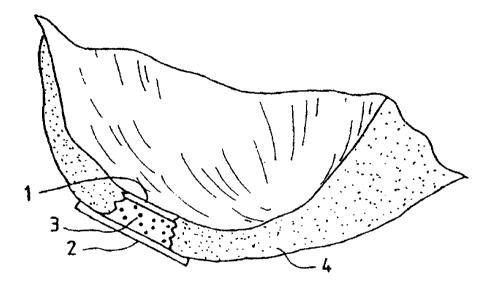
1015202530CA 02264138 1999-02-26wo 98/118469 PCTIUS97Il5258Method, Instruments and Kit for Autologous TransplantationField of the InventionThe instant invention concerns the ï¬eld of chondrocyte transplantation, bone andcartilage graï¬ing, healing, joint repair and the prevention of arthritic pathologies. Inparticular methods for the preparation of the graft site, instruments for such preparation andfor the autologous transplantation of cells to the prepared graft site.Background of the InventionMore than 500,000 arthroplastic procedures and total joint replacements areperformed each year in the United States. Approximately the same number of similarprocedures are performed in Europe. Included in these numbers are about 90,000 total-kneereplacements and around 50,000 procedures to repair defects in the knee per year in Europe.The number of procedures are essentially the same in the U. S. (In: Praemer A., Fumer S.,Rice, D.P., Musculoskeletal conditions in the United States, American Academy ofOrthopaedic Surgeons, Park Ridge, Ill., 1992, 125). A method for regeneration-treatment ofcartilage would be most useful, and could be performed at an earlier stage of joint damage,thus reducing the number of patients needing artificial joint replacement surgery. With suchpreventative methods of treatment, the number of patients developing osteoarthritis wouldalso decrease.Techniques used for resurfacing the cartilage structure in joints have mainlyattempted to induce the repair of cartilage using subchondral drilling, abrasion and othermethods whereby there is excision of diseased cartilage and subchondral bone, leavingvascularized cancellous bone exposed (Insall, J., Clin. Orthop. 1974,101,61; Ficat R.P. er al,Clin Orthop. 1979, 144, 74', Johnson L.L., ln: Qgerative Arthroscopy, McGinty J.B.. Ed.,Raven Press, New York, 1991, 341).Coon and Cahn (Science 1966, 153, 1116) described a technique for the cultivationof cartilage synthesizing cells from chick embryo somites. Later Cahn and Lasher (PNASUSA 1967, 58, 1131) used the system for analysis of the involvement of DNA synthesis asa prerequisite for cartilage differentiation. Chondrocytes respond to both EFG and FGF bygrowth (Gospodarowicz and Mescher, J. Cell Physiology 1977, 93, 117), but ultimatelyl10I5202530CA 02264138 1999-02-26WO 98108469 PCTIUS97I15258lose their differentiated function (Benya et al., Cell 1978, 15, 1313). Methods for growingchondrocytes were described and are principally being used with minor adjustments byBrittberg, M. et al. (New Engl. J. Med. 1994, 331, 889). Cells grown using these methodswere used as autologous transplants into knee joints of patients. Additionally, Kolettas et al.(J. Cell Science 1995, 108, 1991) examined the expression of cartilage-speciï¬c moleculessuch as collagens and proteoglycans under prolonged cell culturing. They found that despitemorphological changes during culturing in monolayer cultures (Aulthouse, A. et al., InVitro Cell Dev. Biol., l989,25,659; Archer, C. et al., J. Cell Sci. 1990,97,36l; Héinselmarm,H. et al., J. Cell Sci. 1994,lO7,l7; Bonaventure, J. et al., Exp. Cell Res. l994,2l2,97), whencompared to suspension cultures grown over agarose gels, alginate beads or as spinnercultures (retaining a round cell morphology) tested by various scientists did not change thechondrocyte - expressed markers such as types II and IX collagens and the largeaggregating proteoglycans, aggrecan, versican and link protein did not change (Kolettas, E.et al., J. Cell Science l99S,108,l99l).The articular chondrocytes are specialized mesenchymal derived cells foundexclusively in cartilage. Cartilage is an avascular tissue whose physical properties dependon the extracellular matrix produced by the chondrocytes. During endochondral ossiï¬cationchondrocytes undergo a maturation leading to cellular hypertrophy, characterized by theonset of expression of type X collagen (Upholt, W.B. and Olsen, R.R., In: CmjlggeMolecular Aspects (Hall, B & Newman, S, Eds.) CRC Boca Raton 1991, 43;Reichenberger, E. et al., Dev. Biol. 1991, 148, 562; Kirsch, T. et al., Differentiation, 1992,52, 89; Stephens, M. et al., J. Cell Sci. 1993, 103, 1111).Excessive degredation of type II collagen in the outer layers or articular surfaces ofjoints is also caused by osteoarthritis. The collagen network is accordingly weakened andsubsequently develops ï¬brillation whereby matrix substances such as proteoglycans are lostand eventually displaced entirely. Such ï¬brillation of weakened osteoarthritic cartilage canreach down to the calcified cartilage and into the subchondral bone (Kempson, G.E. et al.,Biochim. Biophys. Acta 1976, 428, 741; Roth, V. and Mow. V.C., J. Bone Joint Surgery,1980, 62A, 1102; Woo, S.L.-Y. et al., in Handbook of Bioengineering (R. Skalak and S.Chien eds.), McGraw-Hill, New York, 1987, pp. 4.1-4.44).Ix)I015202530CA 02264138 1999-02-26wo 93/03459 PCT/US97/15258Descriptions of the basic development, histological and microscopic anatomy ofbone, cartilage and other such connective tissues can be found for example in Wheater,Burkitt and Daniels, Functional Histology, 2"â Edition, (Churchill Livingstone, London,1987, Chp. 4). Descriptions of the basic histological anatomy of defects in bone, cartilageand other connective tissue can be found for example in Wheater, Burkitt, Stevens andLowe, Basic Histogathology, (Churchill Livingstone, London, 1985. Chp. 21).Despite the advances in cultivating chondrocytes, and manipulating bone andcartilage, there has not been great success with the attempts to transplant cartilage orchondrocytes for the repair of damaged articulating surfaces. The teachings of the instantinvention provide for effective and efficient means of promoting the transplantation ofcartilage and/or chondrocytes into a defect in an articulating joint or other cartilage coveredbone surface, whereby cartilage is regenerated to fix the defect. The instant invention alsoprovides for surgical instruments which are designed prepare the graft site so as to facilitatethe efï¬cient integration of grafted material to the graft site.Brief Summary of the InventionThe instant invention provides a method for the effective treatment of articulatingjoint surface cartilage by the transplantation of chondrocytes in a suitable matrix, to asurface to be treated, with a hemostatic barrier and a cellâfree covering-patch comprising;first placing a hemostatic barrier proximal to the surface to be treated, placing chondrocytesin a suitable matrix upon the surface to be treated distal to the hemostatic barrier, coveringthe surface to be treated with a cellâfree covering-patch. A hemostatic banier, as will befurther described below, is a barrier which inhibits or prevents the penetration ofvascularizing cells and tissue into the grafted material. In particular, the instant methodprovides for a hemostatic ba.rrier that is a resorbable, semi-perrneable material whichinhibits or prohibits vascular inï¬ltration through the barrier. In one embodiment thehemostatic barrier contains collagen. Cell-free, is used herein as in the art, and means amaterial that is substantially free from intact cells which are capable of further cell division.promulgation or biological activity. In a preferred embodiment, a cell-free material is freefrom all intact nucleated cells. In one embodiment, the instant method encompasses the useof a cellâfree covering patch which contains a semiâperrneab1e collagen matrix. In one202530CA 02264138 1999-02-26wo 93/03459 PCTIUS97ll5258preferred embodiment of the method, the porous surface of the cell-free coveringâpatch isdirected towards the implant material.The instant invention further provides for the autologous transplantation of collagenor chondrocytes to a graft site, wherein the graft site has first been prepared by surgicalmanipulation to better accept the grafted material. In one embodiment. the graft site issculpted such that the walls of the graft site are contoured in an undulating pattern such thatthe grafted material, when placed within the graft site and expanded to contact the graft sitewall, there will be resistance against removal or expulsion of the entire graft from the graftsite. The instant invention further provides for surgical instruments designed to sculpt thegraft site as taught by the method of the invention.The invention further provides for a kit for cartilage and/or chondrocytetransplantation onto the surface of an articular joint wherein said kit comprises a hemostaticbarrier, cellâfree semi-permeable covering-patch, and organic glue. In a furtherembodiment, the kit can optionally further provide one or more surgical instruments whichcan be used to sculpt the graft site in accordance with the methods of the instant invention.Brief Description of the DrawingsThe present invention will be better understood by examining the following figureswhich illustrate certain properties of the instant invention wherein:Figure 1A is a drawing showing a typical articulating end of a bone. Typically, thebone material is covered on the articulating surface with a cartilaginous.Figure 1B shows an example of where a defect or injury to the cartilaginous capoccurs (gap in cartilage), and such a defect can be treated directly, enlarged slightly, orsculpted to accept the grafted material by surgical procedures prior to treatment.Figure 1C shows how the hemostatic barrier (numbered 1) is placed within thedefect in the cartilage cap to inhibit or prevent vascularization into regenerating cartilage,from the underlying bone. The chondrocytes to be implanted into the defect cavity are thenlayered on top of the hemostatic barrier.Figure 2 is a drawing showing the treated defect (gap in cartilage) in thecartilaginous cap covered by a cellâfree semi-penneable material (numbered 2) which isused to form a cap/patch or bandage over the defect site. This cap is fixed in place, either10I5202530CA 02264138 2002-06-17sutured to the edge of the cavity into healthy cartilage, or otherwise attached. This cap iscovering the defective area of the joint into which the cultured chondrocytes/cartilagetransplant has been placed, or will be placed under the partially attached cap.Figure 3A is a diagram illustrating the differential response to compression andshearing forces by left side and right side cartilage with subsequent zone of demarcation.Figure 3B illustrates the graft site, after the defect has been sculpted to haveundulating walls.Figure 3C illustrates the sculpted graft site with the hemostatic barrier (1),transplanted material (3), and cell-free covering-patch (2) in place within the articularsurface cartilage (4).Figure 4A illustrates one embodiment of the surgical device of the instant inventionshowing cutting teeth (5) and protruding placement pin (6). The crossâsection illustrationsto the right show two possible configurations of the cutting blades.Figure 4B illustrates a second embodiment of the surgical device of the instantinvention.Figure 5 is a diagram illustrating the modiï¬ed differential response to compressionand shearing forces by harder cartilage and softer cartilage after sculpting the graft site.Figure 6A is an MRI image of a pig knee showing cartilage defect in left (medial)condyle.Figure 6B is an MRI image of the same pig knee three months after treatment.Detailed Description of the lnventionThis invention concerns the use of certain products that inhibit the formation ofvascular tissue, for instance such as capillary loops projecting into the cartilage beingestablished, during the process of autologous transplantation of chondrocytes into defects inthe cartilage. The formation of vascular tissue from the underlying home will tend to projectinto the new cartilage to be formed leading to appearance of cells other than themesenchymal specialized chondrocytes desired.The contaminating cells introduced by the vascularization may give rise toencroachment and over-growth into the cartilage to be formed by the implantedchondrocytes. One of the types of commercial products which can be used in -this inventionis Surgicalâ (Ethicon Ltd., UK) which is absorbable after a period of 7 â 14 days. The use ofthis material in the method of the instant invention is contrary to the normal use of a1015202530CA 02264138 1999-02-26WO 98108469 PCTIUS97/ 15258hemostatic device, such as Surgicel° as it is described in the package insert from EthiconLtd.Surprisingly, we have found that in a situation where you wish to inhibit re-vascularization into cartilage, a hemostatic material will act like a gelâlike artiï¬cialcoagulate. If red blood cells should be present within the full-thickness defect of articularcartilage that is capped by such a hemostatic barrier, these blood cells will be chemicallychanged to hematin, and thus rendered unable to induce vascular growth. Thus a hemostaticproduct used as a re-vascularization inhibitory barrier with or without fibrin adhesives, suchas for example the Surgicelâ, is effective for the envisioned method as taught by the instantinvention. Another part of this invention is the use of a cell-free component, that is used asa patch covering the defective area of the joint into which the culturedchondrocytes/cartilage are being transplanted, using autologous chondrocytes for thetransplantation. The method of the invention also contemplates the use of suitable allogenicchondrocytes or xenogenic chondrocytes for the repair of a cartilage defect.Thus the instant invention teaches methods for effective repair or treatment ofcartilage defects in articular joint bone surfaces which comprises administering an agent ordevice to block vascular invasion into the cartilage site to be repaired, and also providingfor a cell-free barrier which will isolate the repair site and keep transplanted cells in place.Thus the instant invention also provides for a kit comprising a hemostatic barriercomponent for insertion into the site to be repaired, such that there is effective inhibition ofvascularization into the site to be repaired; and once the chondrocytes to be transplanted areplaced into the site to be repaired, a cell-free semi-pemieable barrier is capped over therepair site such that the transplanted chondrocytes are held in place, but are still able to gainaccess to nutrients.Certain aspects of the invention have been exemplified using an in vitra system tostudy the behavior of the chondrocytes when in contact with a certain product or acombination of certain products that inhibit the formation of vascular tissue. This in vitrotesting predicts the ability of certain tested materials to inhibit vascularization, as will occurin viva where capillary loops project into the cartilage being established during the processof autologous transplantation of chondrocytes into defects in the cartilage.Suitable hemostatic products will be characterized by having the ability to inhibitthe growth, or invasion of vascular tissue, osteocytes, fibroblasts etc. into the developingcartilage. A suitable hemostatic material will achieve the goal of the method of the instantIOI5202530CA 02264138 1999-02-26wo 93/03459 PCT lUS97I15258invention in that vascular and cellular invasion into developing cartilage should beprevented in order to optimize the formation of cartilage and achieve repair of the full-thickness of any defects in the articular cartilage. Ideally, the hemostatic barrier will bestable for an extended period of time sufficient to allow for full cartilage repair, and then beable to be resorbed or otherwise broken down over time. One material identified as suitableis called Surgicelâ W1912 (an absorbable hemostat containing oxidized regenerated sterilecellulose; Lot GG3DH, Ethicon Ltd. UK ). Another example of a suitable material isBioGide® (a commercially available type I collagen matrix pad; Geistlich Sohne,Switzerland).Suitable organic glue material can be found commercially, such as for exampleTisseel"â or Tissucol° (ï¬brin based adhesive; Immuno AG. Austria), Adhesive Protein (Cat.#A-2707, Sigma Chemical, USA), and Dow Corning Medical Adhesive B (Cat. #895â3,Dow Corning, USA).The surgical instruments contemplated by the instant invention can be manufacturedfrom metal and/or plastic suitable for making single-use disposable, or multi-use reusablesurgical instruments. The cutting instrument may contain cutting teeth that are fully circularor ï¬at, or anything in between. As cartilage is a relatively soft material it may beadvantageous to manufacture hardened plastic cutting edges which will be able to sculptcartilage without being able to damage bone. Such cutting instruments can be manufacturedto incorporate openings for administration of ï¬uid, suction removal of cutting debris andï¬uid, and fiber optic threads for illumination and visualization of the defect site.Certain aspects of the instant invention may be better understood as illustrated bythe following examples, which are meant by way of illustration and not limitation.Example 1In order for the Surgicel °ââ to be used according to the invention for preventingdevelopment of blood vessels into autologous implanted cartilage or chondrocytes,Surgicelâ was first treated with a fixative, such as glutaric aldehyde. Brieï¬y, Surgicelâ wastreated with 0.6% glutaric aldehyde for 1 minute, followed by several washings to eliminateglutaric aldehyde residues that may otherwise be toxic to tissue. Alternatively, theSurgicelâ was treated with the ï¬brin adhesive called Tisseel® prior to treatment withglutaric aldehyde as described in Example 2. It was found that the Surgicelâ ï¬xated forinstance with a ï¬xative such as glutaric aldehyde, washed with sterile physiological saline71015202530CA 02264138 1999-02-26wo 93/03459 PCT/US97/ 15258(0.9%) and stored in refrigerator, does not dissolve for 1 to 2 months. Generally, Surgical isresorbed in a period between 7 and 14 days. This time would be too short, because a longertime is needed in preventing the development of blood vessels or vascularization as suchfrom the bone structure into the implanted cartilage before the implanted chondrocytes havegrown into a solid cartilage layer getting its nutrition requirements from the neighboringcartilage. In other words sufficient inhibition of the vascularization is needed for a longertime such as for instance one month. Therefore, the product should not be absorbedsigniï¬cantly prior to that time. On the other hand resorption is needed eventually. Hence,the organic material used as an inhibiting barrier shall have these capabilities, and it hasbeen found that the Surgicalâ treated in this manner provides that function.Example 2The Surgicelâ was also coated with an organic glue. in this example the glue usedwas Tisseelâ but others can also be used. This product, together with the Surgicelâ producesa useable barrier for the particular purpose of the invention. Any other hemostat or vascularinhibiting barrier could be used. The Tisseelââ was mixed as described below. The Surgicel°was then coated with Tisseel° by spraying the Surgicelâ° material on both sides until soaked.The Tisseel° (ï¬brin glue) was then allowed to solidify at room temperature. Immediatelyprior to completed solidiï¬cation, the coated Surgicelâ was then placed in 0.6% glutaricaldehyde for 1 minute and then washed with sterile physiological (0.9%) saline. The pHwas then adjusted by PBS and/or with NaOH until pH was stable at 7.2 to 7.4. Afterwardsthe thus treated Surgicelw was then washed in tissue culture medium such as minimumessential medium/F12 with 15 mM Hepes buffer.As mentioned in this example we have used Tisseel® as the ï¬brin adhesive to coatthe Surgicelâ. Furthermore the ï¬brin adhesive or glue may also be applied directly on thebottom of the lesion towards the bone, on which the Surgieel® is glued. The in vitro systemused, in lieu of in vivo testing, consisted of a NUNCLONTM Delta 6-well sterile disposableplate for cell research work (NUNC, lnterMed, Roskilde, Denmark). Each well measuresapproximately 4 cm in diameter.In the invention the ï¬brin adhesive can be any adhesive which together with theï¬brin component will produce a glue that can be tolerated in humans (Ihara, N, et al., BurnsIncl. Therm. Inj., 1984, 10, 396). The invention also anticipates any other glue componentthat can be used in lieu of the fibrin adhesive. In this invention we used Tisseelâ orl0l5202530CA 02264138 1999-02-26PCT/US97/15258WO 98/08469Tissucol® (Immuno AG, Vienna, Austria). The Tisseel® kit consists of the followingcomponents:Tisseelâ, a lyophilized, virus-inactivated Sealer, containing clottable protein,thereof: ï¬brinogen, Plasma frbronectin (CIG) and Factor XIII, and Plasminogen.Aprotinin Solution (bovine)Thrombin 4 (bovine)Thrombin 500 (bovine)Calcium Chloride solutionThe Tisseelâ kit contains a DUPLOJECTâ Application System. The ï¬brin adhesiveor the two-component sealant using Tisseelo Kit is combined in the following manneraccording to the Immuno AG product insert sheet:Example 3Chondrocytes were grown in minimal essential culture medium containing HAMF12 and 15 mM Hepes buffer and 5 to 7.5% autologous serum in a C02 incubator at 37°Cand handled in a Class 100 laboratory at Verigen Europe A/S, Symbion Science Park,Copenhagen, Denmark. Other compositions of culture medium may be used for culturingthe chondrocytes. The cells were trypsinized using trypsin EDTA for 5 to 10 minutes andcounted using Trypan Blue viability staining in a Btlrker-Tiirk chamber. The cell count wasadjusted to 7.5 x 10⁵ cells per ml. One NUNCLONTM plate was uncovered in the Class 100laboratory.The Surgicelâ hemostatic barrier was cut to a suitable size ï¬tting into the bottom ofthe well in the NUNCLONW tissue culture tray. In this case a circle, of a size ofapproximately 4 cm (but could be of any possible size) and placed under aseptic conditionson the bottom in well in a NUNCLONTâ Delta 6-well sterile disposable plate for cellresearch work (NUNC, lnterMed, Roskilde, Denmark). The hemostatic barrier to be placedon the bottom of the well was preâtreated as described in Example 1. This treatment delaysthe absorption of the Surgicel significantly. This hemostatic banier was then washedseveral times in distilled water and subsequently several times until non-reactedglutaraldehyde was washed out. A small amount of the cell culture medium containingserum was applied to be absorbed into the hemostatic barrier and at the same time keepingthe hemostatic barrier wet at the bottom of the well.Approximately 10° cells in 1 ml culture medium were placed directly on top of the910202530CA 02264138 1999-02-26W0 98/084659 PCTIUS97I 15258hemostatic barrier, dispersed over the surface of the hemostatic banier pre-treated with0.4% glutaraldehyde as described above. The plate was then incubated in a CO, incubator at37°C for 60 minutes. An amount of 2 to 5 ml of tissue culture medium containing 5 to 7.5%serum was carefully added to the well containing the cells avoiding splashing the cells byholding the pipette tip tangential to the side of the well when expelling the medium. Itappeared that the pH of the medium was too low (pH -6.8). The pH was then adjusted to7.4 to 7.5. The next day some chondrocytes had started to grow on the hemostatic barrier,arranged in clusters. Some of the cells had died due to the low pH exposure prior to theadjustment of the pH. The plate was incubated for 3 to 7 days with medium change at day3.At the end of the incubation period the medium was decanted and cold refrigerated2.5% glutaraldehydc containing 0.1M sodium salt of dimethylarsinic acid, (also calledsodium cacodylate, pH is adjusted with HCl to 7.4), was added as ï¬xative for preparation ofthe cell and supporter (hemostatic barrier) for later preparation for electron microscopy.Example 4Chondrocytes were grown in minimal essential culture medium containing HAMF12 and 15 mM Hepes buffer and 5 to 7.5% autologous serum in a C03 incubator at 37°Cand handled in a Class 100 laboratory at Verigen Europe A/S, Symbion Science Park,Copenhagen, Denmark. Other compositions of culture medium may be used for culturingthe chondrocytes. The cells were trypsinized using trypsin EDTA for 5 to 10 minutes andcounted using Trypan Blue viability staining in a Bï¬rker-Turk chamber. The cell count wasadjusted to 7.5 x 10⁵ cells per ml. One NUNCLONTâ plate was uncovered in the Class 100laboratory.The Surgicel" (for use as the hemostatic barrier) was treated with 0.6% glutaricaldehyde for one minute as described in Example 1, and washed with 0.9% sterile sodiumchloride solution or, preferably, with a buffer such as a PBS buffer or the culture mediumsuch as MEM/F12, because pH after the glutaric aldehyde treatment is 6.8 and shouldpreferably be 7.0 to 7.5. The Tisseel° was applied on both side of the Surgicel°° using theDUPLOJECTQ system, thus coating both sides of the Surgicel®, the patch intended to beused, with fibrin adhesive. The glue is left to dry under aseptic condition for at least 3 to 5minutes. The "coated" hemostatic ban'ier was placed on the bottom of the well in aNUNCLONTM Delta 6-well sterile disposable plate for cell research work. A small amount101015202530CA 02264138 1999-02-26wo 93/03459 PCTIUS97/15258of tissue culture medium containing serum was applied to be absorbed into the hemostaticbarrier. Approximately 10° cells in 1 ml tissue culture medium containing serum wasplaced directly on top of the Hemostat, dispersed over the surface of the hernostatic barrier.The plate was then incubated in a CO: incubator at 37°C for 60 minutes. An amount of 2 to5 ml of tissue culture medium containing 5 to 7.5 % serum was carefully added to the wellcontaining the cells avoiding splashing the cells by holding the pipette tip tangential to theside of the well when expelling the medium. After 3 to 6 days, microscopic examinationshowed that the cells were adhering to and growing into the Surgicel° in a satisfactory waysuggesting that Surgicelâ did not show toxicity to the chondrocytes and that thechondrocytes grew in a satisfactory manner into the Surgicel°.The plate was incubated for 3 to 7 days with medium change at day 3. At the end ofthe incubation period the medium was decanted and cold refrigerated 2.5% glutaraldehydecontaining O.lM sodium salt of dimethylarsinic acid, also called sodium cacodylate, pH isadjusted with HCl to 7.4, was added as fixative for preparation of the cell and supporter(hemostatic barrier) for later preparation for electron microscopy.Example 5Chondrocytes were grown in minimal essential culture medium containing HAMF12 and 15 mM Hepes buffer and 5 to 7.5% autologous serum in a CO, incubator at 37°Cand handled in a Class 100 laboratory at Verigcn Europe A/S, Syrnbion Science Park,Copenhagen, Denmark. The cells were trypsinized using trypsin EDTA for 5 to 10 minutesand counted using Trypan Blue viability staining in a Biirker-Turk chamber. The cell countwas adjusted to 7.5 x 10⁵ to 2 x 10" cells per ml. One NUNCLONTâ plate was uncovered inthe Class 100 laboratory.It has been found that the Bio-Gideâ can be used as a resorbablc bilayer membranewhich will be used as the patch or bandage covering the defective area of the joint intowhich the cultured chondrocytes are being transplanted as well as the hemostatic barrier.The Bio-Gideâ is a pure collagen membrane obtained by standardized, controlledmanufacturing processes (by E.D. Geistlich Sohne AG, CH-6110 Wolhusen). The collagenis extracted from veterinary certiï¬ed pigs and is carefully puriï¬ed to avoid antigenicreactions, and sterilized in double blisters by 7-irradiation. The bilayer membrane has aporous surface and a dense surface. The membrane is made of collagen type I and type IIIwithout further cross-linking or chemical treatment. The collagen is resorbed within 24lll0l5202530CA 02264138 1999-02-26wo 98I08469 PCT/US97ll5258weeks. The membrane retains its structural integrity even when wet and it can be ï¬xed bysutures or nails. The membrane may also be "glued" using ï¬brin adhesive such as Tisseel®to the neighboring cartilage or tissue either instead of sutures or together with sutures.The Bio-Gideâ was un-covered in a class 100 laboratory and placed under asepticconditions on the bottom of the wells in a NUNCLONTâ Delta 6âwell sterile disposableplate for cell research work, - either with the porous surface of the bilayer membrane facingup or with the dense surface facing up. Approximately 10° cells in 1 ml tissue culturemedium containing serum was placed directly on top of the BioâGide°, dispersed eitherover the porous or the dense surface of the Bio-Gideâ. The plate was then incubated in aC02 incubator at 37°C for 60 minutes. An amount of 2 to 5 ml of tissue culture mediumcontaining 5 to 7.5 % serum was carefully added to the well containing the cells avoidingsplashing the cells by holding the pipette tip tangential to the side of the well whenexpelling the medium.On day 2 after the chondrocytes were placed in the well containing the BioâGide°the cells were examined in a Nikon Inverted microscope. It was noticed that somechondrocytes had adhered to the edge of the Bio-Gide. It was of course not possible to beable to look through the Bio-Gideâ itself using this microscope.The plate was incubated for 3 to 7 days with medium change at day 3. At the end ofthe incubation period the medium was decanted and cold refrigerated 2.5% glutaraldehydecontaining 0.1M sodium salt of dimethylarsinic acid, also called sodium cacodylate, pH isadjusted with HCl to 7.4, was added as ï¬xative for preparation of the cell and the Bio-Gide° supporter with the cells either cultured on the porous surface or the dense surface.The Bio-Gideâ patches were then sent for electron microscopy at Department of Pathology,Herlev Hospital, Denmark.The electron microscopy showed that the chondrocytes cultured on the densesurface of the Bio-Gideâ did not grow into the collagen structure of the Bio-Gideâ, whereasthe cells cultured on the porous surface did indeed grow into the collagen structure andfurthennore, showed presence of proteoglyeans and no signs of fibroblast structures. Thisresult shows that when the collagen patch, as for instance a Bio-Gideâ patch is sewn as apatch covering a cartilage defect the porous surface shall be facing down towards the defectin which the cultured chondrocytes are to be injected. They will then be able to penetratethe collagen and produce a smooth cartilage surface in line with the intact surface, and inthis area a smooth layer of proteoglyeans will be built up. Whereas, if the dense surface ofl21015202530CA 02264138 1999-02-26WO 98/08469 PCTIU S97/ 15258the collagen is facing down into the defect the chondrocytes to be implanted will notintegrate with the collagen, and the cells will not produce the same smooth surface asdescribed above.Example 6Chondrocytes were grown in minimal essential culture medium containing HAMF12 and 15 mM Hepes buffer and 5 to 7.5% autologous serum in a CO: incubator at 37°Cand handled in a Class 100 laboratory at Verigen Europe A/S, Symbion Science Park,Copenhagen, Denmark. The cells were trypsinized using trypsin EDTA for 5 to 10 minutesand counted using Trypan Blue viability staining in a Bï¬rker-Tiirk chamber. The cell countwas adjusted to 7.5 x 10â to 2 x 10° cells per ml. One NUNCLONTâ plate was uncovered inthe Class 100 laboratory.The Bio-Gideâ used as a resorbable bilayer membrane may also be used togetherwith an organic glue such as Tisseelqâ with additional, signiï¬cantly higher content ofAprotinin than normally found in Tisseelâ°, as described in the product insert. By increasingthe content of Aprotinin to about 25,000 KIU/ml, the resorption of the material will bedelayed by weeks instead of the normal span of days.To test this feature in vitro, the Tisseelâ is applied to the bottom of the well of theNUNCLONTâ plate, and allowed to solidify incompletely. A collagen patch such as a Bio-Gide® is then applied over the Tisseel® and glued to the bottom of the well. Thiscombination of Bio-Gideâ and Tisseelâ is designed to be a hcmostatic barrier that willinhibit or prevent development or infiltration of blood vessels into the chondrocytctransplantation area. This hybrid collagen patch can now be used for both as a hemostaticbarrier at the bottom of the lesion (most proximal to the surface to be repaired) but also as asupport for cartilage formation because the distal surface can be the porous side of thecollagen patch and thus encourage infiltration of chondrocytes and cartilage matrix. Thusthis hybrid collagen patch can also be used to cover the top of the implant with the collagenporous surface directed down towards the implanted chondrocytes and the barrier formingthe top. The hybrid collagen patch, with elevated Aprotinin component may also be usedwithout any organic glue such as Tisseel° and placed within the defect directly, adhering bynatural forces. Thus the collagen patch can be used both as the hcmostatic barrier, and thecell-free covering of the repair/transplant site, with the porous surfaces of the patchesoriented towards the transplanted chondrocytes/cartilage. Another variant would use a1310l5202530CA 02264138 1999-02-26wo 93/03459 PCT/US97l15258collagen patch which consists of type II collagen (ie. from Geistlich Sohne AG, CHâ6l 10Wolhusen).Thus the instant invention provides for a hybrid collagen patch where said patch is acollagen matrix with elevated levels of aprotinin component, preferably about 25,000KIU/ml, in association with an organic matrix glue, where the collagen component issimilar to the Bio-Gideâ resorbable bilayer material or Type II collagen, and the organicglue is similar to the Tisseel° material. In another embodiment, the hybrid collagen patchdoes not use any organic glue to adhere to the site of the repair.Example 7Because of the weakened structure of osteoarthiitie cartilage, adherence of culturedautologous chondrocytes transplanted to a graft site in defective cartilage may be inhibited,thus creating a marginal zone (zone of demarcation) between the newly implantedcartilage/chondrocytes and the surrounding established cartilage. This marginal zone will bemost pronounced if the graï¬ site is prepared for the graft by creating straight, smooth wallscut in a linear fashion. The shearing and compression forces across such a marginal zone (asillustrated in Figure 3A) will exert great force to dislodge the graft when the graft site is cutin a linear fashion. This marginal zone, and differential movement of materials along thiszone will inhibit confluent healing between the grafted material and the surroundingmaterial. This marginal zone shearing is exacerbated when the hardness of the abuttingmaterial is different. In many cases the graft material is softer than the surroundingmaterial, however, in some instances of osteoarthritis disease, the surrounding cartilagemay in fact be softer than the implanted chondrocytes/cartilage.Therefore, in order to solve this problem, the method of the invention teaches theuse of surgical instruments to sculpt the walls of the graft site such that the walls are non-linear, and thus provide for undulated surfaces which will reduce marginal zone shearingand provide anchorage for grafted material. It is also possible to shape the graft site suchthat the diameter of the site proximal to the bone surface is of a greater dimension then theopening distal to the bone and at the surface of the cartilage to be repaired such that there isa âreverse funnelâ effect. A narrowed opening at the surface will aid in reducing marginalzone shearing and the expulsion of graï¬ material from the graft site. A preferredembodiment describes the sculpting of the walls of the graft site in an fashion similar to athreaded opening for receiving a belt or screw (as illustrated in Figure 3B), thus providingl4I0202530CA 02264138 1999-02-26WO 98/08469 PCTIUS97l15258mechanical resistance to the compression and or ejection of the grafted material from thegraft site which can be described as âmaleâ and âfemaleâ threading.The surgical instruments contemplated by the instant invention can be manufacturedï¬'om metal and/or plastic suitable for making singleâuse disposable, or multi-use reusablesurgical instruments. As cartilage is a relatively soft material it may be advantageous tomanufacture hardened plastic cutting edges which will be able to sculpt cartilage withoutbeing able to damage bone. Such cutting instruments can be manufactured to incorporateopenings for administration of ï¬uid, suction removal of cutting debris and ï¬uid, and ï¬beroptic threads for illumination and visualization of the defect site. In certain embodiments ofthe instrument, the base of the instrument may have protruding point or pin-like structurewhich will assist in guiding and placing the instrument in the graft site. Of course such a pinwould be designed to minimized damage to the underlying bone.While the cutting surface of the instrument may be single toothed. or multi-toothed,or describe a screw-like pattern such as that in a metal tap used to generate threaded holesin metal parts, the characteristic required of the cutting instrument is that the resultingsculpted sides of the graft site is undulated, and non-linear. For example, in certainembodiments, the cutting edge of the instrument can be shaped similar to that shown inFigure 4A, or as in Figure 4B. The cutting edge maybe ï¬at, or circular in that it wrapsaround the diameter of the cutting instrument. Many other shapes can be designed toaccomplish the purpose of the method of the invention to create an interface which providesfor mechanical resistance to differential reaction to compression and shearing forces on thetransplanted material and the surrounding material.Example 8A four month old mixed Yorkshire breed pig was subjected to general anesthesiaand placed on its back. The pig was washed and draped in a surgical suite at HarringtonArthritis Research Center, Phoenix, Arizona. The entire surgical procedure was performedaseptically. The left hind-leg and adjacent abdomen and inguinal area was cleaned withiodine. The knee joint was localized, and the patella localized. A medial incision wasperformed approximately 3 cm from the posterior part of the patella and the severalsubcutis, muscle layers and ligaments was cut approximately in order to get access to themedial femoral condyle. Using a circular cutter a lesion was prepared in the white cartilageon the medial part of the medial condylc, leaving a 0.5 to 1 cm margin to the edge of theISl015202530CA 02264138 1999-02-26W0 98108469 PCTIUS97/1 5258cartilage covering the posterior-medial part of the condylc (left condyle, Figure 6A). The0.5 to 1 cm defect was placed in a caudal weight bearing part of the medial condyle. Theentire surgical procedure was done without tourniquet on the left femur. The different layersand skin was sutured appropriately.On day 3 the animal was again brought to the surgical suite and positioned as aboveon the operating table and given general anesthesia. The left hind leg, abdomen andinguinal region was ionized as described above. Sutures were cut and the area opened. Itwas noticed that a moderate hematoma was present in the knee joint. The blood clot wasremoved and the defect inspected. There was a blood clot in the defect which was removed.A sterile surgical instrument designed with a male thread cutting edge, with a sizecorresponding to, or slightly bigger than the circumference of the lesion was carefullyscrewed down into the defect. A BioGide°â pad was cut to a size equal to the bottom of thedefect. The first glue used. called Adhesive Protein (A-2707, Sigma Chemical, USA) wasapplied on the dense side of the trimmed hemostatic barrier pad, and the pad was placeddense side down into the bottom of the lesion, using it as a barrier as described above. Itwas found that this glue did not seem to dry very fast. The slight bleeding from the bottomof the defect stopped immediately. A second BioGide" was cut somewhat bigger incircumference than the lesion and was placed with dense side up (thus the porous side downtowards the graft) as described above.This nonâcellular covering-pad was then sutured over the cavity, leaving one edgeopen, where the chondrocyte to be explanted could be injected into the graft site. Thesurrounding part of the edge of the pad was covered with the second glue, Dow CorningMedical Adhesive B (Cat. #895-3, Dow Corning, USA). This second glue dried much fasterand more efficiently than the first glue. It was found that during this particular procedure,the ï¬rst glue had not dried sufficiently to hold the hemostatic barrier in place when suturingof the cap was attempted. The main barrier formed on the proximal surface of the graft sitewas by the glue itself.Using a 1 ml syringe and a 16 gauge needle. the chondrocyte cell suspension (about0.6 ml) was drawn up into the barrel of the syringe. A 23 gauge short needle was switchedfor the 16 gauge needle, and the cell suspension was injected under the sutured covering-patch into the graft site (about 10 x 10° cells). The open edge of the cap was then gluedprior to removal of the needle, and the needle carefully withdrawn. No leakage of cells wasseen. The wound was sutured and as above, no tourniquet was used, no bleeding wasl61015202530CA 02264138 1999-02-26WO 98/08469 PCT IU S97/15258observed. The final skin layers were sutured. No protrusion of the skin occurred aftersuturing, which indicates that there was no hematoma. Postoperative recovery wasuneventful.As expected, the grafted chondrocytes produced cartilage matrix sufï¬cient to repairthe defect made in the articular cartilage surface of the knee joint of the test pig. Figure 6Ais an MRI image of a pig knee showing the cartilage defect created in the knee (leftcondyle, the medial condyle), and Figure 6B is an MRI image of the same pig knee threemonths after treatment showing repair of the defect.Example 9A kit comprising the components useful for practicing the method of the invention,will allow for the convenient practice of the method of the invention in a surgical setting. Ina preferred embodiment, a kit of the invention will provide sterile components suitable foreasy use in the surgical environment, and will provide a suitable hemostatic barrier, suitablecovering patch, and if needed organic glue. A kit of the invention may also provide sterile,cellâfree matrix material suitable for supporting autologous chondrocytes that are to beimplanted into an articular joint surface defect. In one embodiment, a kit of the inventioncontains a Surgicel° hemostatic barrier and a Bio-Gideâ covering patch with suitablecoating of Tisseel"° organic glue, where the Surgicel° and Bio-Gideâ have been treatedaccording to the teachings of the invention to increase the time till resorption. In instanceswhere Tisseel® is preâcoated, in one embodiment the 'lâisseel® is supplemented withadditional aprotinin to increase time till resorption.In another preferred embodiment, the hemostatic barrier and covering-patch are botha semi-permeable collagen matrix which is treated to extend the time till resorption of thematerial. It is also possible to provide 'Iâisseelââ glue in enhanced form as a separatecomponent to be applied as needed because of the inherent variability and uniquecircumstances every repair/transplantation procedure will encounter.A further embodiment of a kit of the invention will include a surgical instrument asdescribed in Example 7 above, or suitable variations thereof.It will be appreciated by persons skilled in the art that numerous variations and/ormodifications may be made to the invention shown in the specific embodiments withoutdeparting form the spirit and scope of the invention as described.