Note: Descriptions are shown in the official language in which they were submitted.
CA 02270460 2006-04-06
1
An Intravaginal Set For Treatment of Prolapse of Urogenital
Organs and Urinary Stress Incontinence
BACKGROUND OF THE INVENTION
The present invention relates to an intravaginal set, used in
the case of prolapse of the urogenital organs and urinary
stress incontinence, or during the period when a therapeutic
intravaginal insert for treatment of static disorders of the
urogenital organs and urinary stress incontinence is not
currently inserted and the method of the treatment with
usage of this intravaginal set, and application thereof.
CA 02270460 2006-04-06
2
Aging and past parturitions result in weakening and
elongation of the perineum muscles leading to the prolapse
of urogenital organs and other anatomical disorders.
Once extended, muscles become weaker and weaker which
subsequently results in progress of prolapse up to
transvaginal eversion of the uterus. As the vaginal canal is
the "locus minoris resistentiae" in the pelvic floor, walls
of the vagina may become the ring of hernia (cystocele,
uretrocele and rectocele). In the course of progressive
prolapse of urogenital organs, discomfort in lower abdominal
part intensifies, from a feeling of "heaviness" to one of
pain, and urinary stress incontinence becomes apparent.
Prolapse of the urogenital organs causes a decrease in the
distance between the uterine cervix and the vaginal inlet,
descent of the anterior wall of the vagina along with the
urinary bladder and urethra (the cysto-urethral angle becomes
more obtuse) , and dislocation of the urethra to outside an
operation range of the intra-abdominal pressure.
This results in impaired blood outflow from the urogenital
organs due to venous constriction (low-pressure blood
vessels).
In less advanced cases of the urogenital prolapse, special
therapeutic exercises are recommended to strengthen the
pelvic floor muscles.
CA 02270460 2006-04-06
3
The physical exercises may only increase the efficiency of
the cross-striated muscles, i.e. muscles dependent on the
person's own physical characteristics, whereas the
urogenital organs equilibrium is independent of the patient's
muscle responses since it is controlled by the autonomous
nervous system. Smooth muscles build the urinary bladder,
urethra, internal sphincter muscle of urethra, and muscles
in uterine ligaments fix the uterus in its normal position.
Significant progress in the treatment of static disorders of
the urogenital organs has been made by development of a
therapeutic insert (patent specification No. RP 138406),
consisting of a hollow ball with a string attached freely
outside the ball, and a smaller ball placed freely inside the
hollow ball. This device generates mechanical impulses
stimulating contractions of surrounding muscles, both
smooth, and cross-striated. A metal ball placed for free
movement in the intravaginal therapeutic insert generates
mechanical impulses by hitting its cap as a result of
translocation to the center of gravity while the patient is
walking. The mechanical impulse stimulates muscular
contraction. Regular muscular exercise results ir_ muscle
hypertrophy and an increase in muscular force. The
therapeutic insert may be used twice a day, over a period of
about 30 minutes.
Prolonged application longer than 30 minutes leads to
muscular overstrain and abdominal pain. Use of the
therapeutic insert every day over a period of three months
achieved satisfactory therapeutic results in the treatment of
minor- prolapse of the urogenital organs (grade I), while in
CA 02270460 2006-04-06
4
more advanced cases (grades II, III, IV) no improvement was
observed.
The most probable explanation of this result is that in the
case of more advanced urogenital prolapse the uterus lowers
during intervals between use of the therapeutic device,
resulting in unfavorable effects (isometric contraction,
passive congestion).
When conservative therapy is ineffective, the reconstructive
surgery is performed to restore the normal position of the
urogenital organs. Different strategies of therapeutic
management are used, depending on symptoms, age and progress
of the illness. From among around 200 modifications applied
to correct the position of the urogenital organs, the basic
management consists in shortening of muscles and ligaments.
Such therapeutic management ameliorates the position of the
organs, but it does not restore the normal muscular function
and thus a lasting and complete recovery.
In the case of contraindications for surgery, a device-
prostheses may be used to ensure a normal position of the
urogenital organs.
One such device is the well-known intravaginal disc for
support of a lowered or prolapsed uterus. This disc is
applied in a manner such that its ring surrounds the uterine
cervix, thus preventing uterus prolapse by extension of the
vaginal wall in the area of the posterior vaginal fornix. A
disadvantage of this design includes difficulties in
application and removal. These problems have limited use of
CA 02270460 2006-04-06
this device. Also, as the disc is placed in the upper part of
the vagina, it does not transmit the contraction of the
levator ani muscle. Thus, the uterus is not elevated. The
British patent application discloses a plastic planar arc,
5 the wider arm of which rests on the pubic symphysis, while
the second, narrow arm presses the urethra against the
urinary bladder. The maximum time for intravaginal
application is 2 hours, which limits the usefulness of this
device. The mode of action of this device is local
compression of the urethra, which may lead to inflammation
and decubitus ulcers.
There is also a well-known disposable vaginal pack (made in
Germany), which is placed in the vagina for a period of up
to 8 hours. Its mode of action is a mild compression of the
urethra and adjustment to the vagina after soaking with water
before intravaginal application. This vaginal pack, however,
does not provide the desired corrective functions, and may
cause an inflammatory state in the case of prolonged
intravaginal presence. Soaking with vaginal secretion may
result in distension of the vagina leading to progressive
prolapse of the uterus and urinary bladder.
OBJECT OF THE INVENTION
The object of the invention is to provide an intravaginal set
and a method of treatment using the set to obtain permanent
optimal positioning of the uterus and urinary bladder.
CA 02270460 2006-04-06
6
BRIEF DESCRIPTION OF THE INVENTION
An intravaginal set is provided to be used in the treatment
of prolapse of urogenital organs and urinary stress
incontinence, or in the period of intervals in women when
the intravaginal therapeutic insert for treatment of static
disorders of the urogenital organs and urinary stress
incontinence is not currently inserted. The therapeutic
insert comprises a hollow plastic ball with string attached
and freely moving outside the ball, a smaller ball is placed
to be freely movable inside the hollow plastic ball, which
smaller ball has a weight adequately adjusted to generate
mechanical impulses stimulating alternate contractions of
the muscles. The set also includes a subset of intravaginal
corrective inserts and an intravaginal measuring subset for
determining the size of the insert. The subset of
intravaginal corrective inserts has at least two balls of
different diameters, ranging between the minimal and maximal
woman patent vaginal diameter. Each ball is preferably
hollow, and each ball has the loosely hanging string. Each
ball is preferably made of medical material, such as
polycarbonate or methyl methacrylate, while the intravaginal
measuring subset comprises at least two balls made of metal
or plastic with graduated diameters corresponding to
graduated diameters of the balls of subset of intravaginal
corrective inserts. The measuring balls, instead of having
the loosely hanging string, have a rigidly mounted,
preferably linearly scaled, slat for measurement of optimal
diameter and depth of localization of the insert in the
vagina, depending on actual and individual anatomical
conditions of urogenital organs of the woman being treated.
CA 02270460 2006-04-06
The invention also is directed to a method of treatment of
prolapse of urogenital organs and urinary stress
incontinence or in the period of intervals in women when the
intravaginal therapeutic insert for treatment of static
disorders of the urogenital organs and urinary stress
incontinence is not currently inserted, said method being
realized by means of an intravaginal set of inserts,
characterized by selecting the appropriate size of the
corrective insert from a subset of intravaginal corrective
inserts, consisting of at least two balls with a different
increasing diameter, ranging between the minimal and maximal
woman vaginal diameter, each ball being preferably hollow,
and each ball has a loosely hanging string and each ball is
made preferably of medical material, such as polycarbonate
or methyl methacrylate. Selecting a ball is realized by
means of an intravaginal measuring subset comprising at least
two metal or plastic balls having graduated diameters
corresponding to graduated diameters of the balls from the
subset of the intravaginal corrective inserts, which balls
instead of having the loosely hanging string have a rigidly
mounted linearly scaled slat for measurement of optimal
diameter and depth of location of the corrective insert in
the vagina, depending on actual and individual anatomical
conditions of the urogenital organs of the woman being
treated, by the selection of appropriate optimal diameter
and depth of location of the corrective insert in the vagina
by approximations by using the balls from the measuring
subset, so that contraction of the levator ani muscle will
cause the elevation of the insert and the elevation of the
insert will cause the elevation of the uterus and/or
correction of the cysto-urethral angle, and during the
progress of said treatment, the sizes of successive applied
corrective inserts are adjusted by analogous selection of
CA 02270460 2006-04-06
8
appropriate optimal diameter and depth of location of the
measuring ball in the vagina, and advantageously by carrying
out the exercises of the pelvic floor muscles of the woman
being treated lying in prone or genucubital position in the
intervals between successive replacements of the corrective
inserts.
An application of an intravaginal set in the treatment of
prolapse of urogenital organs and urinary stress
incontinence or in the period of intervals in women when the
intravaginal therapeutic insert for treatment of static
disorders of the urogenital organs and urinary stress
incontinence is not currently inserted. A method is realized
by means of an intravaginal set of inserts from which the
appropriate size corrective insert is selected from a subset
of intravaginal corrective inserts, consisting of at least
two balls of different diameter, ranging between the minimal
and maximal woman vaginal diameter, each ball being
preferably hollow and having a loosely hanging string and
each ball is made preferably of medical material, such as
polycarbonate or methyl methacrylate. The selection is
realized by means of an intravaginal measuring subset
comprising at least two metal or plastic balls of different
diameters corresponding to the diameters of the balls from
the subset of the intravaginal corrective inserts, each of
which balls of the measuring subset instead of the loosely
hanging string has a rigidly mounted linearly scaled slat for
measurement of optimal diameter and depth of localization of
the corrective insert in the vagina, depending on actual and
individual anatomical conditions of urogenital organ of the
woman being treated. Selection of appropriate optimal
diameter and depth of location of th corrective insert in the
vagina is by approximations using the balls from the
CA 02270460 2006-04-06
9
measuring subset, with the corrective insert selection, so
that contraction of the levator ani muscle will cause the
elevation of the insert and the elevation of the insert will
cause the elevation of the uterus and/or correction of the
cysto-urethral angle. During the course of treatment the
sizes of successive applied corrective inserts are adjusted
by anagogic selection of appropriate optimal diameter and
depth of location of the measuring ball in the vagina, and
advantageously by carrying out exercises of the pelvic floor
muscles of the treated woman lying in a prone or genucubital
position in the intervals between successive replacements of
the corrective inserts.
An application of the corrective intravaginal insert enables
maintaining of the insert in the vagina for an indefinite
period of time. The insert diameter, selected by means of
the measuring device, should ensure permanent correction of
uterine and bladder placement, so that the wall of the vagina
is not too tense, and that a mass of lowered organs does not
cause prolapse of the insert in the standing position.
Correction of the placement of the urogenital organs and
reduction of the isometric contraction of muscles supporting
the urogenital organs result in restoration of normal blood
supply, allowing the muscles to strengthen and also
preventing progressive lowering of the urogenital organs.
Correction of the cysto-urethral angle enables control of
the micturition.
CA 02270460 2006-04-06
The corrective insert from the set being the subject of the
invention is used in the intervals between applications of
the therapeutic insert and exerts a beneficial effect on the
muscles by removal of the isometric contraction and better
5 preparation for dynamic contraction following application of
the therapeutic insert. An alternating application of the
therapeutic insert and the corrective insert enables the
treatment of the more advanced cases of the urogenital
prolapse and improves results of the therapy up to 800.
The corrective insert applied alternately with the
therapeutic insert is properly adjusted, thus it supports
the uterus in its optimal placement and, due to its
spherical shape, it may move and turn freely in different
directions, thus preventing decubital ulceration.
BRIEF DESCRIPTION OF THE DRAWINGS
The invention will now be described by way of example in more
detail with reference to the drawings, wherein:
FIG. 1 shows the subset of the corrective intravaginal
inserts from the set being the subject of the invention;
FIG. 2 shows the measuring subset from the set being the
subject of the invention;
CA 02270460 2006-04-06
11
FIG. 3 shows a simplified sagittal cross-section of the
female pelvis with the cysto-urethral angle enlarged almost
up to 180° when the micturition is started in a
controlled or uncontrolled way;
FIG. 4 shows a sagittal cross-section of the female pelvis
with the corrective insert from the set being the subject of
the invention causing correction of the cysto-urethral angle
so that the resulting angle is a right or obtuse angle. At
this angle, the mechanism of urethral closure being most
effective; and
FIG. 5 depicts the method of measuring the diameter of the
vagina for determination of optimal location and size of the
corrective insert.
DETAILED DESCRIPTION OF THE INVENTION
The intravaginal set of the invention includes subset of
corrective intravaginal inserts (FIG. 1) being preferably
hollow balls I of different diameter, ranging between the
minimal and maximal vaginal diameter. Each ball 1 has an
attached loosely hanging string 2 and is made preferably of
a medically acceptable inert plastic, such as polycarbonate
or methyl methacrylate.
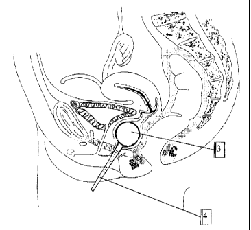
The set also includes an intravaginal measuring subset (FIG.
2) containing measuring balls 3 made preferably of metal or
plastic with graduated diameters corresponding to the
graduated diameters of the balls 1 of the subset of
CA 02270460 2006-04-06
12
corrective intravaginal inserts of FIG. 1. The balls 3
instead of having the loosely hanging string 2, each has a
rigidly mounted, linearly scaled slat 4. The slat 4 is
preferably scaled for measurement of diameter and optimal
location of the corrective insert in the vagina in the case
of urinary stress incontinence.
FIG. 3 shows a simplified sagittal cross-section of the
female pelvis with the cysto-urethral angle enlarged almost
up to 180°, and where the micturition is controlled or
involuntary.
FIG. 5 shows the method of measuring the diameter of the
vagina using the ball 3 from the measuring subset for
determination of depth of location and size of the
corrective insert. The appropriate anatomical size is
determined using the balls 3 from the measuring subset.
Determination is accomplished by checking as to whether an
inserted measuring insert is or is not pushed out by the
descending organs. If it is not pushed out, a greater insert
diameter should be applied. In the case of expulsion, the
insert is too large and a smaller diameter insert should be
used.
The application of the corrective intravaginal insert
involves determination of the optimal diameter of the insert
and intravaginal application of the corrective intravaginal
insert for an indefinite period of time.
CA 02270460 2006-04-06
I3
FIG. 4 shows a sagittal cross-section of the female pelvis
with the corrective insert of properly selected diameter of
the ball 1 by means of the measuring subset. The string 2 is
used for removal of the insert.
The mode of action of the insert is as follows. The insert
upon being placed in the vagina is supported on the levator
ani muscle. It results in forward and upward shift of the
lowered anterior wall of the vagina and elevation of the
urethra and urinary bladder. An elevation of the urethra
restores its normal position in relation to the posterior
wall of the urinary bladder (reduced cysto-urethral angle
shown on FIG. 4). In the case of concomitant uterine
prolapse, the insert causes elevation of the uterus.
Use of the corrective insert of the set of the invention
results in fast pain relief. The device also enables a
controlled micturition due to correction of the cysto
urethral angle "a" without the necessity to remove the insert
for micturition.
Displacement of the uterus to the "pure zone" enables
complete removal of inflammation. Relaxation of muscles and
fasciae restores normal blood supply of the organs of the
pelvis minor. It is most probable that the corrective insert
improves results of the treatment with the therapeutic
insert. Alternating application of the therapeutic insert
and the corrective insert enables the treatment of the more
advanced cases of urogenital descent and improves results of
the therapy.
CA 02270460 2006-04-06
14
The insert can be easily applied and removed. It is supported
on the posterior wall of the vagina at the level of the
levator ani muscle of anus and thus it prevents excessive
extension of the vaginal walls.
The corrective insert from the set being the subject of the
invention supports the prolapsed uterus and blocks the
urethra in the case of urinary stress incontinence, i.e.,
inadequate urethral occlusion at increased intra-abdominal
pressure. An optimal adjustment of insert diameter to the
vagina allows the muscles to regenerate by reduction in
uterine and cystic pressure. The corrective insert supports
these organs, and after regeneration, the muscles will
support the uterus and urinary bladder again, but in a
corrected position.
The inventive method of the treatment of prolapse of the
urogenital organs and urinary stress incontinence in women
with usage of the intravaginal set lies in that the
appropriate size corrective insert is selected from the
subset of corrective intravaginal inserts, consisting of at
least two balls of different diameter, ranging between the
minimal and maximal vaginal diameter, each ball being
preferably hollow. Each ball has a loosely hanging string and
is made of medical material, such as polycarbonate or methyl
methacrylate. There is an intravaginal measuring subset that
contains at least two metal or plastic balls with diameters
corresponding to the diameters of the balls of the corrective
intravaginal subset which, instead of the loosely hanging
string, each ball has a rigidly mounted, scaled slat for
measurement of optimal diameter and localization of the
insert in the vagina, depending on individual anatomical
CA 02270460 2006-04-06
conditions of urogenital organ in the woman being treated by
selection of the appropriate diameter and depth of vaginal
location by means of approximations using the meaning balls
from the measuring subset, so that contraction of the
5 levator ani muscle causes elevation of the insert and
elevation of the insert causes elevation of the uterus,
and/or correction of the cysto-urethral angle. During the
progress of treatment the sizes of successive applied inserts
are adjusted by selection of appropriate diameter and depth
10 of localization in the vagina and it is beneficial to carry
out exercises of the pelvic floor muscles while laying on
prone or genucubital position between the successive
replacements of the insert.
The size of the applied insert is gradually decreased under
medical supervision using the measuring device and ensures
permanent correction of uterine and bladder placement by
correction of the cysto-urethral angle "a", so that the
muscle fibers diffused in surrounding tissue may restore the
lowered organs to primary placement.
In the case of significant urogenital prolapse, the uterus is
at the same level as the arms of the levator ani muscle,
increasing distance between them.
In this case, the corrective intravaginal insert causes an
elevation of the uterus, while contractions of the levator
ani muscle elevates the insert and, indirectly, the uterus.
The corrective insert may fall out at muscular contraction,
when its diameter is too small. It is beneficial to make the
CA 02270460 2006-04-06
16
test for contraction of the levator ani muscle, following an
adjustment of the corrective insert by the measuring device.
Exercise of the levator ani muscle with the usage of the
corrective insert leads to improved efficiency of the
levator ani muscle, vaginal stenosis and elevation of the
uterus.
It is necessary to check the position of the insert and the
uterus within 2 weeks from the beginning of the recommended
exercise.
In advanced cases of urogenital prolapse in which the uterine
cervix is placed below the arms of the levator ani muscle,
an exercise of the muscle with the usage of the corrective
insert elevates the uterus with a decrease in distance
between both arms; the insert elevates, resulting in uterine
elevation.
Progress in therapy may be monitored using the measuring
device for examination of so-called pelvic floor thickness.
That is, following insertion, the measuring device is
slightly pulled downward to rest it on the arms of the
levator ani muscle and the distance from the pubic symphysis
is read from the scaled slat.
CA 02270460 2006-04-06
17
An assessment of the therapeutic effect includes repeated
measurements of optimal diameter of the corrective insert.