Note: Descriptions are shown in the official language in which they were submitted.
CA 02273616 1999-06-02
METHOD FOR PARALLEL SCREENING OF ALLELIC VARIATION
STATEMENT AS TO FEDERALLY SPONSORED RESEARCH
This invention was made with Government support awarded by the National
Institutes of Health, grants HG01633-01 and HG00185-01. The Government may
have
certain rights in this invention.
INTRODUCTION
Background
1 o Genetic linkage maps show the relative locations of specific DNA markers
along
a chromosome. Any inherited physical or molecular characteristic that differs
among
individuals and is easily detectable in the laboratory is a potential genetic
marker. DNA
sequence polymorphisms are useful markers because they are plentiful and easy
to
characterize precisely. Many such polymorphisms are located in non-coding
regions
and do not affect the phenotype of the organism, yet they are detectable at
the DNA
level and can be used as markers. Examples include restriction fragment length
polymorphisms (RFLPs), which reflect sequence variations in DNA sites or
differences
in the length of the product, which can be cleaved by DNA restriction enzymes,
variable
number of tandem repeat (VNTR) sequences, which are short repeated sequences
2o that vary in the number of repeated units, single nucleotide polymorphisms
(SNPs),
and the like.
The "linkage" aspect of the map is a measure of how frequently two markers are
inherited together. The closer the markers are to each other physically, the
less likely
a recombination event will fall between and separate them. Recombination
frequency
thus provides an estimate of the distance between two markers. The value of
the
genetic map is that an inherited trait can be located on the map by following
the
inheritance of a DNA marker present in affected individuals, but absent in
unaffected
individuals, even though the molecular basis for the trait may not yet be
understood.
Genetic maps have been used to find the exact chromosomal location of several
3o important disease genes, including cystic fibrosis, muscular dystrophy,
sickle cell
disease, Tay- Sachs disease, fragile X syndrome and many others.
There is currently a substantial effort being put into sequencing the genome
of
a variety of organisms, including many viruses, bacteria, and eukaryotic
organisms.
-1-
CA 02273616 1999-06-02
Recent work has generated genetic maps of every human chromosome, and more
refined maps are continuously being developed. This information makes it
possible to
perform whole genome screening for genetic mapping in a number of different
species.
When combined with statistical methods such as sib pair analysis,
s affected-pedigree-member analysis, or efficient Lod score analysis, whole
genome
screening is a powerful tool with which to identify genes.
One tool showing considerable promise for genome-wide analysis is the nucleic
acid array, reviewed by Ramsay (1998) Nat. Biotech. 16:40-44. These arrays
contain
dense collections of nucleic acids, either PCR products or oligonucleotides,
usually of
known sequence, that have been either synthesized or printed at fixed spatial
locations
on suitable substrates, such as nylon filters or glass slides. When labeled
DNA or RNA
samples are hybridized to the arrays, the abundance of specific sequences in
solution
can be quantitated based on the fluorescent or radioactive signal intensity at
the
position of the complementary probe. While recent interest has been directed
toward
~5 the use of arrays for monitoring global gene expression, arrays can also be
used for
rapid detection of sequence variation.
An emerging class of marker for genetic analysis of the single nucleotide
polymorphism, and other simple polymorphisms, e.g. deletions, double
nucleotide
polymorphisms, etc. SNPs are generally biallelic systems, that is, there are
two alleles
2o that a population may have for any particular marker. This means that the
information
content per SNP marker is relatively low when compared to microsatellite
markers,
which may have upwards of 10 alleles. SNPs also tend to be very population-
specific;
a marker that is polymorphic in one population may not be very polymorphic in
another.
SNP markers offer a number of benefits that will make them an increasingly
2s valuable tool. SNPs, found approximately every kilobase (see Wang et al.
(1998)
S ,~r'ence 280:1077-1082), offer the potential for generating very high
density genetic
maps, which will be extremely useful for developing haplotyping systems for
genes or
regions of interest, and because of the nature of SNPs, they may in fact be
the
polymorphisms associated with the disease phenotypes under study. The low
mutation
3o rate of SNPs also makes them excellent markers for studying complex genetic
traits.
In principle, any base that differs among allelic sequences could serve as a
marker for linkage analysis. Single-base differences between allelic single
copy
sequences from two different haploid genomes have been estimated to occur
about
_2_
'i CA 02273616 1999-06-02
once per 300 by in an outbred Western European population. This calculates to
a total
of about 10' potential markers for linkage analysis per haploid genome. Only a
tiny
fraction of these nucleotide differences contribute to mapping using current
methods.
There is, therefore, substantial interest in developing new methods that
utilize the
available genomic information more efficiently and can provide information
concerning
multi-gene traits. Such methods could be valuable, not only for gene mapping,
but also
for genetic diagnosis and risk assessment. Allelic variation can be used for
strain
identification, in population genetics, linkage analysis and recombination
studies.
Relevant literature
The complete genome sequence of a number of organisms may be found at the
National Center for Biotechnology Information,
http:llwww.ncbi.nlm.nih.govlEntrezl
Genome%rg.html. The availability of sequences of genes of the human genome is
discussed in Schuler (1996) Science 274:540. The complete sequence of the
genome
~5 of S. cer~evisiae is available at several Internet web sites, and is
discussed in Goffeau
et al. (1996) Science 274:546.
A number of methods are available for creating microarrays of biological
samples, such as arrays of DNA samples to be used in DNA hybridization assays.
Exemplary are PCT Application Serial No. W095/35505, published December 28,
20 1995; U.S. patent no. 5,445,934, issued August 29, 1995; and Drmanac et
al., Science
260:1649-1652. Yershov et al. (1996) Genetics 93:4913-4918 describe an
alternative
construction of an oligonucleotide array. The construction and use of
oligonucleotide
arrays is reviewed by Ramsay (1998) supra.
Methods of using high density oligonucleotide arrays are known in the art. For
2s example, Milosavljevic et al. (1996) Genomics 37:77-86 describe DNA
sequence
recognition by hybridization to short oligomers. The use of arrays for
identification of
unknown mutations is proposed by Ginot (1997) Human Mutation 10:1-10.
Detection of known mutations is described in Hacia et al. (1996) Nat. Genet.
14:441-447; Cronin et al. (1996) Human Mut. 7:244-255; and others. The use of
arrays
3o in genetic mapping is discussed in Chee et al. (1996) Science 274:610-613;
Sapolsky
and Lishutz (1996) Genomics 33:445-456; etc. Shoemaker et al. (1996) Nat.
Genet.
-3-
CA 02273616 1999-06-02
14:450-456 perform quantitative phenotypic analysis of yeast deletion mutants
using
a parallel bar-coding strategy.
Quantitative monitoring of gene expression patterns with a complementary DNA
microarray is described in Schena et al. (1995) Science 270:467. DeRisi et al.
(1997)
Science 270:680-686 explore gene expression on a genomic scale. Wodicka et al.
(1997) Nat. Biotech. 15:1-15 perform genome wide expression monitoring in
S. cerevisiae.
SUMMARY OF THE INVENTION
Methods are provided for detection and analysis of allelic variation between
two
closely related genomes, through parallel hybridization analysis. Detectable
allelic
variations may be substitutions, insertions or deletions of one or more
nucleotides in
length. A map of allelic variation can be generated with the subject methods,
and used
for genetic linkage analysis, determination of chromosomal regions having low
diversity
~5 or high diversity, forensic studies, etc. By identifying the parental
origin of DNA
sequences in offspring, the locations of segregating loci can be determined in
parallel.
The subject methods have broad applicability to the analysis of variation and
of the
inheritance of multigenic or quantitative trait loci.
The provided methods utilize genomic DNA from two closely related sources.
2o DNA samples from both sources are cleaved to generate short fragments. The
fragments are end-labeled, and then hybridized to a high density
oligonucleotide array.
Hybridization patterns for the two samples are detected, normalized and
compared.
Those positions on the array that correspond to sequences with allelic
variation
between the two samples will show decreased hybridization efficiency for one
of the
25 samples relative to the other.
' CA 02273616 1999-06-02
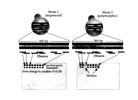
BRIEF DESCRIPTION OF THE DRAWINGS
Figure 1 is a schematic illustrating the detection of allelic variation using
high-
density arrays.
Figure 2 is a comparison of hybridization patterns for two strains of
S. cerevisiae.
Figure 3 is a schematic showing the inheritance of markers (3 chromosomes)
in one tetrad from a cross between YJM789 and S96. The genotypes of the
segregants are given in Table I.
Figure 4 is a schematic showing the inheritance of DNA in 10 segregants.
Figure 5 is a graph showing the probability of random segregation for the
entire
yeast genome.
Figure 6A, 6B and 6C are flow charts illustrating an exemplary data analysis
for
use with the subject methods.
DESCRIPTION OF THE SPECIFIC EMBODIMENTS
Methods are provided for the rapid detection of allelic variation. Genomic DNA
from two related samples are compared by hybridization to a high density DNA
array.
DNA samples from both sources are cleaved chemically or enzymatically to
generate
short fragments. The fragments are end-labeled, and then hybridized to a high
density
oligonucleotide array. Hybridization patterns for the two samples are
visualized,
normalized and compared. Probes that correspond to sequences with allelic
variation
between the two samples will show decreased hybridization efficiency for one
of the
samples relative to the other. A map of allelic variation can be generated
with the
subject methods, and used for genetic linkage analysis, determination of
chromosomal
regions having low diversity or high diversity, forensic studies, etc.
Knowledge of genetic variation is important for understanding why some people
are more susceptible to disease or respond differently to treatments.
Variation can
also be used to determine which genes contribute to multigenic or quantitative
traits
such as increased yield or pest resistance in plants or for understanding why
some
3o strains of a microbe are exceptionally virulent. Genetic variation can also
be employed
for identification purposes, both in microbiology and in forensics, for
studies of
recombination, and in population genetics. Rapid and cost effective ways to
analyze
variation are clearly needed. The methods of the present invention allow
genetic
-5-
CA 02273616 1999-06-02
variation in any two isolates of a species to be scanned, mapped and scored
directly
and efficiently without allele-specific PCR, without creating new strains or
constructs,
and without knowing the specific nature of the variation.
One of the most important uses for variation is to map genetic differences
within
a species. The chromosomal location of such variation provides a means of
identifying
individuals, and of tracing inheritance for genetic mapping. The information
derived
from genetic mapping studies has a wide range of uses. For example, mapping is
useful in agricultural species for tracing the genes associated with a
particular
phenotype. In human studies it is used for determining loci associated with
traits such
~o as disease predisposition.
Within a species, there are genetic sites that are polymorphic, i.e. within a
population, more than one nucleotide (G, A, T, C) is found at a specific
position. Allelic
variation, as used herein, refers to polymorphisms in genomic DNA sequence
between
two individuals. Allelic variation may be substitution, addition or deletion
of one or
~5 more nucleotides at a particular site. Frequently the detected variation
will be a point
mutation, or single nucleotide polymorphism. However, small deletions,
additions, and
multiple nucleotide variations are also detected.
The subject methods are also used to determine which genes or regions of
genes are conserved, and which contain variable regions. Such information is
useful,
2o for example, in the design of vaccines where it is desirable to use
epitopically
conserved antigens; or in the choice of targets for drug screening.
Alternatively,
information about variable regions of the genome may indicate those loci that
differ
between pathogenic and non-pathogenic strains.
The source of genomic DNA is two strains or individuals from one species or
two
25 closely related species, where partial sequence information is available
for one of the
genomes. There should be a high degree of sequence identity between the two
DNA
samples, such as one would expect to find between individuals in a species.
The
percent of sequence identity will usually be at least about 99%, more usually
at least
about 99.5%, and may be at least about 99.9%, or higher.
3o The complete genome is used, or predetermined portions thereof, e.g.
isolated
chromosomes, messenger RNA fractions, BACs, YACs, cosmids, EST libraries, etc.
One of the samples may, but does not necessarily, comprise a complete genome
sequence, while the other sample comprises a pre-determined subpopulation of
the
CA 02273616 1999-06-02
genome. Where the complete genome is used in screening, it will preferably be
obtained from a prokaryote, virus, or lower eukaryotes, e.g. fungi,
protozoans, plants
having a small genome, etc.
The sample complexity, i.e. the length of sequence that will be analyzed, will
usually be less than about 1 O9 bp, preferably less than about 108 bp, more
preferably
less than about 5 x 10' by in size, and may be less than about 1.5 x 10' bp. A
viral
genome will usually be greater than 103 nucleotides in length, while a
bacterial genome
will usually be greater than 1 OS by in length. Larger genomes, e.g, having a
complexity
of greater than about 10' bp, or greater than about 108 bp, may be separated
into
samples of lower complexity for analysis.
Partial sequence characterization of target regions in one of the samples is
required. Dispersed nucleotide sequences of at least about 16 nucleotides,
usually at
least about 20 nucleotides and preferably at least about 25 nucleotides
throughout the
region to be analyzed are desirable. Known sequences may be dispersed
throughout
the genome, chromosome or locus of interest, usually spaced not more than
about
10,000 nucleotides apart, more usually not more than about 1000 nucleotides
apart,
and preferably not more than 500 nucleotides apart.
A number of organisms have sufficient sequence information to meet these
requirements, including organisms with complete known genome sequences, e.g.
2o Aquifex aeolicus; Archaeoglobus fulgidus; Bacillus subtilis; Bornelia
burgdorferi;
Escherichia coli; Haemophilus influenzae; Helicobacter pylori ;
Methanobacterium
thennoautorrophicum; Methanococcus jannaschii; Mycoplasma genitalium;
Mycoplasma pneumoniae; Saccharomyces cerevisiae; Synechocystis PCC6803; and
organisms with substantial sequence and mapping information known, e.g.
Arabidopsis
thaliana; Caenorhabditis elegans; Drosophila melanogaster; Homo sapiens;
Leishmania major, Mus musculus; Oryza sativa; Saccharomyces cerevisiae; Zea
mays,
etc.
The two DNA samples are prepared initially in accordance with conventional
methods, e.g. lysing cells, removing cellular debris, separating the DNA from
proteins,
lipids or other components present in the mixture and then using the isolated
DNA for
cleavage. See Molecular Cloning, A Laboratory Manual, 2nd ed. (eds. Sambrook
et al.) CSH Laboratory Press, Cold Spring Harbor, NY 1989. Usually, at least
about
CA 02273616 1999-06-02
0.5 Ng of DNA will be employed, more usually at least about 5 Ng of DNA, while
less
than 50 Ng of DNA will usually be sufficient.
The nucleic acid samples are cleaved to generate probes. It will be understood
by one of skill in the art that any method of random cleavage will generate a
distribution
of fragments, varying in the average size and standard deviation. Usually the
average
size will be at least about 12 nucleotides in length, more usually at least
about 20
nucleotides in length, and preferably at least about 35 nucleotides in length.
Where
the variation in size is great, conventional methods may be used to remove the
large
and/or small regions of the fragment population.
It is desirable, but not essential to introduce breaks randomly, with a method
which does not act preferentially on specific sequences. Preferred methods
produce
a reproducible pattern of breaks. Methods for introducing random breaks or
nicks in
nucleic acids include reaction with Fenton reagent to produce hydroxyl
radicals and
other chemical cleavage systems, integration mediated by retroviral integrase,
partial
digestion with an ultra-frequent cutting restriction enzymes, partial
digestion of single
stranded with S1 nuclease, partial digestion with DNAse I in the absence or
presence
of Mn++, etc.
The fragmented nucleic acid samples are denatured and labeled. Labeling can
be performed according to methods well known in the art, using any method that
2o provides for a detectable signal either directly or indirectly from the
nucleic acid
fragment. In a preferred embodiment, the fragments are end-labeled, in order
to
minimize the steric effects of the label. For example, terminal transferase
may be used
to conjugate a labeled nucleotide to the nucleic acid fragments. Suitable
labels
include biotin and other binding moieties; fluorochromes, e.g. fluorescein
2s isothiocyanate (FITC), rhodamine, Texas Red, phycoerythrin,
allophycocyanin, 6-
carboxyfluorescein (6-FAM), 2',7'-dimethoxy-4',5'-dichloro-6-
carboxyfluorescein (JOE),
6-carboxy-X-rhodamine (ROX), 6-carboxy-2',4',7',4,7-hexachlorofluorescein
(HEX),
5-carboxyfluorescein (5-FAM) or N,N,N',N'-tetramethyl-6-carboxyrhodamine
(TAMRA),
and the like. Where the label is a binding moiety, the detectable label is
conjugated
3o to a second stage reagent, e.g. avidin, streptavidin, etc. that
specifically binds to the
binding moiety, for example a fluorescent probe attached to streptavidin.
Incorporation
of a fluorescent label using enzymes such as reverse transcriptase or DNA
polymerase, prior to fragmentation of the sample, is also possible.
_g_
CA 02273616 1999-06-02
Each of the labeled genome samples is separately hybridized to an array of
oligonucleotide probes. Hybridization of the labeled sequences is accomplished
according to methods well known in the art. Hybridization can be carried out
under
conditions varying in stringency, preferably under conditions of high
stringency, e.g.
s 6X SSPE, 65°C, to allow for hybridization of complementary sequences
having
extensive homology, usually having no more than one or two mismatches in a
probe
of 25 nucleotides in length, i.e. at least 95% to 100% sequence identity.
High density microarrays of oligonucleotides are known in the art and are
commercially available. The sequence of oligonucleotides on the array will
correspond
to the known target sequences of one of the genomes, as previously described.
Arrays
of interest for the subject methods will generally comprise at least about 103
different
sequences, usually at least about 104 different sequences, and may comprise
105 or
more different sequences. The length of oligonucleotide present on the array
is an
important factor in how sensitive hybridization will be to the presence of a
mismatch.
15 Usually oligonucleotides will be at least about 12 nt in length, more
usually at least
about 15 nt in length, preferably at least about 20 nt in length and more
preferably at
least about 25 nt in length, and will be not longer than about 35 nt in
length, usually not
more than about 30 nt in length.
Methods of producing large arrays of oligonucleotides are described in U.S.
2o Patent no. 5,134,854 (Pirrung et al.), and U.S. Patent no. 5,445,934 (Fodor
et al.) using
light-directed synthesis techniques. Using a computer controlled system, a
heterogeneous array of monomers is converted, through simultaneous coupling at
a
number of reaction sites, into a heterogeneous array of polymers.
Alternatively,
microarrays are generated by deposition of pre-synthesized oligonucleotides
onto a
25 solid substrate, for example as described in International Patent
application
WO 95/35505.
Microarrays can be scanned to detect hybridization of the labeled genome
samples. Methods and devices for detecting fluorescently marked targets on
devices
are known in the art. Generally such detection devices include a microscope
and light
so source for directing light at a substrate. A photon counter detects
fluorescence from
the substrate, while an x-y translation stage varies the location of the
substrate. A
confocal detection device that may be used in the subject methods is described
in U.S.
Patent no. 5,631,734. A scanning laser microscope is described in Shalon et
al. (1996)
_g_
CA 02273616 1999-06-02
Genome Res. 6:639. A scan, using the appropriate excitation line, is performed
for
each fluorophore used. The digital images generated from the scan are then
combined
for subsequent analysis. For any particular array element, the ratio of the
fluorescent
signal from one Nucleic acid sample is compared to the fluorescent signal from
the
s other Nucleic acid sample, and the relative signal intensity determined.
Methods for analyzing the data collected by fluorescence detection are known
in the art. Data analysis includes the steps of determining fluorescent
intensity as a
function of substrate position from the data collected, removing outliers,
i.e. data
deviating from a predetermined statistical distribution, and calculating the
relative
binding affinity of the targets from the remaining data. The resulting data
may be
displayed as an image with the intensity in each region varying according to
the binding
affinity between targets and probes.
The images from the two or more genome samples from the two strains, or
progeny from crosses of the two strains are compared to determine feature
signals
~5 showing a bimodal distribution pattern, i.e. that detect allelic variation.
A flow chart of
the data analysis process is provided in Figure 6A, 6B and 6C. Referring to
FIG. 6A
(steps 1 and 2), the system is initialized by requesting the user to enter the
names of
the sample CEL files and their genotypes (if known). The CEL files contain the
quantitated feature intensities from the scanned images. The feature
intensities from
2o the CEL files are adjusted with a monotonic, variance-stabilizing
transformation. At
step 3, the overall signal strength of each image are estimated as the mean of
a subset
(initially all) of the features.
Next, the expected signal response for each feature is determined using the
data from the CEL files. First (step 6), for each feature, a single regression
line is fit
2s to the overall signal strengths of the images (x axis) and their
corresponding adjusted
feature intensities (y axis). This determines the expected signal response and
variance
for the feature given the signal strength of the image. However, this assumes
that the
signal response is the same for all samples (i.e. there is only one genotype).
Next,
separate lines are fit for each genotype in parallel (step 7) to model the
expected signal
3o responses if there are actually multiple genotypes. Samples whose genotypes
are
unknown are assigned to the genotype that minimizes the variance of the
resulting fits.
An F-test is used to distinguish between these models. If the same signal
model is
rejected, genotypes are assigned to each sample whose genotype is unknown
along
-10-
CA 02273616 1999-06-02
with the probability that the genotype is correct. This probability is
computed using the
expected signals and variances from the regression fits at the sample's
overall signal
strength based on a t-distribution. For example, Pr(G1)=P(G1)/(P(G1)+P(G2))
where
G1 is the assigned genotype, G2 is the other genotype, and P(X) is the
probability of
observing the signal given the expected signal and variance for genotype X.
The
overall signal strengths are re-estimated using only the features that have
the same
signal response, regardless of genotype, and this process is repeated until
this subset
of features does not change significantly.
Then the chromosome location of every feature corresponding to a marker is
1o determined (step 13). Any features that appear more than once in the genome
are
excluded from the analysis. Next, the meiotic breakpoints are determined for
each
sample. The marker genotype probabilities along each chromosome are used to
determine these sites by maximum likelihood (step 14). Additional breakpoints
are
added only if they substantially increase the log likelihood ratio, which
tests the model
containing an additional breakpoint against the current one.
If the data are to be used for mapping purposes, the breakpoints are used to
reassign the inherited genotypes, eliminating noise at step 15. Then, the
genotypes
for each marker from all of the samples are compared. The probability of
observing
the genotypes by chance is computed from the given genotypes. This information
is
2o then displayed by chromosome to indicate which regions of the genome were
inherited
non-randomly.
Genetic linkage markers are polymorphic sequences distributed throughout a
genome. Using the subject methods, polymorphisms are detected as a sequence
difference between the compared genomes. A wide variety of polymorphic markers
may be identified for any given genome. The subject methods may be used in
mapping genes by use of family studies, segregating tetrads, pairs of
relatives that
have a genetically influenced trait of interest, etc. "Affected relative pair"
methods are
useful when the penetrance of the allele that confers the trait is low or age-
dependent,
or when the trait is multigenic or quantitative, e.g. height and build.
Disease-
3o susceptibility genes are particularly relevant. By determining where on the
genetic map
a small set, including two, of "affected" relatives have inherited identical
sequences
from a common source, and disregarding other family members, a highly
efficient
strategy for extracting linkage information from a pedigree is provided. The
resulting
-11-
' CA 02273616 1999-06-02
identity-by-descent maps from multiple pairs of similarly-affected relatives
can be
combined and the composite map searched for loci where genotypic concordance
between affected relatives occurs more frequently than would be expected by
chance.
With a sufficiently large number of affected relative pairs, such an analysis
can reveal
the positions of genes that contribute even a slight susceptibility to the
trait. The
procedure may also find wide application in routine screening for shared
genetic risks
in families.
The subject methods find application in following segregation of traits
associated
with breeding of plants and animals, the association of particular regions in
the
1o genomic map with particular traits, especially traits associated with
multiple genes, the
transmission of traits from ancestors or parents to progeny, the interaction
of genes
from different loci as related to a particular trait, and the like. While only
two sources
may be involved in the comparison, a much larger sampling may also be used,
such
as 20 or more sources, where pairwise comparisons are made between the various
sources. Relationships between the various sources may vary widely, e.g.
grandparents and grandchildren; siblings; cousins; and the like.
The subject methods may also be used for the ordered mapping of genomic
libraries. Typically, the term "genomic library" is defined as a set of
sequence
fragments derived from one or more genome molecules. Such molecules may be
2o whole chromosomes, subsets thereof, plasmids, or other similar large
polynucleotides.
Specifically, the methods of the present invention are useful for mapping high
molecular weight polynucleotides including chromosomal fragments, cosmids,
yeast
artificial chromosomes (YACs), etc.
Mapping techniques typically involve the identification of specific genetic
markers on individual nucleic acid fragments from a genomic library.
Comparison of
the presence and relative position of specific markers on fragments generated
by
different cleavage patterns allows for the assembly of a contiguous genomic
map, or
"contig". Methods of genomic mapping are provided, using the allelic variant
detection
methods already described. Polymorphic sites are identified on the individual
3o fragments of a genomic library using the methods described above. Sites
that
demonstrate a bimodal distribution pattern are used as genetic markers, and a
contig
of the particular library is then assembled. The exact sequence of variants
can be
-12-
CA 02273616 1999-06-02
determined by various methods known in the art, e.g. PCR amplification
followed by
sequence determination of the amplification product.
When repeated on separate fragments from the library, each fragment will
generally produce a distinctive hybridization pattern. These hybridization
patterns may
be compared with hybridization patterns from differentially generated
fragments.
Where a specific marker is present in both fragments, it is an indication of
potential
overlap between the fragments. Two fragments that share several of the same
markers, i.e. overlapping fragments, will show similar hybridization patterns
on the
oligonucleotide array. The greater the similarity or correlation between two
fragments,
1o the higher the probability that these fragments share an overlapping
sequence. By
correlating the hybridization pattern of each fragment in the library against
each other
fragment in the library, a single contiguous map of the particular library can
be
constructed.
In practice, each fragment is correlated to each other fragment, and a
correlation score is given based upon the number of probes which cross-
hybridize with
a marker of both the first and second fragment. High scores indicates high
overlap.
For example, the comparison of two identical sequences would produce a
correlation
score of 1. Similarly, sequences sharing no overlapping sequence would ideally
produce a correlation score of 0. In practice, sequences that do not overlap
will
2o generally have correlation scores above zero, due to potential non-specific
hybridizations, e.g. single base mismatches, background hybridization,
duplicated
sequences, which may provide some baseline correlations between otherwise
unrelated fragments. As a result, a cutoff may be established below which
correlation
scores are not used. The precise cutoff may vary depending upon the level of
nonspecific hybridizations for the particular application.
The methods described herein are useful in a variety of applications. For
example, as is described above, these methods can be used to generate ordered
physical maps of genomic libraries, as well as genetic linkage maps which can
be used
in the study of genomes of varying sources. The mapping of these genomes
allows
3o further study and manipulation of the genome in diagnostic and therapeutic
applications, e.g. gene therapy, diagnosis of genetic predispositions for
particular
disorders and the like.
-13-
CA 02273616 1999-06-02
In addition to pure mapping applications, the methods of the present invention
may also be used in other applications. For example, the methods described
herein
are used in the identification of the source of a particular sample. This
application
would include forensic analysis to determine the origin of a particular tissue
sample,
s such as analyzing blood or other evidence in criminal investigations,
paternity
investigations, etc. Additionally, these methods can also be used in other
identification
applications, for example, taxonomic study of plants, animals, bacteria,
fungi, viruses,
etc. This taxonomic study includes determination of the particular identity of
the
species from which a sample is derived, or the interrelatedness of samples
from two
1o separate species. Where a hybridization pattern from both the sample and
the source
are identical or highly similar, it is indicative that the sample was derived
from the
source. Where the sequences captured from the sample and known source share a
large number of identical sequences, it is indicative that the sample is
related to the
known source. However, where the sample and source share few like sequences,
it is
15 indicative of a low probability of interrelation.
Precise levels of interrelation to establish a connection between source and
sample will typically be established based upon the interrelation which is
being proved
or disproved, the identity of the known source, the precise method used, and
the like.
Establishing the level of interrelation is well within the ordinary skill in
the art. For
2o example, in criminal investigations, a higher level of homology between
sample and
known source sequences will likely be required to establish the identity of
the sample
in question. Typically, in the criminal context, interrelation will be shown
where there
is greater than 95% marker identity, preferably greater than 99%, and more
preferably,
greater than 99.9% identity. For other identification applications,
interrelation between
2s sample and known source may be established by a showing of greater than 50%
identity, and typically greater than 75% identity, preferably greater than 90%
identity,
and more preferably greater than 95 to 99% identity.
For convenience, kits may be supplied which provide the necessary reagents
in a convenient form and together. For example kits could be provided that
include
3o chips containing an appropriate microarray for the subject to be analyzed,
terminal
transferase, DNAse I, biotin labeled nucleotides, and/or fluorochrome labeled
avidin.
Other components such as automated systems for determining and interpreting
the
-14-
CA 02273616 1999-06-02
hybridization results, software for analyzing the data, or other aids may also
be
included depending upon the particular protocol which is to be employed.
EXPERIMENTAL
Detection of Allelic Variation in S cerevisiae
Strain Selection. To maximize the amount of allelic variation that could be
detected, two distantly-related S. cer~evisiae strains, S96 (MATa ho lys5 man,
isogenic
with S288c, and YJM789 (MATa ho::hisG lys2 pdr5 MAL) were chosen for this
study.
The S. cerevisiae genome sequence is from strain S288c and 88% of the S288C
1o genome is derived from EM93, which was isolated from a rotting fig near
Merced,
California in 1938. YJM789 is isogenic with YJM145, a segregant of a clinical
isolate
of S. cerevisiae. YJM145 has been characterized genetically, and the ultimate
source
of its parent (human lung) differs significantly from that of S288c in that
the strains
were isolated from different environments, at different times and in different
geographic
locations. S288c and YJM789 were considered to be unrelated, and therefore
likely
to exhibit considerable allelic variation.
To determine the frequency of allelic variation in YJM789, a library of YJM789
genomic DNA was constructed and partially sequenced. Genomic DNA was isolated
from strain YJM789 and sheared to 1000-basepair insert sizes using a re-
circulating
2o point-sink flow shearing device (Oefner et al. (1996) Nucleic Acids Res 24,
3879-86).
Fragments were cloned into an M13 sequencing vector and the sequence was
determined for 696 clones using dye-primer chemistry in cycle-sequencing
reactions
on ABI 377 sequencing machines (Dietrich et al. (1997) Nature 387:78-81). The
sequences were called using phred basecaller software (see
http:llchimera.biotech.washington.edulUWGCl~oolslphred.htm), which produces a
quality measurement for each base (-10 x Iog10 (probability of an error)).
Using this
quality measurement, 122258 bases were sequenced with > 99% confidence. The
YJM789 sequences were compared to the fully sequenced strain of S. cenevisiae
using
the cross match program (see
http:llchimera.biotech.Hrashington.edulUWGCltoolsl
3o phrap.htm). Discrepancies between the YJM789 and S288c sequences were then
classified by quality and assigned into coding and non-coding regions using
the phred
basecaller. In most cases, since only a single trace was available and no
alignments
-15-
CA 02273616 1999-06-02
were performed, regions of the traces that did not show high quality were
excluded
from the analysis.
When high quality sequence (>99.7% accurate) from YJM789 was aligned with
that of S288c, 466 cases of allelic variation were observed with a frequency
of one
every 160 bases. Most were single-base pair polymorphisms, but small
insertions and
deletions were also observed. Large deletions were not readily identified by
this
shotgun sequencing approach because of the difficulty associated with aligning
the
sequence fragments using automated methods. A small bias (10%) toward non-
coding
regions was observed. 288 of the 466 cases of allelic variation in sequences
with
>99.97 accuracy were from coding regions (61 %). 8.637 Mb of the estimated
13.2 Mb
yeast genome is annotated as coding sequence by SGD (65%). These data
suggested
that if some fraction of the existing allelic variation could be rapidly and
reproducibly
detected, a dense genome-wide genetic map could be constructed.
High-density oligonucleotide arrays. Commercially available high-density
arrays
containing a large number of oligonucleotide probes from genomic DNA sequence
have been designed and used to monitor genome-wide gene expression in yeast.
For
oligonucleotide probes, hybridization is dependent on the absence of
mismatches in
the corresponding target sequence (Conner et al. (1983) Proc Natl Acad Sci U S
A
80:278-82), and thus it was hypothesized that these arrays could serve in the
rapid
detection of allelic variation in yeast (Figure 1 ). These arrays contain 20
or more
25mer oligonucleotide probes derived from the sequence of each annotated open
reading frame in the yeast genome.
Figure 1 shows a schematic for detection of allelic variation using high-
density
arrays. A minimum of 20 25-base oligonucleotide probes was chosen from yeast
genomic sequence for every annotated open reading frame in the yeast genome.
Probes were arranged on the array in a way that generally reflected their
position in the
genome. All probes were from predicted coding regions with a bias toward the
1000
bases at the 3' ends of genes. When YJM789 DNA fragments containing
polymorphic
3o regions (*) are hybridized to the array localized decreases in signal
intensity are
observed if a probe complementary to this region is found of the array.
In addition to probes designed to be perfectly complementary to regions of
yeast
coding sequence (designated perfect match or PM probes), probes containing a
single
-16-
CA 02273616 1999-06-02
base mismatch (MM) in the central position of the oligonucleotide were also
synthesized in a physically adjacent position. The mismatch probes serve as
local
background and non-specific hybridization controls (Wodicka et al. (1997)
Nature
Biotechnology 15:1359-1367).
The probes were synthesized in a spatially-addressed fashion using a
combination of photolithography and solid-phase chemistry (Fodor et al. (1991)
Science 251:767-73), on a series of five 1.64 cmz arrays. Each array contains
more
than 65,000 synthesis features, with each feature consisting of more than 10'
copies
of the specific oligonucleotide probe. The collection of five arrays contains
a total of
157,112 different 25mer probe pairs.
Excluding the rDNA and CUP1 repeats, the largest gap is 41,325 bases wide
at position 510,000 on Chromosome XII. This region contains three tandem
repeats
of the ASP3 gene and an adjacent gene of unknown function, a region of
ribosomal
DNA and a Ty-1 element. Probes complementary to this region are present on the
array but were ignored in the analysis, as were all non-unique probes. Though
some
probes spatially overlap one another, the collection of five arrays covers
21.8% of the
non-repetitive regions of the yeast genome.
Detecting allelic variation using high density oligonucleotide arrays. Due to
the
2o high-degree of genomic coverage (22%), it was expected that a significant
fraction of
the allelic variation in YJM789 could be detected using the arrays. To test
this,
genomic DNA from S96 and YJM789 was isolated, fragmented and biotin-labeled.
Yeast cells were grown in YEPD to late log phase at 30°C. Genomic
DNA was
purified using Qiagen genomic DNA 100 Ng columns according to the
manufacturer's
protocol. Zymolyase and protease digestion times were extended from 30 to 45
minutes. DNA was re-suspended in 400 NI TE, reprecipitated, and re-suspended
in
30NI deionized H20. Yeast genomic DNA (10 Ng ) was digested in 0.15 Units
DNAse
I (Gibco BRL PCR grade) in 1 X One-Phor-All buffer (Pharmacia) containing 1.5
mM
CoCl2 for 5 minutes at 37°C. The reaction was stopped by heating the
samples to
100°C for 15 minutes. Digestion was checked by examining 1 NI of the
reaction
product on a 2% agarose gel containing 1:10000 SYBR-II green (Molecular
Probes,
Eugene, OR). The procedure was repeated if the majority of the product was not
digested to a size of less than 100 bases (it was observed that the
reproducibility of the
-17-
' CA 02273616 1999-06-02
reaction was highly sensitive to contaminants in the DNA preparation, such as
EDTA).
The DNA fragments were labeled by incubating the samples with 25 U terminal
transferase (Boehringer Mannheim) and 1 nmole Biotin-N6-ddATP (NEN) for one
hour
at 37°C. The entire sample was hybridized to the array in a 200 ul
volume containing
6X SSPE (Accugene), 0.005 % Triton-X 100 detergent, 20 Ng fragmented denatured
Salmon Sperm DNA (Gibco-BRL) and 1 nmole of a 3'-biotin control
oligonucleotide that
hybridizes to the border features on the array.
Samples were heated to 100°C for 10 minutes, and then cooled on ice
before
being applied to the array. Samples were hybridized for 2 hours at
42°C. The arrays
1o were washed, stained with phycoerythrin-streptavidin (Molecular Probes) and
scanned
at an emission wavelength of 560 nm at 7.5 NM resolution using an Affymetrix
GeneChip Scanner as previously described (Wodicka et al., supra.)
After hybridization, the arrays were washed, stained with a phycoerythrin-
streptavidin conjugate and scanned with a laser confocal scanning device that
detects
and records the amount of fluorescence at approximately three million physical
locations. Scanned images of arrays hybridized with S96 and YJM789 DNA were
collected. For illustration, the images from the arrays hybridized with S96
and YJM789
DNA were colored red and green, respectively. The two images were
electronically
superimposed on one another and a portion of the array is shown in Figure 2.
Regions
2o in yellow indicate probes that hybridized roughly equally to genomic DNA
from the two
parental strains, while regions in red are locations of allelic variation
where S96 DNA
hybridized to a greater extent than DNA from YJM789. Isolated red spots
covering one
to five probe features are caused by short polymorphic stretches in the YJM789
sequence at these elements on the array. A few large deletions were also
evident.
Some green spots, usually in the mismatch (MM) row, may be due to YJM789 DNA
hybridizing more strongly with the S96 mismatch sequence. An example of this
sort
is shown in Figure 2. The fact that the two scanned images can be superimposed
demonstrates the reproducibility of the experiment, a feature critical to the
analysis of
a large number of scanned images obtained with different DNA samples and
generated
3o at different times using different arrays.
Figure 2 is a comparison of hybridization patterns for two strains of S.
cer~evisiae. DNA samples from YJM789 and S96 were labeled and hybridized to
two
separate sets of arrays. The array hybridized with DNA from S96 was colored
red
-18-
CA 02273616 1999-06-02
digitally; the image from the array hybridized with YJM789 DNA was colored
green and
the two scanned images were merged. Only a fraction of the array is shown.
Probes
which hybridized S96 DNA more efficiently than YJM789 DNA are red while probes
that
hybridize to both DNA types with equal intensity are yellow. Some yellow
features are
brighter than others, because some oligonucleotides hybridize more
efficiently. These
differences in hybridization signal intensity are reproducible and do not
adversely affect
the analysis. The figure close-up shows a region in which one of the mismatch
features is bright green. Shotgun sequencing of YJM789 demonstrated that the
actual
sequence of YJM789 was complementary to the sequence of the oligonucleotide in
the
mismatch row and not to that in the perfect match row.
To collect a robust set of markers, two additional hybridizations of each
parental
strain DNA sample were performed and the hybridization intensity for each
probe in the
scanned image was quantitated. Grids were aligned to the scanned images using
the
known feature dimensions of the array. The hybridization intensities for each
of the
~5 elements in the grid were determined using the 75th percentile method in
the
Affymetrix GeneChipa software package.
Markers were selected recursively by analyzing the scanned images of 20 array
sets hybridized with different DNA samples (3 samples from each parental
strain, and
14 samples from haploid progeny derived from sporulation of a YJM789/S96
diploid,
2o described below and in Table 1 using software written for this purpose.
The overall array hybridization intensity (n for each hybridization (20
altogether)
was determined by calculating the mean PM signal intensity using all features
that
showed little normalized variation across all hybridizations (non-markers),
determined
recursively as described below. Then for each feature on the array, a
regression line
25 of PM on I for each hybridization was determined by the least squares
method first
under the null hypothesis that the S96 and YJM789 samples had the same
response,
and then under the alternative hypothesis that the S96 samples had a higher
signal
than the YJM789 sample (i.e. a marker). The models were compared with the F-
test
and the identical signal model was rejected in favor of a marker with
alpha=0.01.
30 3808 of the probes on the array were estimated to have a 99% or higher
probability of being a marker based on their exhibiting a consistent bimodal
distribution
for all hybridizations. These markers were expected to be from probes whose
complementary sequence is completely absent in YJM789 or whose complementary
-19-
CA 02273616 1999-06-02
sequence contained a base change in the central region of the oligonucleotide
probe.
25% of the polymorphisms detected by sequencing and having a corresponding
probe
on the array were found in the set of 3808 markers. In these cases, the base
change
was almost invariably in the central 10 bases of the complementary 25mer
probe.
Excluding the rDNA repeat on Chromosome XII, the average marker spacing for
this set of 3808 markers was 3510 bp. 14 gaps were observed with the largest
gaps
(59 kb) centered near position 150400 on Chromosome III. Gaps were often found
near regions with low probe density, for example, near repeated elements in
the
genome but in some cases, probe density was adequate, suggesting that the gap
might be due to a high level of conservation or to a recent common origin of
the region
between the two strains.
Meiotic recombination breakpoints and segregation of markers. To determine
whether the set of chosen probes constituted a robust set of markers usable
for linkage
~5 analysis, meiotic inheritance was examined. An S96/YJM789 diploid was
sporulated
and DNA from four segregants of one tetrad was isolated and hybridized to the
arrays.
The data was analyzed and a score (S96 or YJM789) and a confidence value, p,
was
assigned to each of the 3808 markers for each hybridization.
It was expected that half the markers would be scored as having an S96 origin
2o and half would be scored as YJM789; that in most cases each marker would
segregate
with a ratio of 2:2 in the four segregants; and that crossovers would be
observed about
once per every 290 kb (1 cM = 2.9 kb for chromosomes XIII, XIV and XV). The
locations of the markers, and each marker's score (S96 or YJM789) are shown
for
three chromosomes (Figure 3).
25 Figure 3 shows the inheritance of markers (3 chromosomes) in one tetrad
from
a cross between YJM789 and S96. The location of the marker on the chromosome
is
indicated below. Markers that exhibit the YJM789 hybridization pattern are
colored red
while markers that exhibit the S96 hybridization pattern are colored green.
The
probable locations of cross-over events are shown for each segregant. The
genotypes
30 of the segregants are given in Table I.
For the three chromosomes (about 2.8 million bases), 21 cross-overs were
observed at an average of 1 per 268 kb, close to the expected value (1 per 290
kb).
For the entire genome 97 cross-overs were observed (90 expected).
-20-
CA 02273616 1999-06-02
1051 of the markers had a high p value (less than 5% probability of an error)
for
all four segregants. p is the probability of observing a signal for a
particular marker
using a t distribution, based on the estimated variance and expected signal of
that
feature for all hybridizations examined. For this set, the number of markers
scored as
having an S96 origin was approximately equal to those having an YJM789 origin
(2080
were YJM789 and 2124 were S96 in origin). Of these, 95.9% segregated 2:2. For
this
group, some of the markers segregating 3:1, or 4:0 are probably the result of
non-
reciprocal recombination events. Gene conversion occurs in yeast at
frequencies
ranging from 0.5 % to 30% per locus per tetrad, in agreement with these
results.
1o For the remaining markers, p for at least one of the segregants was too low
to
estimate the frequency of gene conversion. The average number of markers
segregating 2:2, for the entire set of 3808 markers for the tetrad was 78.3%.
These
data suggest that the probability of mis-scoring a marker for the set of data
examined
here was approximately 5%, but that the probability that a marker will be mis-
scored
for a particular hybridization is strongly correlated with its p value and is
thus
predictable. In studies of single marker events such as gene conversion, or
for high-
resolution mapping, increased confidence in individual marker quality could be
obtained by repeating those hybridizations that gave overall low confidence
scores for
the set of markers. Regardless, a very clear inheritance pattern was
discerned,
2o indicating that linkage analysis could be performed using this set of
markers.
Mapping multiple simple traits with high-density arrays. The YJM789 strain and
the S96 strain are phenotypically distinguishable. It was predicted that the
genomic
regions encoding the molecular bases for these differences could be identified
by
hybridizing DNA from segregants of a cross between the two strains to the
array and
analyzing the inheritance of alleles. YJM789 (MAT a) carries a mutation in the
lys2
gene on Chromosome II and contains an insertion in the homothallic mating type
locus
(ho::hisG) on Chromosome IV. S96 carries a mutation in the lys5 gene
(Chromosome
VII) and a deletion in the homothallic mating-type-switching locus (ho) that
is
3o distinguishable by PCR from the mutation carried by YJM789. The ho alleles
of
YJM789 and S96 were scored by performing PCR using primers PR49 (SEQ ID N0:1)
(5' AAACCTAATGTGACCGTCGC 3') and PR50 (SEQ ID N0:2) (5'
-21-
CA 02273616 1999-06-02
CCAACCATCAAGAGAAGAACC 3') on genomic DNA, and checking the size of the
products by agarose gel electrophroresis.
In addition, relative to S96, YJM789 is hyper-sensitive to multiple drugs,
including cycloheximide. Cycloheximide hypersensitivity segregated 2:2 in 99
tetrads
of a cross between S96 and YJM789 indicating that a single locus is
responsible for
the phenotype. Analysis of other crosses between the YJM789 parent strain and
an
S288c background strain mapped this cycloheximide hypersensitive mutation to
between ade2 and his3. This map location suggested allelism with pdr5, one of
the S.
cerevisiae multidrug resistance gene homologs.
A test cross was performed between YJM789 (MATa lys2 ho:hisG cyh) and S96
(MATa lys5 ho). After mating, the S96/YJM789 diploid was sporulated and the
segregants of 99 tetrads were classified. Yeast strains were routinely grown
in YEPD
medium; sporulation medium and defined medium for scoring auxotrophs were
prepared as previously described (Sherman et al. Methods in Yeast Genetics:
~5 Laboratory Manual (Cold Spring Harbor Laboratory, Cold Spring Harbor, NY,
1974)).
Segregants were complementation tested to distinguish lys2 from lys5.
Cycloheximide
sensitivity was scored by inability to grow on YEPD plates containing 0.5
Ng/ml
cyclohexamide (added after autoclaving).
Of the 396 segregants examined, 17 segregants were identified that were MATa
20 lys2 LYS5 ho cyh. DNA from some of these segregants (ten) was prepared and
hybridized to the arrays and the hybridization patterns were analyzed until
all five loci
could be unambiguously assigned to a specific genetic interval.
The loci could have been mapped using any segregant as long as the genotype
was known, however, segregants with similar phentypes were chosen to simplify
the
25 analysis.. The probability of an interval segregating 10 to 0 randomly (a
false positive)
was estimated to be about 40% for each outcome. No false positives were
observed
with 10 segregants and therefore no additional hybridizations were performed.
This
conservative estimate of probability, which does not take into account
recombination
hotspots, or interference, was calculated by dividing the genome size (12 Mb),
by the
3o average interval, (29 kb for 10 segregants using 1 cM = 2.9 kb for yeast)
and then
multiplying by the probability of 10 events having the same outcome. In
general, up
to 13 segregants (or more if the trait is non-Mendelian) may need to be
examined to
have a 95% probability of identifying a single region as responsible for a
trait.
-22-
CA 02273616 1999-06-02
Figure 4 shows the position of all markers (tick marks), the marker's score
(color) and the probable parental origin (YJM789 or S96) as a solid bard in
pink or dark
green below the ticks for ten segregants and as well for the tetrad
(segregants 1 a to
d, described earlier). To determine the probable parental origins, a software
routine
was written that calculated the locations of recombination breakpoints for
each of the
segregants for the entire genome using a maximum likelihood method. For each
marker, the probability that a signal is from S96 was computed as
P(S96)/[P(S96)+P(YJM789)], where P(X) is the probability of observing the
signal as
described earlier. The maximum likelihood breakpoints were recursively added
to each
chromosome using these probabilities. The log probability of a breakpoint (and
two
breakpoints initially and then at chromosome ends) between each pair of
markers was
tested against the log probability of no breakpoint. The breakpoints) that
maximized
this likelihood were accepted if the lag likelihood was greater than 30. This
procedure
was repeated for each new sub-interval created by a breakpoint to 500 by
resolution.
~5 This method allowed aberrantly-segregating markers (caused by gene-
conversion events or by other mis-scoring) to be ignored. The number of
segregants
inheriting a YJM789 or an S96 region was tabulated for every point along the
genomic
map (Figure 5). The y-axis (log base 10) indicates the probability of random
segregation calculated using a binomial distribution. The names and locations
of open
2o reading frames inside the intervals with the lowest probability of random
segregation
(10 out of 10 = (1/2) 10 ) are shown and are shaded in gray, except for those
surrounding HO. The empirical and theoretical segregation distributions are
shown
in the inset. Of the 413 total intervals (continuous chromosomal regions of
inheritance
across all segregants), 377 were at least 50 cM from all mapped loci. The
histogram
25 shows the number of these intervals observed with each S96:YJM789
segregation
ratio. The curve is the expected number of intervals for each ratio, according
to the
binomial distribution.
Only five regions on Chromosomes II, III, IV, VII and XV showed a low
probability of random segregation (probability = 0.001 per region). Four of
these
3o regions correlate well with the known positions of LYS2 (Chr II, 469702),
MAT (Chr III,
198278), LYS5 (Chr VII, 215281 ), and HO (Chr IV, 46272). The mating type
locus
(MATJ was mapped to 26 kb interval, even though a 59 kb marker gap was located
-23-
CA 02273616 1999-06-02
adjacent to this locus and the LYS2 gene was mapped to a 11 kb region,
containing
only four candidate genes.
The HO locus was mapped to a 96 kb region, but this interval size was reduced
to 64.5 kb when the data from the tetrad (whose genotype was known) was
included.
The cycloheximide sensitivity could be unambiguously mapped to the remaining
unassigned 57 kb region on Chromosome XV. These data strongly point to PDR5
(Chr
XV, 619838) as the gene responsible for cycloheximide sensitivity, consistent
with
previously-observed genetic linkage to the ade2 and his3 loci, also located on
chromosome XV. To test whether PDR5 was the actual cause of cycloheximide
1o sensitivity, the PDR5 gene was deleted in the S96 genetic background and
the
resulting strain was crossed to YJM789. The deleted strain was unable to
complement
the cycloheximide sensitivity of YJM789. In addition, when YJM789 array
hybridization
data were closely examined, a deletion was identified that covered the PDRS
gene,
providing further evidence that the loss of this gene was the cause of
cycloheximide
sensitivity.
The minimum interval (559541 to 616363) based on maximum likelihood
calculation of chromosomal breakpoint positions for cyh was located just
upstream of
the PDR5 gene (619838-624373) due to a chromosomal breakpoint being assigned
to a position 3 kb upstream of PDRS for one segregant (86c). While several
markers
2o both upstream and downstream of PDRS show S96 inheritance for this
segregant,
markers from PDR5 itself were of the YJM789 pattern. The misassignment of the
chromosome breakpoint is most probably due to a gene conversion event near the
breakpoint.
In this work 3808 genetic markers were identified in a natural isolate of
S. cenevisiae and these markers were used to map five genetic loci in this
strain with
a resolution ranging from 3 to 35 cM by examining only 10 segregants. The
number
is low because every marker is informative. It is likely, however, that up to
14
segregants (or more if the trait was non-Mendelian) might need to be examined
to have
so a high probability of only identifying a single region as responsible for a
trait.
The set of 3808 markers constitutes about 4.7% of the estimated variation in
the
strain. At this resolution (approximately 1.0 cM) the map marker density
exceeds that
of the traditional yeast genetic map (2600 markers) assembled over a period of
40
-24-
CA 02273616 1999-06-02
years. Even more variation might be detected using different arrays designed
specifically for the purpose of mapping. Currently arrays can be synthesized
at
densities of 2.5 X10⁵ sequences/cm2 but improvements in technology promise
even
denser oligonucleotide arrays. Even at 2.5 x 10⁵ different sequences/cm2, a
set of six
arrays could contain probe pairs for all non-duplicated regions of the yeast
genome.
One advantage of the approach described here is simplicity. The entire set of
2560 markers can be scored in one day without amplification steps or enzymatic
manipulation. Other methods commonly used for scoring markers involve the
prior
amplification of the selected fragments of DNA containing the allele
beforehand. This
same inexpensive direct labeling method employed here could be used to
identify and
score the inheritance of alleles in metazoans.
The amount of effort expended could also be reduced by using pooling
strategies to reduce the number of hybridizations that would need to be
performed to
map genes. The MAL gene was mapped with 45 kb (13 cM) resolution by examining
~5 10 segregants. This interval could be narrowed by examining more segregants
but not
necessarily by performing more hybridizations. Multiple loci could be mapped
with one
hybridization of a pooled DNA sample. This adaptation will be important for
the
analysis of multigenic quantitative trait loci in which DNA from a large
number of
affected individuals (or strains) will need to be examined to demonstrate
linkage.
2o For the set of experiments reported here, DNA from haploid strains was
hybridized to the arrays, effectively making the signal at a position
equivalent to what
would be observed for the homozygous diploid. However, because high-density
arrays
can be used to detect subtle changes in gene expression (as low as 20%), 50%
differences in signal at individual probe features in the heterozygote are
also
25 detectable. In organisms with short generation times, the sensitivity could
be
enhanced by performing several backcrosses.
The data presented herein demonstrates that polymorphic strains of a species
whose genome sequence is known can be studied using powerful new technologies.
The ability to work with polymorphic natural isolates allows researchers to
access a
3o virtually unlimited pool of strains or individuals having different
interesting heritable
characteristics. The analysis of the genetic diversity in populations is
likely to be an
increasingly important area of research as the number of completed genome
sequences grows.
-25-
' CA 02273616 1999-06-02
TABLE I
Strain Genotype Method of Construction
or reference
S96 ho lys5 gal2 SUC2 mal Isogenic with S288c
YJM145 HO gal2 pdr5 MAL SUC2
YJM789 lys2 ho::hisG pdr5 lys2
MATa
(isogenic derivative of
YJM145)
1a ho MAL pdr5 MATa segregant of YJM789/S96
1 b ho::hisG mal lys5 MATa segregant of YJM789/S96
1 c ho mal lys2 pdr5 MATa segregant of YJM789/S96
1d ho::hisG MAL lys2 lys5 MATasegregant of YJM789/S96
100c ho MAL lys2 pdr5 MATa segregant of YJM789/S96
28a ho MAL lys2 pdr5 MATa segregant of YJM789/S96
64d ho mal lys2 pdr5 MATa segregant of YJM789/S96
86c ho MAL lys2 pdr5 MATa segregant of YJM789/S96
~5 79c ho MAL lys2 pdr5 MATa segregant of YJM789/S96
69b ho MAL lys2 pdr5 MATa segregant of YJM789/S96
54d ho MAL lys2 pdr5 MATa segregant of YJM789/S96
50c ho MAL lys2 pdr5 MATa segregant of YJM789/S96
85a ho MAL lys2 pdr5 MATa segregant of YJM789/S96
20 26d ho mal lys2 pdr5 MATa segregant of YJM7891S96
-26-
CA 02273616 1999-07-27
SEQUENCE LISTING
(1) GENERAL INFORMATION
(i) APPLICANT: The Board of Trustees of the Leland Stanford
Junior University
(ii) TITLE OF THE INVENTION: METHOD FOR PARALLEL SCREENING
OF ALLELIC VARIATION
(iii) NUMBER OF SEQUENCES: 2
(iv) CORRESPONDENCE ADDRESS:
(A) ADDRESSEE: Smart & Biggar
(B) STREET: Box 11560, Vancouver Centre, 2200-650 W. Georgia
Street
(C) CITY: Vancouver
(D) STATE: British Columbia
(E) COUNTRY: Canada
(F) ZIP: V6B 4N8
(v) COMPUTER READABLE FORM:
(A) MEDIUM TYPE: Diskette
(B) COMPUTER: IBM Compatible
(C) OPERATING SYSTEM: DOS
(D) SOFTWARE: FastSEQ for Windows Version 2.0
(vi) CURRENT APPLICATION DATA:
(A) APPLICATION NUMBER: CA 2,273,616
(B) FILING DATE: 02-JUN-1999
(C) CLASSIFICATION:
(vii) PRIOR APPLICATION DATA:
(A) APPLICATION NUMBER: US 09/093,947
(B) FILING DATE: 08-JUN-1998
(viii) ATTORNEY/AGENT INFORMATION:
(A) NAME: Smart & Biggar
(C) REFERENCE/DOCKET NUMBER: 81436-4
(2) INFORMATION FOR SEQ ID N0:1:
(i) SEQUENCE CHARACTERISTICS:
(A) LENGTH: 20 base pairs
(B) TYPE: nucleic acid
(C) STRANDEDNESS: single
(D) TOPOLOGY: linear
(ii) MOLECULE TYPE: Other
26a
CA 02273616 1999-07-27
(xi) SEQUENCE DESCRIPTION: SEQ ID N0:1:
AAACCTAATG TGACCGTCGC 20
(2) INFORMATION FOR SEQ ID N0:2:
(i) SEQUENCE CHARACTERISTICS:
(A) LENGTH: 21 base pairs
(B) TYPE: nucleic acid
(C) STRANDEDNESS: single
(D) TOPOLOGY: linear
(ii) MOLECULE TYPE: Other
(xi) SEQUENCE DESCRIPTION: SEQ ID N0:2:
CCAACCATCA AGAGAAGAAC C 21
2 6b