Note: Descriptions are shown in the official language in which they were submitted.
CA 02275218 2007-10-23
POSITIVE FLOW VALVE
Field of the Invention
This invention relates generally to a medical valve, and in particular to a
positive flow valve which,
when connected between a medical implement and a catheter to facilitate fluid
flow therethrough, induces a
positive flow of fluid through a tip of the catheter from the valve upon
disconnection of the medical implement,
thereby eliminating the problem of blood-clogging or clotting in the catheter.
Background of the Invention
The manipulation of fluids for parenteral administration in hospitals and
medical settings routinely
involves the use of connectors and valves for facilitating the movement of
fluids between two points. Fluid
connectors and valves typically employ needles or luers to pierce a septum or
seal covering sterile tubing or
to pierce a septum or seal of a medicament container of fluid. Fluid then
passes from the container or fluid-
filled tubing into a syringe or second set of tubing. Since the ready passage
of fluids through the connectors
and valves is often critical to patient survival, it is imperative that the
connectors and valves function reliably
and repeatedly. Connectors and valves that malfunction during use may be life-
threatening.
Many connectors or valves, especially those employing several mechanical
components, have a
relatively high volume of fluid space within them. There is potential for the
creation of a "dead space" (i.e, an
increase in the fluid containment area which will cause fluid within the
patient to be drawn therein) in the fluid
space during removal or disconnection of the tubing or other medical
irnplements such as conduits, syringes,
IV sets (both peripheral and central lines), piggyback lines, and similar
components which can be used in
connection with a medical valve. Withdrawal of the medical implement creates a
suction force which draws
fluid back toward the valve in a phenomenon known as "backflash." This is
particularly troublesome in the
case where the valve is connected through a catheter to a patient. A suction
force is generated by the
withdrawal of the medical implement which draws blood from the patient into
the catheter. This blood clot and
clog the catheter near its tip, rendering it inoperable, and may even result
in a clot of blood in the patient,
which may prove fatal. Attempts to avoid backflash by coating the inner
surface of the catheter near its tip in
order to prevent blood from sticking to the interior surfaces of the catheter
and clogging it have not been
successful.
The risk of blood clogging of the catheter is significantly heightened where
the inner diameter of the
catheter is small (e.g., 27 gauge). These small catheters have the advantage,
however, that they reduce the
trauma and discomfort caused by insertion into a patient. Because these
catheters have a very small
passage therethrough, even a small suction force may draw sufficient amount of
fluid back through a catheter
toward the valve to introduce blood into the catheter tip, which blood may
clog the catheter's passage. This
back flow is hereinafter referred to as a negative flow.
To avoid negative flow or backflash, healthcare workers presently practice the
method of
disconnecting the valve and simultaneously transferring fluid through the
catheter by manipulating the
CA 02275218 2007-10-23
2
medical implement to induce positive flow. This method is clumsy and
difficult, and may result in an
inaccurate transfer of medicament.
Summary of the Invention
In accordance with the present invention there is provided a positive-flow
medical connector for
controlling a flow of fluid between first and second medical implements. The
medical connector includes a
housing comprising a first end with a first opening adapted to snugly receive
a cannula of a first medical
implement, and a second end with a second opening adapted to be removably
connected to a second
medical implement, the housing defining an interior cavity. The medical
connector also includes a flexible
member comprising a first opening in the region of the first end of the
housing and a second opening in the
region of the second end of the housing, and a fluid passageway within the
flexible member. The medical
connector further includes an interior conduit positioned within the interior
cavity, the conduit in fluid
communication with the fluid passageway of the flexible member. The flexible
member is configured to move
from a substantially closed position, in which a portion of the flexible
member is near the first end of the
housing, to a substantially open position upon insertion of the cannula of the
first medical implement into the
first opening of the housing, and the flexible member is configured to move
from the substantially open
position to the substantially closed position upon withdrawal of the luer of
the first medical implement from the
first opening of the housing, the volume of the fluid passageway within the
flexible member being larger in the
substantially open position than in the substantially closed position so that
fluid contained within the flexible
member upon the withdrawal of the cannula of the first medical implement is
forced toward the second end of
the housing.
The connector is advantageously utilized between a catheter and another
medical implement, and
with which the flow of a fluid between the implement and catheter (and a
patient within which the catheter is
employed). The connector of this invention has several features, no single one
of which is solely responsible
for its desirable attributes.
In general, the positive flow valve of the present invention has the
attributes of safety, positive flow
for eliminating dead space, reliable and repeatable performance, simplicity of
manufacture and use, a seal for
use in establishing fluid flow which need not be pierced with a sharp spike or
cannula, suitability of high
pressure applications, and employment of a valve that is swabbable after use
to provide sterility and has a
fluid tight seal at high pressure.
The present invention is a swabbable, needle-less, positive flow valve that
has a fluid space which
automatically expands upon insertion of a medical implement and contracts upon
withdrawal of the medical
implement. When the valve is connected to a catheter, it induces a positive
flow from the valve to the catheter
tip upon disconnection of the medical implement to avoid the potential
problems of blood-clogging. After use,
the valve is swabbed in the conventional manner with a suitable substance to
maintain sterility. The design of
CA 02275218 2007-10-23
3
the valve avoids accidental needle or spike sticks. The valve is particularly
suited for applications with a
catheter where it is desirable to avoid backflash, but may be used for other
applications as well.
Preferably, the valve includes a housing having a first end adapted for
receiving one end of medical
implement, and having a second end in communication with a catheter. The valve
includes means for
establishing a fluid flow path through the housing and between the medical
implement and the catheter, and
which is also useful in occluding the flow path through the housing and
thereby preventing fluid flow between
the medical implement and catheter.
Preferably, this means comprises a seal movably positioned within the housing.
The seal has a
passage therethrough which defines, in at least one area, a fluid containment
area. The seal has a first end
adapted for engagement by the medical implement. In a first position, the
passage through the seal is closed
at its first end, and in a second position, when the medical implement is
utilized to press the seal distally
within the housing of the valve, the passage through the valve is opened.
Most importantly, when the medical implement is utilized to press the seal
distally and estabiish fluid
flow therethrough, the fluid containment area therein increases in total
volume, thereby retaining a fluid
volume therein. When the medical implement is retracted from the valve, the
seal returns to its position
wherein the passage is closed at the proximal end thereof, and the volume of
the fluid containment area is
reduced. This reduction in fluid containment volume results in a volume of
fluid being forced towards the
catheter (i.e. a positive flow is established).
Brief Description of the Drawings
The preferred embodiments of this invention, illustrating all its features,
will now be discussed in
detail. These embodiments depict the novel and nonobvious method and valve of
this invention shown in the
accompanying drawings, which are for illustrative purposes only. The drawings
include the following Figures,
with like numerals indicating like parts:
Figure 1 is a schematic cross-sectional view of a valve forming a fluid
connection between a syringe
and a catheter.
Figures 2a and 2b illustrate a prior art valve which includes a stylet having
an elongated portion after
use to induce a positive flow.
Figure 3 is a schematic cross-sectional view of a roller-clamp valve which may
be manually
activated to induce a positive flow through a catheter tip from the valve.
Figure 4 is a longitudinal cross-sectional view of the first embodiment of the
positive-flow valve of
this invention before compressing the seal.
Figure 5 is a longitudinal cross-sectional view similar to Figure 4 showing
the valve during
compression of the seal.
Figure 6 is a longitudinal cross-sectional view of the second embodiment of
the positive-flow valve of
this invention before compressing the seal.
CA 02275218 2007-10-23
4
Figure 7 is a longitudinal cross-sectional view similar to Figure 6 showing
the valve during
compression of the seal.
Figure 8 is a longitudinal cross-sectional view of the third embodiment of the
positive-flow valve of
this invention before compressing the seal.
Figure 9 is a longitudinal cross-sectional view similar to Figure 8 showing
the valve during
compression of the seal.
Figure 10 is a longitudinal cross-sectional view of the fourth embodiment of
the positive-flow valve of
this invention before compressing the seal.
Figure 11 is a longitudinal cross-sectional view similar to Figure 10 showing
the valve during
compression of the seal.
Figure 12 is a longitudinal cross-sectional view of the fifth embodiment of
the positive-flow valve of
this invention before compressing the seal.
Figure 13 is a longitudinal cross-sectional view similar to Figure 12 showing
the valve during
compression of the seal.
Figure 14 is a longitudinal cross-sectional view of the sixth embodiment of
the positive-flow valve of
this invention before compressing the seal.
Figure 15 is a longitudinal cross-sectional view similar to Figure 14 showing
the valve during
compression of the seal.
Figure 16 is a longitudinal cross-sectional view of the seventh embodiment of
the positive-flow valve
of this invention before compressing the seal.
Figure 17 is a longitudinal cross-sectional view similar to Figure 16 showing
the valve during
compression of the seal.
Figure 18 is a longitudinal cross-sectional view of the eighth embodiment of
the positive-flow valve of
this invention before compressing the seal.
Figure 19 is a longitudinal cross-sectional view similar to Figure 18 showing
the valve during
compression of the seal.
Figure 20 is a longitudinal cross-sectional view of the ninth embodiment of
the positive-flow valve of
this invention before compressing the seal.
Figure 21 is a longitudinal cross-sectional view similar to Figure 20 showing
the valve during
compression of the seal.
Figure 22 is a longitudinal cross-sectional view of the tenth embodiment of
the positive-flow valve of
this invention before compressing the seal.
Figure 23 is a longitudinal cross-sectional view similar to Figure 22 showing
the valve during
compression of the seal.
CA 02275218 2007-10-23
Figure 24 is a longitudinal cross-sectional view of the eleventh embodiment of
the positive-flow valve
of this invention before compressing the seal.
Figure 25 is a longitudinal cross sectional view similar to Figure 24 showing
the valve during
compression of the seal.
5 Figure 26 is a longitudinal cross sectional view of the twelfth embodiment
of the positive-flow valve
of this invention before compressing the seal.
Figure 27 is a longitudinal cross-sectional view similar to Figure 26 showing
the valve during
compression of the seal.
Figure 28 is a longitudinal cross-sectional view of the thirteenth embodiment
of the positive-flow
valve of this invention before compressing the seal.
Figure 29 is a longitudinal cross-sectional view similar to Figure 28 showing
the valve during
compression of the seal.
Figure 30 is a longitudinal cross-sectional view of the fourteenth embodiment
of the positive-flow
valve of this invention before compressing the seal.
Figure 31 is a longitudinal cross-sectional view similar to Figure 30 showing
the valve during
compression of the seal.
Figure 32 is a longitudinal cross-sectional view of an altemative seal with a
side wall formed with
circular tires.
Detailed Description of the Preferred Embodiments
Figure 1 shows an example of a catheter 50 having a small portion near the tip
52 that is inserted
into the patient, and a valve 54 connected between one end of the catheter and
a medical implement 56. The
problem associated with the creation of "dead space" or a drawing of fluid
from the catheter towards the valve
is illustrated by this Figure. As illustrated therein, when the tip or nose of
the medical implement 56 is
withdrawn from the valve 54, the space previously occupied by the implement 56
becomes "dead space."
This newly created space has a lower pressure than the fluid within the valve,
catheter and patient, such that
fluid is drawn into that space, and thus travels from the patient in the
direction of the dead space. To avoid
blood from being drawn into the catheter, a zero flow or a positive flow,
defined as flow or fluid displacement
directed from the valve through the catheter tip to the patient, must be
effected at the time the medical
implement is withdrawn. For a sufficient margin of safety, a positive flow
toward the patient is desirable.
One way to induce a positive flow in the catheter is illustrated in Figures 2a
and 2b. Here, the
proximal end of a valve 180 is enclosed with a stylet or displacer 182 upon
withdrawal of the medical
implement (not shown). An elongated portion 184 of the stylet 182 takes up at
least a portion of the fluid
space, thereby reducing the volume of the fluid space, and may eliminate the
dead space therein. The
elongated portion 184, however, must be sufficiently long to displace more
fluid than that volume of fluid
which may be drawn from the catheter towards the valve by the withdrawal of
the implement, and hence may
CA 02275218 2007-10-23
5a
be difficult to construct for proper performance. The use of the stylet 182
further requires an additional step
that may be overlooked by the nurse and the stylet 182 may be misplaced or
lost. In addition, this specific
type of valve 180 has many significant drawbacks, among them the fact that it
does not have a seal with a
swabbable surface that can be swabbed after each use for sterility.
The Applicant has recognized that a roller clamp may be used to induce a
positive flow in a medical
valve. The use of a roller clamp in a medical valve 190 to create a positive
flow upon disconnection of a
medical implement (not shown) is illustrated in Figure 3. The roller-clamp
valve 190 is activated manually by
sliding an extemal switch 192 to push a roller 194 against tubing 196 which
connects a medical implement
198 and a catheter (not shown) to cause a positive pressure therein, thereby
creating a positive flow through
the catheter tip (not shown). The flow through the tubing 196 can be opened by
sliding the switch 192 in the
reverse direction.
This valve 190, however, has the same disadvantage of requiring an additional
step of operation as
does the valve with a stylet illustrated in Figures 2a and 2b, and also does
not include a seal having a
swabbable surface. Furthermore, the size of the roller 194 must be
sufficiently large to induce a displacement
of fluid within the tube which is greater than the amount of fluid which may
be drawn by the vacuum force (so
as to generate a positive flow), which may require a bulky valve that is hard
to operate.
First Embodiment
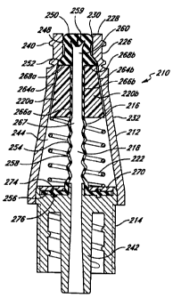
Figures 4 and 5 illustrate a first embodiment of a valve 210 in accordance
with the present invention.
In general, this valve 210 includes a valve body or housing 212, a support
member 214, a seal 216 defining
an inner cavity 218, a pair of clam shells 220a and 220b, and a spring 222.
These components are
assembled, as depicted in Figure 4, without the need for a spike element. The
inner cavity 218 forms an
expandable fluid space inside the valve 210. As discussed below, the clam
shells 220a/220b are constructed
to cause the volume of the fluid space to expand or increase upon insertion of
a medical implement and to
contract or decrease upon withdrawal of the medical implement.
The body or housing 212 has an upper conduit 226 near a proximal end 228,
desirably with a
circular opening 230 that is adapted to receive the medical implement. A side
wall portion 232 is preferably
tapered to cooperate with the clam shells 220a/220b. The body 212 has an upper
ledge 234 formed between
the proximal end 228 and the side wall portion 232. There is desirably a
threaded portion on the housing 212
adjacent the circular opening 230 in the top of the upper conduit 226, as best
seen in Figure 4. Note that
"proximal" is used
CA 02275218 1999-06-14
WO 98/26835 PCT/US97/23258
6
to denote the end of the valve 210 and other components at or near the body
opening 230, while "distal" is used
to denote the opposite end of the vaive.
In the first embodiment, the upper conduit 226 is adapted to receive the tip
or nose 236 of an ANSI
standard syringe 238, as shown in phantom in Figure 5. It is, however,
contemplated that the outer diameter of
the upper conduit 226 can be of any size to accommodate the attachment of
other connector devices thereto.
Advantageously, the proximal end of the upper conduit 226 can be equipped with
a locking mechanism to facilitate
locking of the vaive 210 to a variety of connector devices. For example,
referring to Figure 4, the threaded portion
of the housing 212 are preferably provided such that the housing 212 can be
locked into any compatible Luer-Lock
device known to those with skill in the art. The housing 212 of the first
embodiment according to this invention
includes conventional Luer-Lock threads 240 on the outer diameter of the upper
conduit 226.
The support member 214 has at its distal end the inner conduit 242 which may
be connected to a terminal
end of a catheter (not shown). The support member 214 serves as a support and
attachment device for the seal
216 by holding the seal 216 in place inside the internal cavity 244 of the
housing 212. The inner conduit 242 and
inner cavity 218 of the seal 216 present a continuous passageway for fluid
during use.
The seal 216 is prepared from a resilient material that is flexible, inert,
and impermeable to fluid, such as
silicon. The seal 216 has a seal cap 248 with a generally flat top surface
250, a shoulder 252, a side wall 254,
and a base 256. The side wall 254 advantageously is comprised of wall portions
258 which deform in an accordion-
like fashion and assist in the reformation of the seal 216 to close the
housing opening 230 upon withdrawal of the
syringe 238. During compression of the seal 216, the wall portions 258 expand
outwardly in the radial direction.
The interior of the seal 216 is hollow to provide the inner cavity 218, as
best seen in Figure 4. There are preferably
gaps between the wail portions 258 which facilitate deformation and
reformation of the seal 216. The shoulder 252
engages the upper ledge 234 provided in the upper conduit 226 of the housing
212 such that the upper ledge 234
confines the movement of the shoutder 252 toward the opening 230 to prevent
the seal 216 from being blown
through the opening 230 under hiah pressure in the inner cavity 218 of the
seal 216.
The seai cap 248 reseals the valve 210 at the opening 230, with the top
surface 250 of the seal 216
approximately flush with or slightly above or below the opening 230 upon
removal of the medical implement 238.
Preferably, the seal cap 248 substantially fills the opening 230 in the top of
the upper conduit 226. After assembly,
the top surface 250 of the seal cap 248 is essentially flush with the opening
230, so that the seal cap 248 can
be swabbed with alcohol or other disinfectant without leakage of the
disinfectant into the valve 210. Therefore,
it is preferable that the top surface 250 be exposed so that it may be swabbed
with a disinfectant.
To provide a fluid-tight seal at the opening 230 and to eliminate the need for
a spike element to induce
fluid flow upon insertion of a medical implement, the seal cap 248 has a
unique shape and includes a precut slit 259,
also having a unique shape. The seal cap 248 desirably has an oval or
elliptical shape with a major axis having a
length larger than the inner diameter of the circular opening 230 such that
the oval seal cap 248 substantially fills
the opening 230 in the top of the upper conduit 226 in the decompressed state.
The precut slit 259 in the seal
cap 248 is squeezed shut by the circular opening 230 in the decompressed
state, as seen in Figure 4. In its resting
CA 02275218 1999-06-14
WO 98/26835 PCT/US97/23258
7
state, the precut slit 259 is open. During compression of the seal 216 by
insertion of a medical implement such
as the syringe 238, as illustrated in Figure 5, the precut slit 259 returns to
its resting state and opens, as the seal
cap 248 is allowed to stretch in the portion of the upper coinduit 226 which
has a larger inner diameter. Fluid is
thus allowed to pass through the slit 259. Note that the terms "compressed
state" and "decompressed state" are
used conveniently to refer to campression and decompression of the seal 216 by
insertion and withdrawal of the
medicai implement 238 along the longitudinal axis of the seal 216. The terms
do not relate to the radial compression
of the seal cap 248 by the opening 230 of the housing 212.
To further assist in creating a fluid-tight seal in the decompressed state,
the seal 216 of Figure 4
advantageously includes the enlarged, internai, pressure respoinsive member
260 which is integrai with the seal cap
248. The pressure responsive member 260 enables the valve 210 to maintain a
fluid-tight seal even at very high
pressures sometimes experienced in medical applications, particuiarly when the
valve 210 is connected to a patient's
artery.
As shown in Figures 4 and 5, the clam shells 220a1220b are desirably identical
pieces disposed opposite
one another symmetrically inside the valve body 212. They are preferably made
of a firm material such as a hard
plastic. The external surface 264a1264b of each clam shell 220a/220b is
tapered to cooperate with the tapered
side wall portion 232 of the housing 212, and is configured to slide along the
side wall portion 232 during
compression and decompression. The internal surfaces 266a(266b of the clam
shells 220a/220b cooperate with one
another to squeeze a portion of the seal side wall 254, preferably adjacent
the shoulder 252, to form a constricted
portion 267 of the seal 216. The proximal ends 268af268b of the ciam shells
220a1220b engage the shoulder 252
of the seal 216 to facilitate movement of the clam shells 220a1220b with the
compression of the seal 216. The
internal surfaces 266a/266b preferably are shaped to cause the constricted
portion 267 to be substantially circular.
In this embodiment, each internal surface 266a/266b has a semi-circular,
longitudinal groove that squeezes the seal
216.
The spring 222 is disposed between the distaf ends of the clam shells
220a/220b and the base 256 of the
seal 216, but desirably a hard retaining disk 270 is provided adjacent the
base 256 of the seal 216 to provide better
support for the spring 222 and the seal 216. In the decompressed state shown
in Figure 4, the spring 222 may
be relaxed or be in slight compression to exert a force on the seal 216
through the clam shells 220a1220b to keep
the seal 216 closed. During insertion of the syringe 238, the spring 222 is
compressed and stores potential energy
from the compression, as illustrated in Figure 5. Upon withdrawal of the
syringe 238, the spring 222 releases the
potential energy and pushes the clam shells 220a1220b proximally to close the
seal 216, as shown in Figure 4. The
spring 222 is preferably not attached or bonded to either the clam shells
220a1220b or the retaining disk 270 for
ease of assembly. Although Figures 4-5 show a helical spring 222, any suitable
spring known to those of skill in
the art may be used.
The seal 216 is desirably relaxed longitudinally in the decompressed state
(Figure 4), and compressed
longitudinally in the compressed state (Figure 5). Alternatively, the seal 216
may be stretched longitudinally in
tension by the spring 222 in the decompressed state and be relaxed or slightly
compressed longitudinal in the
CA 02275218 1999-06-14
WO 98/26835 PCT/US97/23258
8
compressed state. The base 256 of the seal 216 advantageously fits snugly and
securely into a annular groove 274
provided in the retaining disk 270 and an annular groove 276 provided in the
support member 214. The annular
grooves 274,276 form a locking mechanism to support and secure the seal 216
within the cavity 244 of the housing
212.
To illustrate vaive activation, Figure 5 shows the compressed state of the
valve 210 upon insertion of the
syringe 238. A medical implement other than a syringe as known to those of
skill in the art may be used. The nose
236 of the syringe 238 is placed on the seal cap 248 inside the opening 230 of
the housing 212. The application
of pressure on the syringe 238 creates pressure on the seal cap 248, and the
resulting downward pressure
compresses the seal 216. This pushes the seal cap 248 away from the circular
opening 230 and toward the lower
portion of the housing cavity 244 which has a iarger inner diameter, thereby
allowing the precut slit 259 to open.
The downward movement is facilitated by the compression of the spring 222
which stores the potential energy of
compression and by the gaps between the wall portions 258 of the side wall 254
of the seal 216. Fluid is now
able to flow into the syringe 238, or vice versa, depending on whether fluid
is to be withdrawn from the patient
or medication injected into the patient. Figure 5 shows the valve 210 opened
by insertion of the nose 236 of the
syringe 238 into the opening 230. For intravenous applications, the valve 210
can be oriented in the position
diagramed in Figures 4 and 5, or it can be rotated 180' such that fluid flows
in the opposite direction.
In the compressed state shown in Figure 5, the inner cavity 218 of the seal
216 generally contracts
(becomes shorter) as compared to the decompressed state shown in Figure 4. The
constricted portion 267 of the
inner cavity 218, defined by the clam shells 220a/220b, however, expands
(becomes larger) in volume when the seal
216 is in the compressed state. This results from a movement of the clam
shells 220a/220b apart from one another
as they slide along the tapered side wall 232 of the housing 212. The amount
of general contraction of the seal
216 in relation to the amount of expansion of the constricted portion 267
during compression determine whether
the valve 210 generates a positive, negative, or zero flow upon decompression,
as discussed below.
Upon removal of the syringe 238 from the upper conduit 226, as shown in Figure
4, the seal 216 is free
to move toward its decompressed state, and the clam shells 220a/220b are
pushed proximally toward the opening
230. The movement causes a general expansion of the inner cavity 218 (i.e.,
the cavity increases in length), but
causes a contraction (i.e., reduction in size) of the volume of the
constricted portion 267 of the seal 216. If the
volume change associated with the contraction of the constricted portion 267
equals the volume change associated
with the expansion of the inner cavity 218, the fluid space or inner cavity
will have zero flow. If the increase in
voiume associated with the expansion of the inner cavity 218 is greater than
the reduction in volume associated with
the contraction of the constricted portion 267, there will be a net gain in
fluid space, resulting in an undesirable
negative flow toward the valve 210 through, e.g., a catheter tip (not shown).
If the reduction in volume associated
with the contraction of the constricted portion 267 is greater than the
increase in volume associated with the
expansion of the inner cavity 218, there will be a desirable positive flow
from the valve 210 through the catheter
tip (not shown). Thus, for the valve 210 to be a positive-flow valve requires
that the clam shells be configured to
allow greater expansion of the constricted portion 267 (i.e., an increase in
fluid volume in that area of the seal 216)
CA 02275218 1999-06-14
WO 98/26835 PCTIUS97/23258
9
than the general contraction volume change associated with the expansion of
the inner cavity 218 of the seal 216
upon compression and, hence, greater contraction (i.e., decrease in fluid
volume within that area of the seal) of the
constricted portion 267 than the general expansion (i.e., increase in fluid
volume in that area of the seal) of the seal
216 upon decompression. In other words, for the valve 210 to induce positive
flow upon disconnection of the
medical implement 238 therefrom, the total fluid volume within the valve 210
must decrease. In the instant case,
this decrease in fluid voiume is effectuated by causing the fiuid volume
within the seal to decrease as between its
compressed (when syringe attached) and uncompressed (wheri syringe detached)
states. This reduction or decrease
in available fluid volume within the valve 210 causes fluid tj flow towards
the catheterlpatient, preventing blood
from being drawn into the catheter.
That the valve 210 is advantageouslv configured to tie a positive-fiow valve
210 eliminates any dead space
during decompression of the seal 210 as the syringe 238 is withdrawn, as
illustrated in Figure 4. Furthermore, as
the syringe 238 is withdrawn, the slit 259 remains open until the very end,
i.e., until the seal cap 248 is squeezed
by the circular opening 230 at the top of the upper conduit 226. This further
assists in eliminating dead space and
avoiding backflash. This feature is particularly advantageous in the case
where the valve 210 is connected through
a catheter to a patient, because it prevents blood from being cirawn into the
catheter and clogging it. This invention
therefore eliminates a significant risk by solving the problem of backfiash.
As the seal 216 is free to move to its decompressed state, it essentially
fills the opening 230. The ability
of the seal 216 to return to its original shape and be deformed in its
decompressed state is determined by the
resiliency of the material used to prepare the seal 216. Advantageously, the
ability of the seal 216 to return to its
decompressed state is facilitated by the spring 222 and the gaps between the
wall portions 258 of the seal 216.
The ability of the seal 216 to deform reversibly and return to its
decompressed state is particularly useful because
(1) it immediately stops fluid flow through the valve 210, and (2) it
maintains sterility of the valve.
The ability of the seal 216 to return reversibly to its decompressed state
permits reuse of the valve 210.
Following disconnection, and before reuse, the surface 250 of the seal cap 248
is essentially flush with the opening
230 of the housing 212. Thus, this flush surface 250 can advantageously be
sterilized with alcohol or other surface-
decontaminating substances. The support member 214 and body 212 advantageously
shield both connections from
the surrounding environment to protect the steriiity of the connection.
A cover cap (not shown) can be supplied to fit over the upper conduit 226 as
further protection for the
surface 250 of the seal cap 248 when not in use. Such a cover cap, however, is
not needed to maintain sterility
since the seal 216 may be swabbed with a disinfectant before andtor after each
use. Reversibility of the seal 216
makes the valve 210 particularly attractive as a connector valve to provide
fluid communication between two fluid
lines. Therefore, the present invention provides for placing a first fiuid
line in communication with a second fluid
line using the valve 210 disclosed herein. The reversibility of the valve 210
permits multiple fluid lines to be
successively added, for example, to a fluid line in direct communication with
a patient's vein. Since the valve 210
is easiiy sterilized and sealable, fluid lines can be added and removed
without disconnecting venous contact of the
catheter.
CA 02275218 1999-06-14
WO 98/26835 PCT/US97/23258
The valve body 212 and support member 214 are preferably prepared from a hard
plastic, but it is
additionally contemplated that the valve 210 could be prepared from other
medically inert materials known to those
skilled in the art. Another feature of this invention is that it relies
neither on a needle nor on a spike in order to
establish fluid flow through the valve. This completely eliminates the risk of
skin puncture or fear of puncture during
5 use and manufacture. It also eliminates coring of the seal 216 by a spike
element and all the risks associated
therewith. Further, the fluid flow rate is not limited by the size of a
through passage in a needle or spike, as is
the case in some prior art valves.
As shown in Figure 4, another feature of the invention is that the upper ledge
234 confines the movement
of the shoulder 252 toward the opening 250 to prevent the seal 216 from being
blown through the opening 230
10 under high pressure in the cavity 218. This makes the valve 210
particularly suited for high pressure applications.
Second Embodiment
In a second embodiment of the present invention illustrated in Figures 6 and
7, the valve 310 includes a
valve body or housing 312, a support member 314, a skirt 316, a seal 318, a
resilient member 320, and a pair of
clam shells 322a/322b. The housing 312 is desirabiy similar to the housing 212
of Figure 4 and has a tapered side
wall 324.
Referring to Figures 6 and 7, the second embodiment of the valve 310 has a
bell-shaped skirt 316. The
skirt 316 has an annular ring 328 which is disposed toward an inner conduit
330 of the support member 314. The
skirt 316 creates a shield for the inner conduit 330. This inner conduit 330
is preferably cylindrical in shape and
slightly tapered. The inner conduit may be connected to a terminal end of a
catheter (not shown), which has an
opposite, open end that is generally inserted into a patient. The support
member 314 serves as a support and
attachment device for the seal 318 by holding the seal 318 in place inside the
housing 312.
The support member 314 also serves as a support and attachment device for the
skirt 316. As best seen
in Figure 6, the support member 314 has an edge portion 332 which engages a
ledge 334 of the skirt 316 in
assembly. This attachment secures the skirt 316 in place. The skirt 316
desirably includes a Luer-Lock portion 336
that enables the valve 310 to be removably attached to, for exampie, a fluid
line or catheter connected to a patient.
It is noted that the valve 310 in this embodiment includes a skirt 316
separate from the housing 312 for ease of
assembly. A different embodiment can provide a unitary member which replaces
the housing 312 and skirt 316.
It is therefore contemplated that such an embodiment would fall within the
scope of this invention.
The seal 318 is similar to the seal 210 of Figure 4. The seal 318 is also
preferably silicon and has a
similar seal cap 340 with a precut slit 342, shoulder 344, and pressure
responsive member 348. These components
serve the same function as those of the seal 210. Instead of a side wall
formed with wall portions 258, the seal
318 has a side wall 350 that is generally circular cylindrical and has a
distal portion 352 that is sized to be slip-
fitted with the proximal end 354 of the inner conduit 330 of the support
member 314. During compression of the
seal 318, the side wall 350 simply slides over the proximal end 354 of the
inner conduit 330, forming a fluid-tight
seal therewith. The seal 318 defines an inner cavity 358 above the proximal
end 354 of the inner conduit 330.
The inner cavity 358 forms an expandable fluid space inside the vaive 310. The
inner conduit 330 and inner cavity
CA 02275218 1999-06-14
WO 98/26835 PCT/US97/23258
358 comprise aligned hollow tubes in fluid communication with each other when
the precut slit 342 of the seal 318
opens during compression of the seal 310.
Similar in form and function to the clam shells 220af220b of Figures 4 and 5,
the clam shelrs 322a/322b
are constructed to cause an increase in fluid space upon insertion of a
medical implement into the valve 310 and
a decrease in fluid space upon withdrawal of the medical implement such as a
syringe 362 partially shown in
phantom in Figure 7. The internal surfaces 364a/364b of the clam shells
desirably have longitudinal grooves that
cooperate with one another to squeeze a portion of the seal side wall 350 to
form a constricted portion 366 thereof.
Instead of the spring 222.in Figure 4, the second ernbodiment employs the
resilient member 320 disposed
between the clam shells 322a/322b and the support member 314. The resilient
member 320 advantageously is inert
and impermeable to fluid such as siiicon, and includes wall portions 368 which
deform in an accordion=like fashion
and assist in the reformation of the seal 318 to close the housing opening 370
upon withdrawal of the syringe 362.
The resilient member 320 thus is similar in construction with and serves the
same function as the spring 222 of the
seal 210 of Figures 4 and 5. It is contemplated that a spring (not shown)
similar to the spring 222 of Figure 4 may
be used in place of the resilient member 320, as may other suitable structures
known to those of skill in the art.
As shown in Figures 6 and 7, the resilient member 320 has a base 346. The base
346 fits snugly and
securely within an annular groove 374 provided in the housing 312 and an
annular groove 377 provided in the
support member 314, as shown in Figure 6. The annular grooves 376,377 hence
form a locking mechanism to
support and secure the resilient member 320 within the housing 312. The
shoulder 344 engages an upper ledge 382
provided in an upper conduit 384 of the housing 312 such that the upper ledge
382 confines the movement of the
shoulder 344 toward the opening 370 to prevent the seal 318 from being blown
through the opening 370 under high
pressure in the inner cavity 358 of the seal 318.
The resilient member 320 is desirably relaxed or slightly compressed
longitudinally in the decompressed state
(Figure 6), and compressed longitudinally in the compressed slate (Figure 7).
The resilient member 320 is desirably
not attached or bonded to either of the clam shells 322a/322b or the housing
312.
Figures 7 illustrates compression and Figure 6 illustrates decompression
during valve activation. In the
compressed state, the syringe 362 is placed on the seal cap 340 inside the
opening 370 of the housing 312, and
the application of pressure on the syringe 362 creates pressure on the seal
cap 340. The downward pressure
pushes the seal cap 340 away from the circular opening 370 and toward the
distal lower portion of the housing
312 which has a iarger inner diameter, thereby allowing the precut slit 342 to
open. The side wall 350 slides over
the proximal end 354 of the inner conduit 330, and the resiiient member 320
deforms in an accordion-like manner,
storing potential energy of the compression. Fluid is able to flow into the
syringe 362, or vice versa, depending on
whether fluid is to be withdrawn from the patient or medication injected into
the patient.
The compression of the seal 318 shown in Figure 7 generally causes a
contraction or reduction in the
volume of the inner cavity 358 of the seal 318. The valve 310 has a net gain
in volume of the inner cavity 318,
however, because the general reduction in volume within the inner cavity 358
is less than an increase in volume
within the constricted portion 366 of the inner cavity 358 defined by the clam
shells 322a/322b. The expansion
CA 02275218 1999-06-14
WO 98/26835 PCT/US97/23258
12
results from the movement of the clam shells 322a/322b apart from one another
during compression, facilitated by
the tapered side wall 324 of the housing 312.
Figure 6 illustrates the valve after withdrawal of the syringe 362. The seak
318 returns to its
decompressed state and essentially fills the opening 370, and the clam shells
322a/322b are pushed proximally
toward the opening 370 by the resilient member 320. Because of the contraction
of the inner cavity 358 at the
constricted portion 366 by the clam shells 322a1322h, there is a net loss or
reduction in fluid space, resulting in
a positive flow from the valve 310 through, e.g., a catheter tip (not shown).
The positive-flow valve 310
advantageously eliminates any dead space during decompression of the seal 318.
This is further assisted by the
seal 318 with the slit 342 remaining open until the very end, i.e., until the
seal cap 340 is squeezed by the upper
conduit 384.
In addition, the valve 310 can be reused because the seal 318 can return
reversibly in the decompressed
state. The seal surface 340 is also swabbable for sterility. Other features of
the valve 310 are discussed previously
in connection with the first embodiment of this invention and will not be
repeated.
Third Embodiment
As shown in Figures 8 and 9, a third embodiment of the valve 410 of the
present invention comprises a
valve body or housing 412, a support member 414, a flexible tubing 416, a seal
418, a ring member 420, a pair
of clam shells 422af422b, and a spring 424. The flexible tubing 416 may be
connected to a catheter (not shown)
and, together with the seal 418, defines an inner cavity 426. The inner cavity
426 forms an expandable fluid space
of the valve 410. The clam shells 422a/422b desirably are substantially the
same as the clam shells 220al220b
of Figure 4 and are constructed to cause the fluid space within the vaive 410
to increase upon insertion of a medical
implement and to decrease upon withdrawal of the medical implement such as a
syringe 428 partially shown in
phantom in Figure 9. The housing 412 is desirably simiiar to the housing 212
of Figure 4.
The support member 414 has a hoilow center 430 which supports the flexible
tubing, and a proximal end
432 which encioses a distal end 434 of the housing 412. The support member 414
desirably locks onto the housing
412 via any method known to those of skill in the art. The proximal end 432 of
the support member 414 supports
the spring 424, which in turn supports the clam shells 422a/422b and seal 418.
The seal 418 is prepared from a resifient material that is flexible, inert,
and impermeable to fluid, such as
silicon. Referring to Figure 8, the seal 418 is substantially similar to the
seal 210 of Figure 4, with a portion of
the side wall 438 cut off near the shoulder 440 region. As a result, the side
wall 438 of the seal 418 is
substantially shorter than the side wall 254 of the seal 210 in Figure 4. A
distal end 442 of the side wall 254 is
attached, preferably by adhesive, to a proximal end 444 of the flexible tubing
416. The distal end 442 abuts the
ring member 420 which is disposed between the seal 418 and the clam shells
422a1422h and attached at its inner
surface 446 to a portion of the tubing 416, desirably also by adhesive. Other
suitable means of attachment may
be used. The ring member 420 is desirably made of polycarbon.
The clam shells 422a1422h desirably form a sliding contact at their proximal
ends with the ring member
420 for ease of assembly, but may alternatively be affixed to the ring member
420 by adhesive or similar means.
CA 02275218 1999-06-14
WO 98/26835 PCT/US97/23258
13
The clam shells 422a1422b are desirably the same as the clam shells 220a1220b
of Figure 4, having tapered external
surfaces 450a1450b to cooperate with the tapered side wall portion 452 of the
housing 412 for sliding and grooved
internal surfaces 454a1454b that cooperate with one another to squeeze a
portion of the tubing 416 to form a
constricted portion 456.
The spring 424 is substantially the same as the spring 222 of Figure 4 and
serves the same function, being
disposed between the distal ends of the clam shells 422a1422b and the proximal
end 432 of the support member
414. In the decompressed state shown in Figure 8, the sprinq 424 may be
relaxed or in slight compression to exert
a force on the seal 418 through the clam shells 422a1422b to keep the slit 466
in the seak cap 460 closed. During
insertion of the syringe 428, the spring 424 is compressed and stores
potential energy from the compression, as
illustrated in Figure 9. Upon withdrawal of the syringe 428, ttie spring 424
releases the potential energy and pushes
the clam shells 422a/422b proximally to close the seal 418, as shown in Figure
B. The spring 424 is preferably
not attached or bonded to either the clam shells 422a/422b or the support
member 414 for ease of assembly. The
spring 424 can be a helical spring or any other suitable spring known to those
with skill in the art.
Figure 9 shows the compressed state of the valve 410 upon insertion of the
syringe 428. In the
compressed state, the syringe 428 is placed on the seal cap 460 inside the
opening 464 of the housing 412 and
the application of pressure on the syringe 428 creates pressure on the seal
cap 460. The downward pressure
pushes the seai cap 460 away from the circular opening 464 and toward the
distal end of the housing 412, which
has a larger inner diameter, thereby allowing the precut slit 466 of the seal
cap 460 to open. The resilient tubing
416 and the clam shells 422a1422b also move distally as the spring 424 deforms
in compression, storing potential
energy. Fluid is able to flow into the syringe 428, or vice versa, depending
on whether fluid is to be withdrawn from
the patient or medication injected into the patient.
The compression of the seal 418 shown in Figure 9 generally causes a reduction
in the volume of the inner
cavity 426 formed by the seal 418 and tubing 416. However, because of an
expansion of the constricted portion
456 defined by the clam shells 422a/422b an increase in fluid volume is
created which is greater than the general
reduction in fluid volume within the inner cavity 426, the valve 410 has a net
gain in fluid voiume. The increase
in fluid volume results from the movement of the clam shells 422a1422b apart
from one another during seal
compression, facilitated by the tapered side wall 452 of the hiousing 412 and
resiliency of the tubing 416.
Figure 8 illustrates the valve 410 after withdrawal of the syringe 428. The
seal 418 returns to its
decompressed state and essentially fills the opening 464, and the clam shells
422a1422b are pushed proximally
toward the opening 464 by the spring 424. Because of the contraction of the
inner cavity 426 at the constricted
portion 456 by the clam shells 422a/422b, there is a net loss in fluid space,
resulting in a positive flow from the
vaive 410 through, e.g., a catheter tip (not shown). The positive=flow valve
410 advantageously eliminates any dead
space during decompression of the seal 418. This is further assisted by the
seal 418, with the slit 466 remaining
open until the very end, i.e., until the seal cap 460 is squeezed by upper
conduit 470.
CA 02275218 1999-06-14
WO 98/26835 PCT/US97/23258
14
In addition, the vaive 410 can be reused because the seal 418 can return
reversibly to the decompressed
state. The seal surface 472 is also swabbable for sterility. Other features of
the valve 410 are discussed previously
in connection with the earlier embodiments of this invention and will not be
repeated.
Fourth Embodiment
A fourth embodiment of the present invention is illustrated in Figures 10 and
11. As illustrated therein,
a valve 510, comprises a valve body or housing 512, a support member 514, a
skirt 516, a retaining member 518,
a seal 520, a pair of clam shells 522a1522b, and a resilient member 524. The
valve 510 has several features that
are the same or similar to those of the valve 310 of Figures 8 and 9, having a
similar resilient member 524 and clam
shells 522a/522b. The clam shells 522a/522b have internal surfaces 526a1526b
that cooperate with one another
to squeeze a portion of the seal side wall 528 to form a constricted portion
530 thereof.
The seal 510 is preferably made of silicon and has a seal cap 532 with a
precut slit 534, shoulder 536,
lower lip 538, and pressure responsive member 540 that are similar to the seal
210 of Figure 4. These components
serve the same function as those of the seal 210. The side wall 528 may be
formed with ringed wall portions 258,
as in the seal 210, but Figure 4 shows the side wall 528 that is generally
circular cylindrical. The seal 520 defines
an inner cavity 542 which forms an expandable fluid space inside the valve
510. During compression of the seal
520, the side wall 528 deforms outwardly into a circumferential cusp or bulge
544 in the unconstricted region
between the clam shells 522a1522b and the support member 514. The side wall
528 returns to its decompressed
shape upon decompression of the seal 520. The seal 520 is desirably relaxed
longitudinally in the decompressed
state (Figure 10), and compressed longitudinally in the compressed state
(Figure 11). Alternatively, the seal 520 may
be stretched longitudinally in tension by the resilient member 524 in the
decompressed state and be relaxed or
slightly compressed longitudinal in the compressed state.
Referring to Figure 10, the skirt 516 is a bell=shaped skirt that is similar
to the skirt 316 of Figure 8. The
skirt 516 creates a shield for an inner conduit 548 of the support member 514.
The inner conduit 548 may be
connected to a terminal end of a catheter inot shown) which has an open end
that is generally inserted into a
patient. The support member 514 serves as a support and attachment device for
the seal 520 by holding the seal
520 in place inside the housing 512.
The support member 514 also serves as a support and attachment device for the
skirt 516. Similar to the
valve 310 of Figure 8, the support member 514 shown in Figure 10 has an edge
portion 550 which engages a ledge
552 of the skirt 516 in assembly. This attachment secures the skirt 516 in
place. The skirt 516 desirably includes
a Luer-Lock portion 554 that enables the valve 510 to be removably attached
to, for example, a fluid line or catheter
connected to a patient.
The retaining member 518 is desirably provided to secure the lower lip 538 of
the seal 520 and support
the resilient member 524. The retaining member 518 is held inside the housing
512 by the support member 514,
and is provided for ease of assembling the vaive 510. The retaining member 518
has an annular groove 556, and
the support member 514 has an annular groove 558. The annular grooves 556,558
form a locking mechanism to
support and secure the seal 520 within the housing 512 by engaging the lower
lip 538 snugly with the grooves
CA 02275218 1999-06-14
WO 98/26835 PCT/US97/23258
556,558. It is noted that a different embodiment may provide a unitary member
which replaces the support member
514 and the retaining member 518. It is therefore contemplated that such an
embodiment would fall within the
scope of this invention.
Figure 11 illustrates compression and Figure 10 illustrates decompression
during valve activation. In the
5 compressed state, a medical implement such as the syringe 562 partially
shown in phantom is placed an the seal
cap 532 inside the opening 564 of the housing 512, and the application of
pressure on the syringe 562 creates
pressure on the seal cap 532. The downward pressure pushes the seal cap 532
away from the circular opening
564 and toward the iower portion of the housing 512, which has a larger inner
diameter, thereby allowing the precut
slit 534 to open. The side wall 528 deforms outwardly at the unconstricted
region into a circumferential cusp 544,
10 and the resilient member 524 deforms in an accordion-like manner, storing
potential energy of the compression. Fluid
is able to flow into the syringe 562, or vice versa, depending cin whether
fluid is to be withdrawn from the patient
or medication injected into the patient.
The compression of the seal 520 shown in Figure 11 generally causes a
reduction in the fluid volume of
the inner cavity 542 of the seal 520. The valve 510 has a net gain in volume
of the inner cavity 542, however,
15 because the general reduction in volume within the inner cavity 542 is less
than the increase in volume within the
constricted portion 530 as defined by the clam shells 522a1522b and of the
cusp 544 at the unconstricted region
of the seal 520.
Figure 10 illustrates the vaive 510 after withdrawal of the syringe 562. The
seal 520 returns to its
decompressed state and essentially fills the opening 564, and thie clam shells
522al522b are pushed back up toward
the opening 564 by the resilient member 524. Because of the contraction of the
inner cavity 542 of the seal 520,
there is a net loss in fluid space, resulting in a positive flow from the
valve 510 through, e.g., a catheter tip (not
shown). The positive-flow valve 510 advantageously eliminates any dead space
during decompression of the seal
520. This is further assisted by the seal 520, with the slit 534 remaining
open until the very end, i.e., until the
seal cap 532 is squeezed by the circular opening 564 at the top of the upper
conduit 570.
In addition, the valve 510 can be reused because the seal 520 can return
reversibly in the decompressed
state. The seal surface 572 is also swabbable for sterility. Other features of
the valve 510 are discussed previously
in connection with the earlier embodiments of this invention.
Fifth Embodiment
Figures 12 and 13 show a fifth embodiment valve 610 in accordance with the
present invention, the valve
610 comprising a valve body or housing 612, a seal 614, a ring member 616, and
a spring 618. The housing 612
is similar to the housing 212 of Figure 4, with a circular opening 620, and a
tapered side wall 622, but may have
a straight side wall instead. The seal 614 is similar to the seal 318 of
Figure 8, having a substantially cylindrical
side wall 624 and defining an inner cavity 626 which forms an expandable fluid
space inside the valve 610. The
side wall 624 may have different and variable thickness (not shown). The
components are dimensioned and
configured to cause the fluid space to expand upon insertion of a medical
implement and to contract upon withdrawal
of the medical implement such as a syringe 630 partially showri in phantom in
Figure 13. The distal portion of the
CA 02275218 1999-06-14
WO 98/26835 PCT/US97/23258
16
seal 614 is connected to a fluid iine such as a catheter (not shown), and may
be secured to the housing by means
known to those with skill in the art, such as by the use of a support member
(not shown) simijar to the support
member 214 shown in Figure 15.
The ring member 616 is desirably an annular disk 616 made of a hard plastic
and disposed between a
shoulder 634 of the seal 614 and a proximal end 636 of the spring 618. The
ring member 616 serves as a
constraint for the seal 614 during compression and efficiently transfers the
compressive force to the spring 618,
assisting in the deformation of the seal 614. During decompression, the ring
member 616 efficiently transfers the
spring force to the seal cap 638 of the seal 614 to close the opening 620.
Although the ring member 616
facilitates the deformation and reformation of the seal 614, it is not
necessary for the seal 614 to work. In that
case, the spring 618 will contact the seal cap 638 directly.
The spring 618 is substantially the same as the spring 222 of Figure 4 and
serves the same function, being
disposed between the ring member 616 and a distal end 642 of the housing 612.
In an alternative embodiment,
the distal end 642 may be a separate component from the housing 612 for ease
of assembly. In the decompressed
state shown in Figure 12, the spring 618 may be relaxed or be in slight
compression to exert a force on the seal
614 through the ring member 616 to keep the seal 614 closed. During insertion
of the syringe 630, the spring 618
is compressed and stores potential energy from the compression, as illustrated
in Figure 13. Upon withdrawal of
the syringe 630, the spring 618 releases the potential energy and pushes the
ring member 616 to close the seal 616
as shown in Figure 12. The spring 618 is preferably not fixed with either the
ring member 616 or the distal end
642 of the housing 612 for ease of assembly. The spring 618 can be a helical
spring or any other suitable spring
known to those with skill in the art.
The side wall 624 of the seal 614 is constrained by the ring member 616 and
housing 612, and is
substantially relaxed in the decompressed state. During compression of the
seal 614, the side wall 624 bulges in
the unconstrained region between tne ring member 616 and the distal end 642 of
the housing 612, causing an
increase in the fluid space within the vaive 610. The side wall 624 returns to
its decompressed shape upon
decompression of the seal 614. Alternatively, the side wall 624 may be
stretched in tension by the spring 618 in
the decompressed state and goes through a relaxed position before deforming
under compression to its bulged
condition.
Figure 13 illustrates compression and Figure 12 illustrates decompression
during valve activation. In the
compressed state, the syringe 630 is placed on the seal cap 638 inside the
opening 620 of the housing and the
appiication of pressure on the syringe 630 creates pressure on the seal cap
638. The downward pressure pushes
the seal cap 638 and the ring memner 616 away from the circular opening 620
and toward the lower portion of
the housing 612 which has a larger inner diameter, thereby allowing the precut
slit 646 af the seal cap 638 to open.
The side wall 624 deforms outwardiy and bulges at the unconstricted region, as
the spring 618 is compressed,
storing potential energy of the compression. Fluid is able to flow into the
syringe 630, or vice versa, depending on
whether fluid is to be withdrawn from the patient or medication injected into
the patient. The compression of the
seal 614 shown in Figure 13 results in a net gain in volume of the inner
cavity.
CA 02275218 1999-06-14
WO 98/26835 PCTIUS97/23258
17
Figure 12 illustrates the valve 610 after withdrawai of the syringe 630. The
seal 614 returns to its
decompressed state and essentially fills the opening 620, and the ring member
616 is pushed back up toward the
opening 620 as the spring 618 releases its potential energy. Because of the
contraction of the inner cavity 626
of the seai 614, there is a net loss in fluid space, resulting n a positive
flow from the valve 610 through, e.g., a
catheter tip (not shown). The positive-flow valve 610 advantageously
eliminates any dead space during decompression
of the seal 614. This is further assisted by the seal 614 with the slit 646
remaining open until the very end, i.e.,
until the seal cap 638 is squeezed by the circular opening 620 at the top of
the upper conduit 650 of the housing
612.
In addition, the valve 610 can be reused because the seal 614 can return
reversibly in the decompressed
state. The seal surface 652 is aiso swabbable for sterility. Other features of
the valve 610 are discussed previously
in connection with the earlier embodiments of this invention.
Sixth Embodiment
A sixth embodiment of a valve 710 is illustrated in Figures 14 and 15. The
vaive 710 comprises a valve
body or housing 712 and a seal 714. The housing 712 has an upper conduit 716
near a proximal end with a circular
opening 718 that is preferably adapted to receive a medical impiement. A side
wall portion 720 is orotruded to
facilitate deformation of the seal 714. A distal end 724 of ihe housing 712
forms a lower passage 726 (partially
shown) which supports and constrains a distal portion 728 of the seal 714, and
is connected, for example, to a fluid
line such as a catheter (not shown). Alternatively, a support member (not
shown) may be used to detachably lock
onto the housing 712 and support the seal 714, such as those shown in Figure 4
(214) or Figure 12 (514).
The seal 714 is generally similar to the seal 614 of Figures 12 and 13, and
has a substantially cylindrical
side wall 721, although the side wall 732 may have a slight bulge 733 as shown
in Figure 14. It defines an inner
cavity 734 which forms an expandable fluid space inside the valve 710. In the
decompressed state, the seal 714
is constrained bv the upper conduit 716 and lower passage 7.26 of the housing
712, and is substantially relaxed in
the decompressed state. The components are dimensioned and configured to cause
the fluid space to expand or
increase upon insertion of the medical implement and to cantract or decrease
upon withdrawal of the medical
impiement such as the syringe 730 partially shown in phantom in Figure 15.
During compression of the seal 714,
the side wall 732 bulge in the unconstrained region between the upper conduit
716 and lower passage 726 and the
bulge 738 is substantially round. The side wall 732 return to its decompressed
shape upon decompression of the
seal 714.
Figure 15 illustrates compression and Figure 14 illustrates decompression
during valve activation. In the
compressed state, the syringe 730 is placed on the seal cap 742 of the seal
714 inside the opening 718 of the
housing 712 and the appiication of pressure on the syringe 730 creates
pressure on the seal cap 742. The
downward pressure pushes the seal cap 742 away from the circular opening 718
and toward the protruded portion
720 of the housing 712 which has a larger inner diameter, thereby allowing the
precut slit 746 of the seal cap 742
to open. The side wall 732 deforms outwardly and bulges at the unconstricted
region 738, storing potential energy
of the compression. Fluid is able to fiow into the syringe 730, or vice versa,
depending on whether fluid is to be
CA 02275218 1999-06-14
WO 98/26835 PCTNS97/23258
withdrawn from the patient or medication injected into the patient. The
compression of the seal 714 shown in Figure
15 generates a net gain in volume of the inner cavity.
Figure 14 illustrates the valve 710 after withdrawal of the syringe 730. The
seal 714 returns to its
decompressed state and essentially fills the opening 718. Because of the
contraction of the inner cavity 734 of the
seal, there is a net loss in fluid space, resulting in a positive flow from
the valve 710 through, e.g., a catheter tip
(not shown). The positive-flow valve 710 advantageously eliminates any dead
space during decompression of the
seal 714. This is further assisted by the seal 714 with the slit 746 remaining
open until the very end, i.e., until
the seal cap 742 is squeezed by the circular opening 718 at the top of the
upper conduit 716.
In addition, the valve 710 can be reused because the seal 710 can return
reversibly in the decompressed
state. The seal surface 748 is also swabbable for sterility. Other features of
the valve 710 are discussed previously
in connection with the earlier embodiments of this invention.
Seventh Embodiment
Figures 16 and 17 illustrate a valve 710 in accordance with a seventh
embodiment of the present invention,
the valve 756 comprising a valve body or housing 758 and a seal 760 that are
substantially the same as the housing
712 and seal 714 of Figures 14 and 15, with a distal portion 762 of the seal
760 connected to a fluid line such
as a catheter (not shown). The seal 760, however, is configured to deform upon
compression into a diamond-shaped
cusp 764 instead of a round bulge 738 as illustrated in Figures 14 and 15.
This type of construction may facilitate
deformation and reformation of the seal 760, and may be more easily formed.
The valve activation of this
embodiment is virtually identical to that in Figures 14 and 15, except for the
deformed shape of the seal side wall
770. It is contemplated, therefore, that a seal that may deform into a variety
of shapes other than round and
diamond shapes to achieve positive flow may be employed, as long as the it is
dimensioned and configured to cause
the fluid space of the valve to expand upon insertion of a medical implement
and to contract upon withdrawal of
the medical implement such as the syringe 774 partially shown in phantom in
Figure 28.
Eighth Embodiment
As illustrated in Figures 18 and 19, an eighth embodiment valve 810 of the
present invention is similar to
the embodiments shown in Figures 14-17. The valve 810 also includes a housing
812 having an internal cavity 814
with an upper conduit 816, and a seal 818 disposed inside the internal cavity
814 and having an inner cavity 820
that defines a fluid space. The housing 812 has a distal end 824 which
supports a side wall B26 of the seal 818.
A distal portion 828 of the seal 818 is connected to a fluid line such as a
catheter (not shown). The pressure at
the inner cavity 820 of the seal 818 is P1. Between the housing 812 and the
seal 818 is an enclosed pressure
chamber 832 at pressure P2. The valve activation utilizes the pressure
difference between P2 in the pressure
chamber 832 and P1 in the inner cavity 820 of the sea! 818.
Upon insertion of a medical implement such as a syringe 836 shown in phantom
in Figure 19, the pressure
at the inner cavity 820 of the seal 818 increases from P1 to P3 and the fluid
space inside the seal 818 expands
from the decompressed state of Fioure 18. The expansion of the fluid space
results primarily from a difference in
pressure between P3 and P2. This valve 810 is particularly advantageous in the
case where the side wall 826 of
CA 02275218 1999-06-14
WO 98/26835 PCTIUS97/23258
19
the seal 818 deforms without storing substantial potential energy. For
instance, the side wall 826 of the seal 818
may deform without substantial resistance or resiliency such as a membrane, or
the seal is not constrained
longitudinal by the distal portion 824 of the housing 812 and may siide in and
out of the internal cavity 814 of the
housing 812 through the distal end 824.
Figure 19 illustrates compression and Figure 18 illustrates decompression
during vaive activation. In the
compressed state, the syringe 836 is placed on the seal cap 838 of the seal
818 inside the opening 840 of the
housing 812 and the application of pressure on the syringe creates pressure on
the seal cap 838. The downward
pressure pushes the seal cap 838 away from the circular opening 840 and toward
the lower portion of the housing
812 which has a iarger inner diameter, thereby allowing the precut slit 844 of
the seal cap 838 to open. The entry
of the fluid causes the pressure at the inner cavity 814 of the seal 812 to
increase to P3. As a result, the side
wall 826 deforms outwardly and bulges at the unconstricted region 848.
Potential energy is stored in the change
in pressure differential between the inner cavity 820 and the pressure chamber
832. The side wail 826 of the seal
818 need not deform and store energy, but may do so. Fluid is able to flow
into the syringe 836, or vice versa,
depending on whether fluid is to be withdrawn from the patient or medication
injected into the patient. The
compression of the seal 818 shown in Figure 19 causes a net gain or increase
in fluid volume within the inner cavity.
Figure 18 illustrates the vaive 810 after withdrawal of the syringe 836. The
seal 818 returns to its
decompressed state and essentially fills the opening 840, and the pressure in
the inner cavity 820 returns to P1 and
releases the potential energy. Because of the contraction of the inner cavity
820 of the seal 818, there is a net
loss in fluid space, resulting in a positive flow from the valve 810 through,
e.g., a catheter tip (not shown). The
positive-flow valve 810 advantageously eliminates any dead space during
decompression of the seal 818. This is
further assisted by the seal 818 with the slit 844 remaining open until the
very end, i.e., until the seal cap 838 is
squeezed by the circular opening 840 at the top of the upper conduit 816.
In addition, the vaive 810 can be reused because the seal 818 can return
reversibly in the decompressed
state. The seal surface 854 is also swabbable for sterility. Other features of
the valve 810 are discussed previously
in connection with the earlier embodiments of this invention.
Ninth Embodiment
A ninth embodiment of a valve 910 comprising a housing 912, a support member
914, a skirt 916, a seal
918, and a scissor-like cross member 920, is depicted in Figures 20 and 21.
The housing 912 has an upper conduit
924 with a circular opening 926. The support member 914 has an inner conduit
928 which is connected to a fluid
line such as a catheter (not shown). The seal 918 has a side wall 930
desirably formed of alternating wall portions
932 and defines an inner cavity 934 which forms an expandabde fluid space
inside the valve 910. The cross member
920 is dimensioned and configured to assist in causing the fiuid space to
expand upon insertion of a medical
implement and to contract upon withdrawal of the medical iniplement such as
the syringe 936 partially shown in
phantom in Figure 21.
The cross member 920 has two longitudinal member 940 attached together which
rotates with respect to
one another, and is desirably made of a hard material such as a hard plastic.
The cross member 920 is disposed
CA 02275218 1999-06-14
WO 98/26835 PCT/US97/23258
at a constricted portion 942 of the seal 918 within the inner cavity 934 with
the longitudinal members 940
preferably substantially disposed vertically. The ends 944 of the longitudinal
members 940 are desirably attached
to the side wall 930 as shown in Figure 20. The longitudinal members 940
rotate to a substantially horizontal
orientation upon compression by the insertion of the syringe 936 as shown in
Figure 21. This rotation is referred
5 to as the deformation of the cross member 920. The lonoitudinal members 940
may be attached to rotate freely
with respect to one another. Alternatively, the longitudinai members 940 may
be spring-loaded or attached such that
they rotate under a rotational force but reform to their relaxed position upon
release of the force. Upon withdrawal
of the syringe 936 as shown in Figure 20, the longitudinal members 940 return
to the substantially vertical positions,
referred to as the reformation of the cross member 920. The longitudinal
members 940 are desirably longitudinal
10 plates 940 with sufficient width to expand the constricted portion 942 of
the seal 918 in the substantially horizontal
position but not so wide that they impedes flow therethrough. Alternatively,
they may contain holes (not shown)
through which fluid can pass.
Figure 21 illustrates compression and Figure 20 illustrates decompression
during valve activation. In the
compressed state, the syringe 926 is placed on the seal cap 950 of the seal
918 inside the opening 926 of the
15 housing 912 and the application of pressure on the syringe 936 creates
pressure on the seal cap 950. The
downward pressure pushes the seal cap 950 away from the circular opening 926
and toward the lower portion of
the housing 912 which has a larger inner diameter, thereby allowing the precut
slit 952 of seal cap 950 to open.
The side wall 930 of the seal 918 deforms in an accordion-like manner, and the
cross member 920 deforms and
opens up the constricted portion 922 of the seal 918, storing potential energy
of the compression. Fluid is able to
20 flow into the syringe 936, or vice versa, depending on whether fluid is to
be withdrawn from the patient or
medication injected into the patient. The compression of the seal 918 and
deformation of the cross 920 shown in
Figure 21 qenerally causes a contraction of the volume of the inner cavity 934
of the seal 918. The valve 910 has
a net gain in voiume of the inner cavity 934, however, because the generai
contraction of the inner cavity 934 is
less than by the expansion of the constricted portion 942 pushed apart by the
cross member 920. The expansion
results from the movement of the iongitudinal members 940 of the cross member
920 during compression.
Figure 20 illustrates the valve 910 after withdrawal of the syringe 936. The
seal 918 returns to its
decompressed state and essentially fills the opening 926, and the cross member
920 reforms to allow the constricted
region 942 of the seal 918 to narrow. Because of the contraction of the inner
cavity 934 at the constricted portion
942, there is a net loss in fluid space, resuiting in a positive flow from the
valve 910 through, e.g., a catheter tip
(not shown). The positive-flow valve 910 advantageously eliminates any dead
space during decompression of the
seal 918. This is further assisted by the seal 918 with the slit 952 remaining
open until the very end, i.e., until
the seai cap 950 is squeezed by the circular opening 926 at the top of the
upper conduit 924.
In addition, the valve 910 can be reused because the seal 918 can return
reversibly in the decompressed
state. The seal surface 960 is also swabbable for sterility. Other features of
the valve 910 are discussed previously
in connection with the earlier embodiments of this invention.
Tenth Embodiment
CA 02275218 1999-06-14
WO 98/26835 PCTIUS97/23258
21
Figures 22 and 23 illustrate a valve 1010 in accordance with a tenth
embodiment of the present invention,
the valve 1010 comprising a valve body or housing 1012, a support member 1014
(partially shown), a seal 1016,
a ring member 1018, a resilient reel 1020, and a scissor-like cross member
1022. The support member 1014 has
an inner conduit (not shown) which is connected to a fluid line such as a
catheter (not shown). The seal 1016 has
a seal cap 1028 with slit 1030, shoulder 1032, and pressure responsive member
1034.
The ring member 1018 forms a sliding contact with a distai end 1036 of the
seal 1016 and is preferably
made from a hard plastic. The ring member 1018 desirably has a shoulder 1038
which is constrained by a ledge
1040 of the housing 1012 in the upward direction. The distal end of the ring
member 1018 contacts an upper
flange 1044 of the resilient reel 1020 and facilitates transfer of the
compressive force due to insertion of a medical
implement to cause deformation of the reel 1020. The reel 1020 is made from a
material that is flexible, inert, and
impermeable to fluid, such as silicon. It has a lower flange 1046 that is
supported and secured by the support
member 1014 and a central body portion 1048 that is substantially cylindrical.
The seal 1016, ring member 1018,
and resilient reel 1020 define an inner cavity 1050 which forms an expandable
fluid space inside the valve 1010.
The cross member 1022 is substantially the same of the cross member 920 of
Figures 20 and 21 and is
dimensioned and configured to assist in causing the fluid space to increase
upon insertion of a medical impiement
and to decrease upon withdrawal of the medical implement such as the syringe
1054 partially shown in phantom
in Figure 23. The cross member 1022 has two longitudinal members 1056
rotatably attached together. The cross
member 1022 is disposed adjacent the central body portion 1048 of the reel
1020 within the inner cavity 1050 with
the longitudinal members 1056 preferably pointed toward the vertical direction
and desirably attached to the central
body portion 1048 at its four ends 1058 as shown in Figure 22. The
longitudinal members 1056 rotate to a
substantially horizontal orientation upon compression by the iinsertion of the
syringe 1054 as shown in Figure 23.
This rotation is referred to as the deformation of the cross member 1022. The
longitudinal members 1050 may be
attached to rotate freely with respect to one another. Alternatively, the
longitudinal members 1056 may be spring-
loaded or attached such that they rotate under a rotational force but reform
to their relaxed position upon release
of the force. Upon withdrawal of the syringe 1056 as shown in Figure 22, the
longitudinal members 1056 return
to the substantially vertical positions, referred to as the reformation of the
cross member 1022. The longitudinal
members 1026 are desirably longitudinal plates 1056 with sufiicient width to
open up the central body portion 1048
of the reel 1020 in the substantially horizantal position but not so wide that
they impedes flow therethrough.
Alternatively, they may contain holes (not shown) through which fluid can
pass.
Figure 23 illustrates compression and Figure 22 illustrates decompression
during valve activation. In the
compressed state, the syringe 1054 is placed on the seal cap 1028 inside the
opening 1062 of the housing 1012
and the application of pressure on the syringe 1054 creates pressure on the
seal cap 1028. The downward pressure
pushes the seal cap 1028 away from the circular opening 1062 and toward the
lower portion of the housing 1012
which has a larger inner diameter, thereby allowing the precut slit 1030 to
open. The ring member 1018 moves
toward the support member 1014 and compresses the resilierit reel 1020. The
upper flange 1044 of the resilient
reel 1020 is pushed by the ring member 1018 toward the lower flange 1046. The
central body portion 1048 bulges
CA 02275218 1999-06-14
WO 98/26835 PCT/US97/23258
22
outwardly as the cross member 1022 deforms, storing potential energy of the
compression. Fluid is able to flow
into the syringe 1054, or vice versa, depending on whether fluid is to be
withdrawn from the patient or medication
injected into the patient.
The compression of the seal 1016 and deformation of the cross 1022 shown in
Figure 23 generally causes
a reduction in the voiume of the inner cavity of the seal 1016. The valve 1010
has a net gain in volume of the
inner cavity 1050, however, because the expansion of the central body portion
1048 of the flexible reei 120 causes
an increase in fluid volume which reduction resulting in is greater than the
general contraction of the inner cavity
1050. The expansion results from the movement of the longitudinal members 1056
of the cross member 1022 to
open up the central body portion 104B of the resilient reel 1020 during
compression.
Figure 22 illustrates the valve 1010 after withdrawal of the syringe 1054. The
seal 1016 returns to its
decompressed state and essentially fills the opening 1062, and the cross
member 1022 reforms to ailow the central
body region 1048 of the resilient reel 1022 to narrow. Because of the
contraction of the inner cavity 1050 at the
central body portion 1048, there is a net loss in fluid space, resulting in a
positive flow from the valve 1010
through, e.g., a catheter tip (not shown). The positive=flow vaive 1010
advantageously eliminates any dead space
during decompression of the seal 1016. This is further assisted by the seal
1016 with the slit 1030 remaining open
until the very end, i.e., until the seal cap 1028 is squeezed by the circular
opening 1062 at the top of the upper
conduit 1066 of the housing.
In addition, the valve 1010 can be reused because the seal 1016 can return
reversibly in the decompressed
state. The seaf surface 106B is also swabbable for steriiity. Other features
of the valve 1010 are discussed
previously in connection with the earlier embodiments of this invention.
Eleventh Embodiment
An eleventh embodiment of a vaive 1110 in accordance with the present
invention is illustrated in Figures
24 and 25, and comprises a valve body or housing 1112 and a seal 1114. The
housing 1112 has an upper conduit
1116 near a proximal end with a circular opening 1118 that is preferably
adapted to receive a medical implement
such as a syringe 1120 partially shown in phantom in Figure 25. The housing
1112 has a lower conduit 1124
(partially shown) near a distal end which is connected to a fluid line such as
a catheter (not shown). Disposed
between the upper conduit 1116 and lower conduit 1124 are protruded right and
left side walls 1126a,1126b
connected to resilient ribbed portions 1128a,1128b which allow the side walls
1126a,1126b to be stretched
outwardly and reform inwardly in a substantially horizontal direction. Aside
from the resilient ribbed portions
1128a,1128b, the rest of the housing 1112 is desirably made of a firm materiai
such as a hard plastic.
The seai 1114 is generally similar to the seal 318 of Figure 6 with a simiiar
shoulder 1132, seal cap 1134,
and pressure responsive element 1136. The cylindrical side wall 350 of Figure
6, however, is replaced with a
spreader 1140, which includes two legs 1142a,1142b that extend from the
shoulder 1132 outwardly at distal ends
1144a,1144b that bear against the protruded right and left side walls
1126a,1126b, as best seen in Figure 24.
The distal end 1144a may be attached to the protruded side wall 1126a, and the
distal end 1144b may be attached
to the protruded side wall 11 26b, by adhesives or other available means. An
inner cavity 1150 is formed by the
CA 02275218 1999-06-14
WO 98/26835 PCT/US97/23258
23
seal 1114 and a distal portion 1152 of the housing 1112, and defines a fluid
space of the valve 1110. During
compression of the seal 1114, the spreader 1140 extends further outwardly and
pushes the protruded side walls
1126a,1126b outwardly. The seal 1114 and housing 1112 aire configured and
dimensioned to assist in causing the
fluid space to expand upon insertion of the medical implement 1120 and to
contract upon withdrawal of the medical
implement 1120.
Figure 25 illustrates compression and Figure 24 illustrates decompression
during valve activation. In the
compressed state, the syringe 1120 is placed on the seal cap 1134 inside the
opening 1118 of the housing 1112
and the application of pressure on the syringe 1120 creates pressure on the
seal cap 1134. The downward pressure
pushes the seal cap 1134 away from the circular opening 1118 and toward the
lower portion of the housing 1112
which has a larger inner diameter, thereby allowing the precut slit 1156 oft
he seal cap 1134 to open. The spreader
1140 extends outwardly, stretching the resiiient ribbed portinns 1128a,1128b
and pushing the protruded right and
left side walls 1128a,1126b of the housing 1112 outwardly, storing potential
energy of the compression. Fluid is
able to flow into the syringe 1120, or vice versa, depending on whether fluid
is to be withdrawn from the patient
or medication injected into the patient. The compression of the seal 1114 and
deformation of the spreader 1140
shown in Figure 36 resuits in a net gain in volume of the inner cavity 1150.
Figure 24 illustrates the valve 1110 after withdrawal of the syringe 1120. The
seal 1114 returns to its
decompressed state and essentially fills the opening 1118, and the spreader
1140 and resilient ribbed portions
1128a,1128b reform to allow the protruded right and left siaie waiis
1126a,1126b to move inwardly. Because of
the contraction of the inner cavity 1150, there is a net loss in fluid space,
resulting in a positive fiow from the valve
1110 through, e.g., a catheter tip (not shown). The positive-flow valve 1110
advantageously eliminates any dead
space during decompression of the seal 1114. This is further assisted by the
seal 14 with the slit 1156 remaining
open until the very end, i.e., until the seal cap 1134 is squeezed by the
circular opening 1156 at the top of the
upper conduit 1116.
In addition, the valve 1110 can be reused because the seal 1114 can return
reversibly in the decompressed
state. The seal surface 1160 is also swabbable for sterility. Other features
of the valve 1110 are discussed
previously in connection with the earlier embodiments of this invention.
Twelfth Embodiment
A twelfth embodiment valve 1210 is illustrated in Figures 26 and 27, and
comprises a valve body or
housing 1212, a support member 1214 (partially shown), a seal 1216, a ring
member 1218, and a resilient reel
1226. The housing 1212, support member 1214, and ring member 1218 are
substantially the same as those shown
in Figures 22 and 23. The housing 1212 has an upper conduit 1224 with a
circular opening 1226. The support
member 1214 has an inner conduit (not shown) which is connected to a fluid
line such as a catheter (not shown).
The distal end 1228 of the ring member 1218 contacts ari upper flange 1232 of
the resilient reel 1220 and
facilitates transfer of the compressive force due to insertion of a medical
implement such as a syringe to cause
deformation of the reel 1220. The reel 1220 further includes a central body
portion 1234 and a lower flange 1236
that is desirably supported and secured by the support member 1214.
CA 02275218 1999-06-14
WO 98/26835 PCT/US97/23258
24
The seal 1216 is similar to the seal 1114 of Figures 24 and 25, and has a
similar seal cao 1240 with slit
1242, shoulder 1244, and pressure responsive member 1246. The seal 1246 has a
spreader 1250 that extends from
the shouider 1244 outwardly and forms a circular distal ring 1252 that bears
against the centrai body portion 1234
of the resilient reel 1220, as best seen in Figure 26. The distal ring 1252
may be attached to the central body
portion 1234 by adhesives or other available means. An inner cavity 1254 is
formed by the seal 1216 and a distal
portion 1256 of the resilient reel, and defines a fluid space of the vaive
1210. During compression of the seal 1216,
the spreader 1250 extends further outwardly and pushes the centrat body
portion 1234 of the resilient reel 1220
outwardly. The seal 1216 and resilient reel 1220 are configured and
dimensioned to assist in causing the fluid space
to increase upon insertion of a medical implement and to decrease upon
withdrawal of the medical impiement such
as the syringe 1260 partially shown in phantom in Figure 27.
Figure 27 illustrates compression and Figure 26 illustrates decompression
during valve activation. In the
compressed state, the syringe 1260 is placed on the seal cap 1240 inside the
opening 1226 of the housing 1212
and the application of pressure on the syringe 1260 creates pressure on the
seal cap 1240. The downward pressure
pushes the seal cap 1240 away from the circular opening 1226 and toward the
lower portion of the housing 1212
which has a larger inner diameter, thereby allowing the precut slit 1242 to
open. The ring member 1218 moves
toward the support member 1214 and compresses the resiiient reel 1220. The
upper flange 1232 of the resilient
reel 1220 is pushed by the ring member 1214 toward the lower flange 1236 . The
central body portion 1234
bulges outwardly as the spreader 1250 deforms and pushes the central body
portion 1234 outwardly, storing
potential energy of the compression. Fluid is able to flow into the syringe
1260, or vice versa, depending on whether
fluid is to be withdrawn from the patient or medication injected into the
patient.
The compression of the seal 1216 and deformation of the spreader 1250 and reel
1220 shown in Figure
27 causes an increase in volume of the inner cavity 1254 because of the
expansion of the central body portion 1234
of the fiexible reel 1220. The expansion results from the movement of the
spreaders 1250 to open up the central
body portion 1234 of the resilient reel 1220 during compression.
Figure 26 illustrates the valve 1210 after withdrawal of the syringe 1260. The
seal 1216 returns to its
decompressed state and essentially fills the opening 1226, and the spreader
1250 reforms to allow the central body
region 1234 of the resiiient reel 1220 to narrow. Because of the contraction
of the inner cavity 1254 at the central
body portion 1234, there is a net foss in fluid space, resulting in a positive
flow from the valve 1210 through, e.g.,
a catheter tip (not shown). The positive-flow valve 1210 advantageously
eliminates any dead space during
decompression of the seal 1216. This is further assisted by the seal 1216 with
the slit 1242 remaining open until
the very end, i.e., until the seal cap 1240 is squeezed by the circular
opening 1226 at the top of the upper conduit
1224.
In addition, the vaive 1210 can be reused because the seal 1216 can return
reversibly in the decompressed
state. The seal surface 1266 is also swabbable for sterility. Other features
of the valve 1210 are discussed
previously in connection with the earlier embodiments of this invention.
Thirteenth Embodiment
CA 02275218 1999-06-14
WO 98/26835 PCTIUS97/23258
A thirteenth embodiment valve 1310 in accordance with the present invention is
illustrated in Figures 28
and 29. The valve 1310 comprises a body or housing 1312, a support member 1314
(partially shown), an upper
seal 1316, and a lower seal 1318. The housing 1312 has an upper conduit 1322
near a proximai end with a
circular opening 1324 that is preferably adapted to receive a medical
implement such as a syringe 1326 partially
5 shown in phantom in Figure 40. The body 1312 has an upper side wall 1330
distal to the upper conduit 1322 that
is desirably circular in cross section with a diameter larger than the
diameter of the circular opening 1324. The body
1312 has a lower side wall 1332 distal to the upper side wall 1330 with a
diameter larger than the diameter of
the upper side wall 1330. A middle conduit 1338 is advantageously formed
between the upper side wall 1330 and
lower side wall 1332. The upper side wall 1330 is advantageously tapered from
the upper conduit 1322 to the
10 middle conduit 1338 and the lower side wall 1332 is advan'tageously tapered
from the middle conduit 1338 to a
distal end 1340 of the housing 1312. The middle conduit 1338 has a diameter
larger than the diameter of the upper
conduit 1322 and smaller than the diameter of the distal end 1340 of the
housing 1312.
The support member 1314 has at its distal end an inner conduit (not shown)
which may be connected to
a terminal of a catheter (not showni. The support member 1314 serves as a
support and attachment device for the
15 upper and lower seals 1316, 1318 by holding the seals 1316, 1318 in place
inside the internal cavity 1346 of the
housing 1312.
The upper and lower seals 1316, 1318 are prepared from a resilient material
that is flexible, inert, and
impermeable to fluid, such as silicon. The upper seal 1316 has a seal cap 1350
with a generally flat top surface
1352, a shoulder 1354, a side wall 1356, and a base 1358. The side wall 1356
advantageously is comprised of
20 ringed wall portions 1360 which deform in an accordion=like fashion and
assist in the reformation of the seal 1316
to enclose the housing opening 1324 upon withdrawal of the syringe 1326.
During compression of the upper seal
1316, the diameter of the ringed wall portions 1360 expand outwardly in the
radial direction. The interior of the
upper seal 1316 is hollow to provide an upper inner cavity 1362, as best seen
in Figure 28. The shoulder 1354
engages an upper ledge 1366 provided in the upper conduit 1322 of the housing
1312 such that the upper ledge
25 1366 confines the movement of the shoulder 1354 toward the opening 1324 to
prevent the upper seal 1316 from
being blown through the opening 1324 under high pressure in the upper inner
cavity 1362 of the seal 1316,
The seal cap 1350 of the upper seal 1316 reseals in the valve 1310 at the
opening 1324 with the top
surface 1352 of the seal 1316 flush with or above the opening 1324 upon
removal of the medical impiement 1326.
The seal cap 1350 substantially fills the opening 1324 in the top of the upper
conduit 1322. It is preferred the
top surface 1352 be exposed after assembly so that it may be swabbed with
alcohol or other disinfectant. The seal
cap 1350 of the upper seal 1316 desirably has a unique shape with a precut
slit 1370 such that the seal cap 1350
is squeezed shut by the opening 1324 when assembled and the slit 1370 opens
automatically during compression.
The seal 1316 desirably also includes a pressure responsive member 1372 to
further assist in creating a fluid-tight
seal in the decompressed state.
As shown in Figures 28 and 29, the lower seal 1318 desirably is generally
similar to the upper seal 1316.
The lower seal has a similar seal cap 1380 with a generally flat top surface
1382, a shoulder 1384, and a side wall
CA 02275218 1999-06-14
WO 98/26835 PCT/US97/23258
26
1386. The side wall 1386 defines a lower inner cavity 1390 and may include
similar ringed wall portions Inot
shown). The seal cap 1380 is disposed at the middle conduit 1338 at the
decompressed state and reseals the lower
inner cavity 1390 at the middle conduit 1338 upon removal of the medical
implement 1326. The lower inner cavity
1390 forms a fluid space of the valve 1310, being in fluid communication
through the iower conduit (not shown)
to, e.g., a catheter (not shown). The valve components are configured and
dimensioned to assist in causing the fluid
space to increase upon insertion of the medical implement 1326 and to decrease
upon withdrawal of the medical
implement 1326.
The seai cap 1380 advantageousiy provides a fluid tight seal, having a shape
and a precut slit 1394 similar
to those of the upper seal 1316. The lower seal 1318 also includes desirably a
pressure responsive member 1396
similar to the pressure responsive member 1372 of the upper seal 1316. The
components of the lower seal 1318
are generally larger than those of the upper seal 1316 because of the geometry
of the valve housing 1312.
To illustrate valve activation, Figure 29 shows the compressed state of the
valve 1310 upon insertion of
the syringe 1326. The syringe 1326 is placed on the upper seal cap 1350 inside
the opening 1324 of the housing
1212. The application of pressure on the syringe 1326 creates pressure on the
seai cap 1330, and the resulting
downward pressure compresses the upper seal 1316. This pushes the seal cap
1350 away from the circular opening
1324 and toward the middle conduit 1338 at a region with a larger inner
diameter, thereby allowing the precut slit
1370 to open. The downward movement is facilitated by the compression of the
ringed wall portions 1360 of the
side wall 1356 of the upper seal 1316. The downward force is transferred to
the lower seal 1318 through the base
1358 of the upper seal 1316 which cooperates with the seai cap 1380 of the
lower seal 1318. The application
of the pressure pushes the lower seal cap 1380 away from the middle conduit
1338 and toward the lower portion
of the housing 1312 which has a larger inner diameter, thereby allowing the
precut slit 1394 to open. Fluid is now
able to flow into the syringe 1326, or vice versa, depending on whether fluid
is to be withdrawn from the patient
or medication injected into the patient. Figure 29 shows the valve 1310 opened
by insertion of the syringe 1326
into the opening 1324.
In the compressed state shown in Figure 29, the fluid space generally contract
under pressure from the
decompressed state shown in Figure 28. Upon removal of the syringe 1326 from
the upper conduit 1322, as shown
in Figure 28, the upper and lower seals 1316,1318 are free to move toward
their decompressed states. The
movement normally would cause a general expansion of the fluid space. However,
because of the fluid
communication between the upper inner cavity 1362 and lower inner cavity 1390,
and the ciosing of the precut slit
1394 of the lower seal 1318 upon compression, a decrease in volume results in
the lower inner cavity 1390 of the
valve 1310. The decrease in the fluid space advantageously generates a
positive flow from the valve 1310 through,
e.g., a catheter tip (not shown) to eliminate dead space. Advantageously, any
dead space within the upper inner
cavity 1362 is also minimized since, as the syringe 1326 is withdrawn, the
slit 1370 remains open until the very
end, i.e., until the seal cap 1350 is squeezed by the circular opening 1324 at
the top of the upper conduit 1322.
The elimination of backflash is particularly advantageous in the case where
the valve 1310 is connected through a
catheter to a patient, because it prevents the introduction of blood into the
catheter.
CA 02275218 1999-06-14
WO 98/26835 PCT/US97/23258
27
As the upper seais 1316 is free to move to its decompressed state, it
essentially fills the circular opening
1324. The abiiity of the upper seal 1316 to return reversibly to its
decompressed state, together with the resiliency
of the lower seal 1318, permits the reuse of the valve 1310. Following
disconnection, and before reuse, the surface
1352 of the seal cap 1316 is essentially flush with the opening 1324 of the
housing 1312. Thus, this flush surface
1352 can advantageously be sterilized with alcahol or other surface
decontaminating substances. A cover cap (not
shown) can further be used to fit over the upper conduit to protect the
surface 1352 of the seal cap 1350.
Fourteenth Embodiment
A fourteenth embodiment of a valve 1410 of the present invention is
illustrated in Figures 30 and 32, and
comprises a valve body or housing 1412, a seal 1414, a piston 1416, and a
spring 1418. The nousing 1412 has
an upper conduit 1420 near a proximal end with a circular opening 1422 that is
preferably adapted to receive a
medical implement such as a syringe 1423 partially shown iri phantom in Figure
30. The housing 1412 has a side
conduit 1424 which is connected to a fluid iine such as a catheter (not
shown). Disposed in a lower chamber 1426
of the housing 1412 is the spring 1418 supporting the piston 1416 which bears
against a distal end 1430 of the
seal 1414 disposed in an upper chamber 1432 of the housing 1412. The lower
chamber 1426 of the housing 1412
advantageousiy includes an orifice 1434 for venting the air ttierein to
facilitate movement of the spring 1418. The
upper chamber 1432 and lower cnamber 1426 expand and contract according to the
movement of the piston 1416
under pressure from the seal 1414 and the spring 1418. The housing 1412
advantageously includes a side aperture
1438 additional fluid to be transferred to the patient through the upper
chamber 1432 and side conduit 1424 when
necessary.
The seal 1414 has seal cap 1442 with precut slit 1444, a shoulder 1446, and a
pressure responsive
member 1448. The seai has a side wall 1450 which defines an inner cavity 1452
and has the distal end 1430 that
cooperates with the piston 1416 for efficient transfer of pressure between
them. Near the distal end 1430 of the
seal 1414 is desirably a transverse fluid passaoe 1456 for fluid communication
between the seal 1414 and the upper
chamber 1432. Although Figures 30 and 32 illustrate that the transverse fluid
passage 1456 also facilitates fluid
flow between the side aperture 1438 and the side conduit 1424, it need not do
so if fluid can flow around the seal
1414 in the upper chamber 1432. The upper chamber 1432 and the inner cavity
1450 of the seal 1414 forms the
fluid space of the vaive 1410.
Figure 31 illustrates compression and Figure 30 illustrated decompression
during valve activation. In the
compressed state, the syringe 1423 is piaced on the seal cap 1442 inside the
opening 1422 of the housing 1412
and the application of pressure on the syringe 1423 creates pressure on the
seal cap 1442. The downward pressure
pushes the seal cap 1442 away from the circular opening 1422 and toward the
lower portion of the housing 1412
which has a larger inner diameter, thereby allowing the precut slit 1444 to
open. The side wall 1450 moves further
into the upper chamber 1432 and pushes the piston 1476 downward against the
spring 1418, which is compressed,
storing potential energy of the compression. Fluid is able to flow into the
syringe 1423, or vice versa, depending
on whether fluid is to be withdrawn from the patient or mediication injected
into the patient. The compression of
the seal 1414 shown in Figure 42 generates a net gain or increase in volume of
the fluid space of the valve 1410.
CA 02275218 1999-06-14
WO 98/26835 PCT/US97/23258
28
Figure 30 illustrates the vaive 1410 after withdrawal of the syringe 1423. The
seal 1414 returns to its
decompressed state and essentially fills the opening 1422, and the piston 1416
moves back to its decompressed
position as the spring 1418 releases its potential energy. Because of the
contraction of the upper chamber 1432
of the housing 1412, there is a net loss in fluid space, resulting in a
positive flow from the valve 1410 through, e.g.,
a catheter tip (not shown). The positive-flow valve 1410 advantageously
eliminates any dead space during
decompression of the seal 1414. This is further assisted by the seal 141 with
the slit 1444 remaining open until
the very end, i.e., until the seal cap 1442 is squeezed by the circular
opening 1422 at the top of the upper conduit
1420.
In addition, the valve 1410 can be reused because the seal 1414 can return
reversibly in the decompressed
state. The seal surface 1460 is also swabbable for sterility. Other features
of the valve 1410 are discussed
previously in connection with the earlier embodiments of this invention.
Additional Embodiments
Additional embodiments of the present invention are contemplated without
departing from the spirit and
scope of the present invention. For instance, the volume inside a straight
tubing contracts when the tube is bent.
Thus, one valve embodiment valve may have a fluid space inside a straight
tubing which bends upon insertion of a
medical implement and reforms upon withdrawal of the medical implement,
thereby effecting positive flow.
In addition, many of the ringed side wall of the seals (such as the portions
1360 of the seal 1316 of Figure
28) can be replaced by circular tires 1580 stacked in series one on top of an
adjacent larger=diameter lower tire,
as illustrated in Figure 32. The circular tires 1580 are preferably solid
throughout the diameter of the cross= section
thereof. Like the ringed side wall portions 1360, these circular tires 1580
will deform and reform upon, respectively,
compression and decompression of the seal.
Conclusion
In the embodiments described above, the fluid space inside the valve increases
upon insertion of a medical
impiement in the compressed state and decreases upon withdrawai of the medical
implement in the decompressed
state. In some embodiments, the structure defining the fluid space is
substantially relaxed and does not store
substantial amount of potential energy. Insertion of the medical implement
causes a change in the structure that
allows it to store potential energy. The potential energy is released upon
withdrawal of the medical implement and
the structure returns to a substantially relaxed condition. In other
embodiments, at least some components of the
structure defining the fluid space stores potential energy under strain or
deformation. Upon insertion of a medical
implement in the compressed state, the potential energy in those components is
released and is stored in other
components of the structure or in another form. The stored potential energy in
the compressed state is released
when the medical implement is removed, and the original potential energy is
restored in the structure.
The above presents a description of the best mode contemplated of carrying out
the present invention, and
of the manner and process of using it, in such full, clear, concise, and exact
terms as to enable any person skilled
in the art to which it pertains to make and use this invention. This invention
is, however, susceptible to
modifications and alternate constructions from that discussed above which are
fully equivalent. In particular, many
CA 02275218 1999-06-14
WO 98/26835 PCT/US97/23258
29
of the features of the co=pending applications, seriai nos. anii can be
incorporated into the present invention, and
these applications are incorporated herein by reference. The embodiments
described are meant to be illustrative and
not exhaustive. Consequently, it is not the intention to limit this invention
to the particular embodiments disclosed.
On the contrary, the intention is to cover all modifications anii alternate
constructions coming within the spirit and
scope of the invention as generally expressed by the following claims, which
particularly point out and distinctly claim
the subject matter of the invention.