Note: Descriptions are shown in the official language in which they were submitted.
CA 02287087 1999-10-21
SURGICAL DEVICE FOR TSE COLLECTION OF SOFT TISSUE
Background of the Invention
The diagnosis and treatment of patients with cancerous tumors, pre-
malignant conditions, and other disorders has long been an area of intense
investigation. Non-invasive methods for examining tissue include palpation, X-
ray, MRI, CT, and ultrasound imaging. When the physician suspects that a
tissue
may contain cancerous cells, a biopsy may be done using either an open
procedure or a percutaneous procedure. For an open procedure, a scalpel is
used
by the surgeon to create a large incision in the tissue in order to provide
direct
viewing and access to the tissue mass of interest. The entire mass (excisional
biopsy) or a~part of the mass (incisional biopsy) may then be removed. For a
percutaneous biopsy, a needle-like instrument is used through a very small
incision
to access the tissue mass of interest and to obtain a tissue sample for later
examination and analysis. The advantages of the percutaneous method as
CA 02287087 1999-10-21
-2-
compared to the open method may be significant and may include: less recovery
time for the patient, less pain, less surgical time, lower cost, and less
disfigurement of the patient's anatomy. Use of the percutaneous method in
combination with imaging devices such as X-ray and ultrasound has resulted in
highly reliable diagnoses and treatments.
Generally there are two ways to obtain percutaneously a portion of tissue
from within the body, by aspiration or by core sampling. Aspiration of the
tissue
through a fine needle requires the tissue to be fragmented into pieces small
enough
to be withdrawn in a fluid medium. The method is less intrusive than other
known
sampling techniques, but one can only examine cells in the liquid (cytology)
and
not the cells and the structure (pathology). In core biopsy, a core or
fragment of
tissue is obtained for histologic examination which may be done via a frozen
or
paraffin section.
The type of biopsy used depends mainly on various factors present in the
patient, and no single procedure is ideal for all cases. Core biopsy, however,
is
very useful in a number of conditions and is widely used by physicians.
A number of biopsy devices have been designed and conunercialized for
use in combination with imaging devices. One such biopsy instrument is the
BIOPTY gun, available from C.R. Bard, Inc. and described in U.S. Patents No.
4,699,154 and 4,944,308 as well as in U.S. Reissued Patent No. Re. 34,056. The
BIOPTY gun is a core sampling biopsy device in which the biopsy needle is
spring-powered. However, when using the BIOPTY gun, the breast or organ must
= be punctured and the device is re-inserted each time a sample is taken.
Another
core biopsy device is the TRUE CUT needle manufactured by Travenol
Laboratories. This TRUECUT needle collects a single core of tissue using a
pointed element with a side-facing notch to receive tissue and an outer,
sharpened
sliding cannula to cut the core sample from the surrounding tissue.
Aspiration biopsy devices for obtaining biopsy samples from the body are
described in the following: U.S. Patent 5,492,130; U.S. Patent 5,526,821; U.S.
CA 02287087 1999-10-21
-3-
Patent 5,429,138; and U.S. Patent 5,027,827. These patents describe devices
which use the aspiration method of liquid suspended tissue extraction rather
than
core sampling to extract tissue.
To overcome operator error associated with such devices, and to enable
multiple sampling of the tissue without having to reenter the tissue for each
sample, a biopsy instrument now marketed under the tradename MAMMOTOME
was developed. Embodiments of the invention are described in U.S. Patent No.
,
5,526,822. The MAMMOTOME instrument is a type of image-guided,
percutaneous, coring, breast biopsy instrument. It is vacuum-assisted and some
of
the steps for retrieving the tissue samples have been automated. The physician
uses this device to capture "actively" (using the vacuum) the tissue prior to
severing it from the body. This allows for sampling tissues of varying
hardness.
In the MAMMOTOME biopsy instrument, the cutter is rotated using a motor
drive mounted in the instrument while the surgeon manually moves the cutter
back
and forth by a knob on the outside of the instrument. Thus, the surgeon is
able,
through tactile feedback, to determine whether the blade is effectively
cutting
tissue or if there is a problem, such as binding or stalling. The surgeon may
then
adjust the speed at which the blade is moved through the tissue, stop the
blade, or
back the blade away from the tissue. The device can also be used to collect
multiple samples in numerous positions about its longitudinal axis, without
removing the biopsy needle from the body. These features allow for substantial
sampling of large lesions and complete removal of small ones. In the
MAMMOTOME, a vacuum chamber is attached alongside and fluidly connected
to an elongated, hollow piercer. The vacuum supplied through the vacuum
chamber pulls tissue into the lateral receiving port of the hollow piercer.
For breast biopsies, the devices described so far are most commonly used
in combination with either X-ray or ultrasound imaging to locate suspicious
tissue, although other imaging modalities such as magnetic resonance imaging
are
also available. When using, for example, the MAMMOTOME biopsy device with
an X-ray stereotactic table, the biopsy device is attached to a movable,
mechanical
mounting arm. The patient lies face down on the table and the patient's breast
is
CA 02287087 1999-10-21
-4-
guided through an opening in the stereotactic table. Several X-ray images of
the
breast are taken from different aagles to determine the location of the
calcifications, lesions, etc. which are to be removed from the breast. Next
the
mounting arm is manually repositioned so that the biopsy device is properly
aligned with the breast. Then the mounting arm is manipulated to push piercer
of
the biopsy device into the breast until the tip of the piercer is positioned
alongside
the tissue to be sampled. Additional X-ray images are then made to confirm
that
the port on the distal end of the piercer is in the proper position to collect
the
desired tissue portions. The biopsy device is then used to retrieve one or
more
core samples of tissue. Additional X-ray images are taken to confirm the
removal
of the suspect tissue. Sometimes the biopsy device and mounting arm must be
repositioned during the procedure so that the tip of the piercing element is
in a
new location in order to retrieve more tissue samples. As this brief
description
illustrates, there are many time consuming steps in getting the biopsy device
properly positioned to retrieve the desired tissue. In addition, the
accessibility of
certain parts of the breast may be hindered by the degrees of freedom of the
movement of the mounting arm. Also, the size of the stereotactic table and
associated equipment precludes portability of the system. It is not possible,
for
example, to have a number of patients being*prepared for the procedure in
separate
rooms of a clinic, if there is only one room set-up for doing the procedure.
Having a portable system would allow the surgeon to go from room-to-room and
perform the procedure, and thus allow more patients to be treated in a given
time
period at the clinic.
Biopsy devices are also used with other kinds of X-ray imaging systems
= such as those for which the patient is upright rather than lying down. The
numerous steps described above for locating, confirming, and reconfirming
using
X-ray stereo "snapshots" are also necessary for the upright versions.
The MAMMOTOME biopsy instrument may also be used with real time
handheld imaging devices such as ultrasound imaging devices. When using a
biopsy instrument such as the MAMMOTOME with a handheld ultrasound
imaging device, the surgeon gains the advantage of having real time imaging of
CA 02287087 1999-10-21
-5-
the tissue of interest. Typically the ultrasound imaging device is held in one
hand
and pointed at the tissue being penetrated by the piercer. In order to
facilitate
positioning and manipulation of both the biopsy instrument and the imaging
device, it is normally necessary to attach the biopsy instrument to a
mechanical,
articulating arm which is designed to support the weight of the biopsy
instrument.
In addition, since axial movement of the cutter on the MAMMOTOME is actuated
by hand, the biopsy device must be rigidly supported to allow the surgeon to
actuate the cutter without moving the tip. Alternatively, an assistant may be
used
to help operate the controls for the biopsy device. It would, therefore, be
advantageous to design a handheld core sampling biopsy instrument wherein the
cutter of the instrument was moved using a motor drive which could be actuated
by the touch of a switch. Further, since some of the electrical and vacuum
controls are not on the MAMMOTOME biopsy instrument itself, the biopsy
instrument must be rigidly supported or the surgeon must have an assistant to
actuate the controls. It would, therefore, be further advantageous if the
electrical
and vacuum controls for the biopsy device were positioned in relatively close
proximity either on the instrument or, for example, on an associated
generator.
Automating axial movement of the cutter will, to some extent, eliminate the
tactile
feedback that the surgeon gets from moving the cutter blade manually. It
would,
therefore, be advantageous to provide a method of automatically measuring and
controlling the axial movement of the cutter which could be utilized to, for
example, prevent the cutter from advancing when the port is blocked.
In recent years several patents have issued describing handheld, motorized
devices for the extraction of tissue from the body. Many of these devices are
for
arthroscopic surgery and are not intended for retrieving biopsy core samples
of
tissue for pathological analysis. The motors are for rotationally driving the
cutting/milling end effectors, but not for advancing the end effectors into
the
tissue. Examples of arthroscopic, handheld, motorized devices include the
following U.S. Patents: 4,995,877; 4,705,038; 5,192,292; 5,112,299;
5,437,630; 5,690,660; and 5,320,635.
CA 02287087 1999-10-21
-6-
In U.S. Patent 4,940,061 issued to Terwilliger, et al, on July 10, 1990, a
core sampling, handheld biopsy device_incorporating a battery powered motor
for
driving a means to penetrate and sever tissue is described. The motor axially
drives a cutter to advance the cutter into tissue, thus eliminating the noise
and
jerking associated with mechanical stops of the spring-actuated devices. This
significantly adds to the comfort of both the patient and the surgeon.
However, the
device does not incorporate a vacuum source for obtaining the tissue portion.
As
described in Burbank, et al, '822 and '333, the vacuum greatly facilitates the
capturing of a complete tissue portion within the distal end port on the
piercing
element. Capturing more tissue with each sample reduces the number of. samples
required, and increases the likelihood of obtaining the diseased tissue. The
Terwilliger device in '061 also does not address how to minimize leakage and
spilling of the high volume of fluids present in biopsy procedures.
The surgeon may prefer to use an X-ray imaging system for some patients,
and an ultrasound imager for others. In such situations, it would be desirable
to
use a biopsy instrument which is adaptable to both kinds of imaging systems.
Such an instrument could be used as a handheld instrument or also as an
instrument mounted onto the arm of an X-ray stereotactic table, depending on
the
situation.
It is therefore desirable to provide a more versatile and "patient friendly"
biopsy device than what is currently available. The device should be
particularly
adapted for use without mounting to an X-ray stereotactic table. It should be
a
lightweight, maneuverable, handheld device, so that the surgeon may have the
= option to perform the biopsy procedure in combination with an ultrasound
imaging
device. It is desirable that the device be easily transported from room-to-
room so
that several patients may be prepared for the surgical procedure concurrently,
thus
allowing more patients to be treated in a given time period, and potentially
reducing the overall cost of the surgical procedure. In addition, it is
desirable to
perform a biopsy with fewer steps in order to decrease the overall time of the
procedure. This would be achievable by eliminating the need to set-up and
CA 02287087 1999-10-21
-7-
operate the X-ray stereotactic table. The combination of these factors could
allow
the surgical procedure to be more wideLy available to patients than it is
currently.
It is also desirable to provide a handheld biopsy device which may be held
parallel to the chest wall of the patient, so that suspect tissue masses close
to the
chest wall can be easily sampled. It is desirable that the surgeon be able to
easily
steer the penetrating tip of the handheld device towards the desired tissue to
be
sampled. It is further desired that the surgeon have tactile feedback as the
tissue is
probed by the penetrating tip of the device, to provide the surgeon with clues
regarding the disease state of the tissue encountered. It is also desirable
that the
biopsy device be "patient friendly" by not having noisy or jerky mechanical
actuations during the procedure, and by not having to be used with large
machines
such as an X-ray stereotactic table.
Summary of the Invention
The present invention overcomes problems associated with using a biopsy
instrument which may be used only when mounted to an X-ray stereotactic
system.
In the preferred embodiment, the present invention is a handheld biopsy device
which may be used in combination with another handheld imaging device such as
an ultrasound imaging device. The biopsy instrument is for the collection of
at
least one soft tissue sample from a surgical patient. The biopsy instrument
has a
handpiece which is independently manipulatable by hand movement of the
instrument toward and away from the patient. The biopsy instrument has an
elongated piercer extending from the distal end of the handpiece. The piercer
has
= a piercer lumen through it and a sharpened distal end for entering tissue
when the
handpiece is moved independently by hand toward the surgical patient so as to
cause the sharpened distal end to penetrate tissue. The piercer also has a
port
located prox'imal to the sharpened distal end for receiving a portion of a
tissue
mass when the handpiece is further manipulated independently by hand so as to
position the tissue mass adjacent to the port. The piercer lumen is in fluid
communication with this port.
CA 02287087 1999-10-21
-8-
The present invention also has an elongated cutter with a lumen through it.
This cutter is disposed coaxially and slidably relative to the piercer. The
cutter
has a cutting blade on the distal end for cutting the portion of tissue
protruding
into the port of the piercer when the cutter slides distally past the port. A
portion
of the cut tissue is then deposited within the cutter lumen proximal to the
cutting
blade.
The present invention includes a cutter rotational transmission contained
within the handpiece and operationally connected to the elongated cutter. When
the cutter rotational transmission is actuated, the cutter is rotated about
its
longitudinal axis.
The present invention further includes a cutter axial transmission contained
within the handpiece and operationally connected to the elongated cutter. When
the cutter axial transmission is actuated, the cutter is slid in an axial
direction
relative to the piercer. It is slid in the distal axial direction to cut a
portion of
tissue protruding into the port. It is slid in the proximal axial direction to
retrieve
the cut portion of tissue from the biopsy instrument.
The biopsy device also has a power transmission source which is
operationally engageable with the cutter rotational transmission for rotation
of the
cutter. In the preferred embodiment, the power transmission source is also
operationally engageable with the cutter axial transmission for the
longitudinal
movement of the cutter. A first electric motor is operationally engaged to the
cutter rotational transmission by a first flexible, rotatable shaft. A second
electric
motor is operationally engaged to the cutter axial transmission by a second
flexible, rotatable shaft. The handpiece also includes a holster. The distal
ends
of the first and second rotatable shafts are rotatably mounted in the holster
so that
the first and second shafts are operationally engaged, respectively, to the
cutter
rotational transmission and the cutter axial transmission inside the
handpiece.
In the preferred embodiment of the present invention, a tubular tissue
remover is disposed in the cutter lumen of the cutter. The tissue remover
pushes
CA 02287087 1999-10-21
-9-
the tissue portion out of the distal end of the cutter lumen and onto a tissue
sampling surface of the handle when the cutter is retracted in the proximal
direction. The proximal end of the tissue remover is connected to a first
vacuum
tube which is connected by a first connector to a fluid collection system. The
fluidic contents of the cutter lumen are transported to the fluid collection
system
when the vacuum is actuated. A strainer on the distal end of the remover is
provided to block the tissue portion from entering the remover.
Also in the preferred embodiment, the proximal end of the piercer lumen is
connected by a second vacuum tube which is connected by a second connector to
the fluid collection system. The fluidic contents of the piercer lumen also
are
transported to the fluid collection system when the vacuum of the system is
actuated.
Brief Description of the Drawings
The novel features of the invention are set forth with particularity in the
appended claims. The invention itself, however, both as to organization and
methods of operation, together with further objects and advantages thereof,
may
best be understood by reference to the following description, taken in
conjunction
with the accompanying drawings in which:
Figure 1 is an isometric view of the present invention, a biopsy instrument
which includes a handpiece for the collection of soft tissue;
Figure 2 is an isometric view of the handpiece showing a probe assembly
prior to attachment to a holster;
Figure 3 is an exploded isometric view of the probe assembly;
Figure 4 is an isometric view of the probe assembly of Figure 2 with the
left handle shell removed to reveal the intemal components;
CA 02287087 1999-10-21
-10-
Figure 5 is an exploded isometric view of the holster;
Figure 6A is a top view in section of the probe assembly and a distal
portion of the holster, revealing a cutter in the a first, fully retracted
position;
Figure 6B is a top view in partial section of the distal end of the probe
assembly for when the cutter is in the first position and a port on the distal
end of
a piercer is open;
Figure 7A is a top view in section of the probe assembly and a distal
portion of the holster, revealing the cutter in a third, intermediate
position;
Figure 7B is a top view in partial section of the distal end of the probe
assembly and the port on the distal end of the piercer is open in order to
receive
the tissue portion to be removed from the patient, and a distal blade (shown
with
hidden lines) of the cutter is immediately proximal to the port, corresponding
to
the third position of the cutter shown in Figure 7A;
Figure 8A is a top view in section of the probe assembly and a distal
portion of the holster revealing the cutter in a fourth, fully deployed
position;
Figure 8B is a top view in partial section of the distal end of the probe
assembly and the distal blade (shown with hidden lines) of the cutter is shown
distal to the port on the distal end of the piercer, corresponding with the
fourth
position of the cutter tube shown in Figure 8A;
= Figure 9 is an isometric view of the probe assembly with the left handle
shell removed, showing the cutter in the first position, and a tissue portion
is
shown deposited onto a tissue sampling surface of the handle after the tissue
portion was removed from the distal end of the cutter;
Figure 10 is a partial top view of a second embodiment of the present
invention, wherein a holster upper shell and a probe assembly upper shell have
been removed to reveal the intemal components;
CA 02287087 1999-10-21
-11-
Figure 11 is an isometric view of a holster lower shell and part of a probe
assembly lower shell of the biopsy insErument shown in Figure 10 revealing a
latch
and a holster slot;
Figure 12 is a longitudinal section of the assembled components of Figure
11;
, Figure 13 is an exploded isometric view of a holster of a third embodiment
of the present invention, showing a switch board and a rotation sensor;
Figure 14 is a schematic diagram of a control unit and its relationship to
the other components of the present invention; and
Figure 15 is an enlarged diagram of the display illustrated in Figure 14.
Detailed Description of the Invention
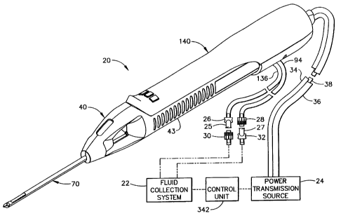
Figure 1 shows a first embodiment of a biopsy instrument comprising a
probe assembly 40, a holster 140, a fluid collection system 22, a control unit
342,
and a power transmission source 24. The probe assembly 40 is detachably
connected to the holster 140. Together they constitute a lightweight,
ergonomically shaped, hand manipulatable portion referred to as a handpiece
20.
The probe assembly 40 includes a piercer 70 extending distally from a hollow
handle 43. The probe assembly 40 is fluidly connected to the fluid collection
system 22 by a first vacuum tube 94 and a second vacuum tube 136. The first
and
second vacuum tubes are detachably connected to the fluid collection system 22
by
a first connector 27 and a second connector 25, respectively. The first
connector
has a male portion 32 and a female portion 28 attached to the first vacuum
tube
94. The second connector 25 has a female portion 30 and a male portion 26
attached to the second vacuum tube 136. The connector portions, 26, 28, 30,
and
32, are attached in this manner to prevent the accidental switching of the
first and
second tubes, 136 and 94, to the fluid collection system 22. The holster 140
includes a first rotatable shaft 34, a second rotatable shaft 36, and a
control cord
CA 02287087 1999-10-21
-12-
38. The first and second rotatable shafts, 34 and 36, are preferably flexible
so
that the operator may easily manipulate the handpiece 20 with one hand. The
control cord 38 operatively connects the handpiece 20 to the power
transmission
source 24 and control unit 342.
Since the handpiece 20 is manipulated by the operator's hand rather than by
an electro-mechanical ann, the operator may steer the tip of the handpiece 20
with
great freedom towards the tissue mass of interest. The surgeon has tactile
feedback while doing so and can thus ascertain, to a significant degree, the
density
and hardness of the tissue being encountered. In addition, the handpiece 20
may
be held approximately parallel to the chest wall of the patient for obtaining
tissue
portions closer to the chest wall then may be obtained when using a instrument
mounted to an electro-mechanical arm. As can be seen in Figure 1, the piercer
70
extends from the distal end of the handpiece 40 and is longitudinally offset
with
respect to the handpiece 40. This offset also facilitates the insertion of the
piercer
70 into the tissue while the axis of the piercer 70 is approximately parallel
to the
plane of the patient's chest wall. As a result, it is possible to extract
tissue
portions which are located close to the chest wall of the patient.
Those skilled in the art may appreciate that a mount or "nest" could be
provided to hold the handpiece 20 securely to the movable arm of an X-ray
stereotactic table or other kind of imaging device which incorporates a
movable
arm for holding a biopsy instrument. This would provide the operator with the
option to use the handpiece 20 to access the tissue mass within the surgical
patient
in much the same manner as was described earlier for using the MAMMOTOME
= instrument. This versatility may be advantageous to the operator, for
example, in
a situation where the handheld imaging device was temporarily not available
for
use, and it would be necessary to use the X-ray stereotactic table.
Figure 2 shows the holster 140 and the probe assembly 40 separated. A
pair of tabs 144 project laterally from each side of a holster upper shell
142, and
insert into right and left undercut ledges, 138 and 139 respectively, of the
hollow
handle 43 of the probe assembly 40. A plurality of indentations 66 are
provided
CA 02287087 1999-10-21
-13-
on the handle 43 to improve the operator's grip on the instrument. A tube slot
162
in the lower shell 156 of the holster 140 provides clearance for first and
second
vacuum tubes, 94 and 136. A first switch 146 , a second switch 148 , and a
third
switch 150 are mounted in the distal portion of the holster 140 so that the
physician can operate the handpiece 20 with a single hand while having the
other
hand free to operate an ultrasonic imaging device or the like. The switches
146,
148, and 150 are provided to operate the power transmission source 24 and the
fluid collection system 22 in conjunction with the control unit 342. A ridge -
152
on the distal end of the holster 140 is provided to assist the operator in
grasping
the handpiece 20 and in operating the switches 146, 148, and 150. The ridge
152
further provides the operator with a tactile reference as to where to properly
grasp
the handpiece 20.
Still in Figure 2, the probe assembly 40 includes a window 58 so that a
portion of the first vacuum tube 94 may be viewed. The first and second vacuum
tubes, 94 and 136, are made from a flexible, transparent or translucent
material,
such as silicone tubing. This enables visualization of the material flowing
through
the tubes. By having the window 58 in the probe assembly 40, the operator can
see the flow in the first vacuum tube 94 without needing to look away from the
tissue into which the piercer 70 is inserted. A transverse opening 68 is
provided in
the distal end of the hollow handle 43 which allows access from either side to
a
tissue sampling surface 64. The tissue extracted from the surgical patient is
retrieved by the operator or an assistant from the tissue sampling surface 64.
Figure 3 is an exploded isometric view of the probe assembly 40. The
handle 43 is formed from a right handle shell 42 and a left handle shell 44,
each
injection molded from a rigid, biocompatible plastic such as polycarbonate.
Upon
final assembly of the probe assembly 40, the left and right handle shells are
joined
together by ultrasonic welding along a joining edge 62, or joined by any of
several
other methods well known in the art. The probe assembly 40 comprises the
piercer 70 which includes an elongated, metallic piercer tube 74 having a
piercer
lumen 80. On the side of the distal end of the piercer tube is a port 78 for
receiving the tissue to be extracted from the surgical patient. Joined
alongside the
CA 02287087 1999-10-21
-14-
piercer tube 74 is an elongated, tubular, metallic vacuum chamber tube 76
having
a vacuum lumen 82. Piercer lumen-80 is in fluid communication with vacuum
lumen 82 via a plurality of vacuum holes 77 (see Figure 6B) located in the
bottom
of the "bowl" defined by the port 78. These holes are small enough to remove
the fluids but not large enough to allow excised tissue portions to be removed
through the first vacuum tube 94 which is fluidly connected to the vacuum
chamber 76. A sharpened, metallic distal end 72 is attached to the distal end
of
the piercer 70. It is designed to penetrate soft tissue such as the breast. In
this
,
embodiment, the sharpened distal end 72 is a three-sided, pyramidal-shaped
point,
although the tip configuration may also have other shapes.
Still referring to Figure 3, the proximal end of the piercer 70 is attached to
a union sleeve 90 having a longitudinal bore 84 through it, a widened center
portion 86, and a transverse opening 88 through the widened center portion 86.
The union sleeve 90 is mounted between the left and right handle shells, 44
and 42
respectively, on a pair of union sleeve ribs 50 projecting from each handle
shell.
An elongated, metallic, tubular cutter 96 is axially aligned within the
longitudinal
bore 84 of the union sleeve 90 and the piercer lumen 80 of the piercer 70 so
that
the cutter 96 may slide easily in both the distal and proximal directions. A
pair of
cutter guides 46 are integrally molded into each of the handle halves, 42 and
44,
to slidably retain the cutter 96 in an coaxially aligned position with the
proximal
end of the piercer tube 74. Cutter 96 has a cutter lumen 95 through the entire
length of the cutter 96. The distal end of the cutter 96 is sharpened to form
a
cutter blade 97 for cutting tissue held against the cutter blade 97 as the
cutter 96 is
rotated. The proximal end of the cutter 96 is attached to the inside of a
cutter gear
= bore 102 of a cutter gear 98. The cutter gear 98 may be metallic or
polymeric,
and has a plurality of cutter gear teeth 100, each tooth having a typical spur
gear
tooth configuration as is well known in the art.
Still in Figure 3, the cutter gear 98 is driven by an elongated drive gear
104 having a plurality of drive gear teeth 106 designed to mesh with the
cutter
gear teeth 100. The function of the drive gear 104 is to rotate the cutter
gear 98
and the cutter 96 as they translate in both longitudinal directions. The drive
gear
CA 02287087 1999-10-21
-15-
104 is preferably made from a metal such as stainless steel. A distal drive
axle
108 projects from the distal end of the drive gear 104 and mounts into an axle
support rib molded on the inside of the left handle shell 44. A gear shaft 110
projects from the proximal end of the drive gear 104 and is supported by a
gear
shaft support rib also molded on the inside of the left handle shell 44. A
left cross
pin 112 is attached to the proximal end of the gear shaft 110 as a means for
rotationally engaging the drive gear 104.
Still referring to Figure 3, a carriage 124 is provided to hold the cutter
gear 98 and to carry the cutter gear 98 as it is rotated in the distal and
proximal
directions. The carriage 124 is preferably molded from a rigid polymer and is
cylindrically shaped with a threaded bore 126 through it and with a carriage
foot
130 extending from its side. The foot 130 has a recess 128 formed into it for
rotatably holding the cutter gear 98 in the proper orientation for the cutter
gear
teeth 100 to mesh properly with the drive gear teeth 106. The carriage 124 is
attached via the threaded bore 126 to an elongated screw 114 which is parallel
to
the drive gear 104. The screw 114 has a plurality of conventional lead screw
threads 116 and is preferably made from a stainless steel. The rotation of the
screw 114 in one direction causes the carriage 124 to move distally, while the
reverse rotation of the screw 114 causes the carriage 124 to move proximally.
In
turn, the cutter gear 98 moves distally and proximally according to the
direction of
the screw rotation, and the cutter 96 is advanced or retracted. In this
embodiment,
the screw 114 is shown with a right hand thread so that clockwise rotation
(looking
from the proximal to distal direction) causes the carriage 124 to translate in
the
distal direction. It is also possible to use a left hand thread for the screw
114 as
long as provisions are made to do so in the control unit 342. A distal screw
axle
118 and a proximal screw shaft 120 project from the distal and proximal ends,
respectively, of the screw 114. The distal screw axle mounts rotatably in a
distal
screw support 48 of the right handle shell 42 while the proximal screw shaft
120
mounts rotatably in a proximal screw support 54, also in the right handle
shell 42.
A right cross pin 122 is attached to the proximal end of the screw shaft 120
as a
rotational engagement means.
CA 02287087 1999-10-21
-16-
Figure 3 also shows the first and second vacuum tubes, 94 and 136
respectively, referred to earlier. The-distal end of the first vacuum tube 94
is
attached to a polymeric vacuum fitting 92 which inserts tightly into the
transverse
opening 88 of the union sleeve 90. This allows the communication of fluids in
the
piercer lumen 80 to the fluid collection system 22. The first vacuum tube 94
is
contained within the hollow handle 43 in an open space above the screw 114 and
drive gear 104, and exits the distal end of the hollow handle through an
opening
57. The second vacuum tube 136 is fluidly attached to the proximal end of an
elongated, metallic, tubular tissue remover 132. The second vacuum tube 136
exits the hollow handle 43 alongside the first vacuum tube 94 out the opening
57.
A strainer 134 is attached to the distal end of the tissue remover 132 to
prevent the
passage of fragmented tissue portions through it and into the fluid collection
system 22. The tissue remover 132 inserts slideably into the tubular cutter
96.
During operation of the biopsy instrument, the tissue remover 132 is always
stationary and is mounted between a pair of proximal supports 52 on the inside
of
the right and left handle shells, 42 and 44 respectively. When the cutter 96
is
fully retracted to the first position, the distal end of the tissue remover
132 is
approximately even with the distal end of the cutter 96. The distal end of the
cutter 96 when at its first, fully retracted position, is slightly distal to a
vertical
wall 69 which is proximal and perpendicular to the tissue sampling surface 64.
In Figure 3, a right access hole 56 is shown in the proximal end of the
right handle shell 43. The right access hole 56 provides access to the
proximal
end of the screw 114 for operational engagement to the power transmission
source
24. Similarly, a left access hole is provided in the left handle shell 44 to
provide
access to the proximal end of the drive gear 104 for operational engagement
with
the power transmission source 24.
The tissue remover 132 has two functions. First, it helps to evacuate fluids
contained in the piercer lumen 80. This is accomplished by the attachment of
the
second vacuum tube 136 to the proximal end of the tissue remover 132. Since
the
distal end of the tissue remover 132 is inserted into the piercer lumen 80,
the
piercer lumen 80 is fluidly connected to the fluid collection system 22.
Second,
CA 02287087 1999-10-21
-17-
the tissue remover 132 removes tissue from the cutter 96 as follows. When a
tissue sample is taken, the cutter 96 advances to the fourth position just
distal to
the port 78, and a severed tissue portion 200 is captured within the cutter
lumen 95
in the distal end of the cutter 96. Then the cutter 96 translates to the first
position
so that the cutter blade 97 is just distal to the tissue sampling surface 64.
At this
position of the cutter 96, the distal end of the tissue remover 132 (which is
always
stationary) is approximately even with the distal end of the cutter 96.
Therefore,
any tissue portion of significant size contained within the cutter lumen 95 is
,
pushed out of the cutter lumen 95 and onto the tissue sampling surface 64, as
is
shown in Figure 9. The tissue portion 200 may then be retrieved by the
operator
or an assistant.
Now turning to Figure 4, an isometric view of the probe assembly 40 with
the left handle shell 44 removed reveals the placement of the components
described for Figure 3. Part of the first vacuum tube 94 has also been removed
for clarity. The carriage 124 is shown in the fully retracted position so that
the
cutter 96 is also at the fully retracted, or first position. The cutter blade
97 is
slightly distal to the vertical wall 69 on the handle 43. The foot 130 of the
carriage 124 is adapted to slide along a carriage guide surface 60 on the
inside
bottom of the hollow handle 43.
As shown in Figure 4, a cutter axial transmission 121 includes the carriage
124, the screw 114, and the screw shaft 120. A cutter rotational transmission
109
includes the drive gear 104, the cutter gear 98, and the gear shaft 110.
Figure 5 is an exploded isometric view of the holster 140 of the first
embodiment of the present invention. A holster upper shell 142 and a holster
lower shell 156 are each injection molded from a rigid, biocompatible plastic
such
as polycarbonate. Upon final assembly, the shells are joined together by
screws
(not shown) or other types of fasteners well known in the art, into a
plurality of
alignment holes 164. A gear drive shaft 180 and a screw drive shaft 182 are
contained within the proximal, enclosed portion of the holster 140. These
shafts
extend from a grommet 176 which has a groove 172 for retainably mounting onto
CA 02287087 1999-10-21
-18-
shell edge 170 of both holster upper and lower shells, 142 and 156,
respectively.
The gronunet 176 rotatably attaches the first rotatable shaft 34 to the screw
drive
shaft 182 and the second rotatable shaft 36 to the gear drive shaft 180. The
first
rotatable shaft 34 rotatably inserts into a left bore 172 of the grommet 176.
The
second rotatable shaft 36 rotatably inserts into a right bore 178. The grommet
176
also provides a strain-relieved attachment of the control cord 38 to the
holster 140.
Still referring to Figure 5, the gear drive shaft 180 is supported rotatably
upon a pair of gear drive mounts 160 formed into a first wall 166 and a second
wall 168 of the inside of the holster shells, 142 and 156. The screw drive
shaft
182 is likewise supported rotatably on screw drive mounts 158. A left coupler
184
is attached to the distal end of the drive gear shaft 180 and has a left
coupler
mouth 192 for rotational engagement with the left cross pin 112 attached to
the
gear shaft 110. When the probe assembly 40 shown in Figure 4 is attached to
the
holster 140, the gear shaft 110 becomes rotatably engaged to the gear drive
shaft
180. This may be seen more clearly in Figure 6A. Similarly, the screw drive
shaft 182 has a right coupler 186 with a mouth 194 which rotatably engages
with
the cross pin 122 of the screw shaft 120. Each of the left and right couplers,
184
and 186, have a coupler flange, 188 and 190, which rotatably insert into
thrust
slots 159 formed into the corresponding portions of the drive mounts 158 and
160.
These coupler flanges, 188 and 190, bear the axial loading of the drive
shafts, 180
and 182.
Still referring to Figure 5, the holster 140 further includes a screw rotation
sensor 198, available from Hewlett-Packard as part number HEDR-81002P, for
providing an electronic signal to the control unit 342 to be described in more
detail
later. In this first embodiment, the rotation sensor 198 is mounted within the
inside of the holster upper shell 142 and in a position directly above the
screw
drive shaft 182. A fluted wheel 199 is attached to the screw drive shaft 182
and
extends in front of a light emitting diode contained within the rotation
sensor 198.
As the fluted wheel 192 rotates, the interrupted light beams are
electronically
detected and transmitted back to the control unit 342 to provide information
about
the rotational speed of the screw drive shaft (cutter tube axial advancement
or
CA 02287087 1999-10-21
-19-
retraction speed), and the number of screw rotations from the beginning of
operation (instantaneous axial position.af the cutter 96). The rotation sensor
leads
196 pass through the grommet 176 and are part of the bundle of conductors
within
the control cord 38.
The holster 140 of the first embodiment of the present invention has the
switches, 146, 148, and 150, mounted on the inside of the holster upper shell
142.
The switches, 146, 148, and 150, are electronically connected to a plurality
of
conductors 193 contained in the control cord 38. In one embodiment, the third
switch 150 operates the fluid communication between the handpiece 20 and the
fluid collection system 22 and also sets the control unit 342 to respond to
various
commands; the second switch 148 operates the movement of the cutter 96 in the
proximal direction and sets the control unit 342 to respond to various
commands;
the firstswitch 146 operates the movement of the cutter 96 in the distal
direction
and sets the control unit 342 to respond to various commands. The functions of
the switches, 146, 148, and 150, are not restricted to what has been described
for
the first embodiment. Also, the physical locations of the switches, 146, 148,
and
150, on the handpiece 20 are not restricted to the locations depicted in
Figure 2.
Other embodiments of the handpiece 20 of the present invention may incorporate
certain ergonomic or other considerations, and the switches, 146, 148, and
150,
may be located elsewhere.
Figures 6A through 8A depict three of the four positions of the cutter 96
during the operation of the present invention as embodied in the prior Figures
1-5.
The three positions are most easily distinguished by observing the relative
= positions of the carriage 124 and the cutter blade 97 of the cutter 96.
In Figures 6A and 6B, the retracted, first position is depicted with the
carriage 124 located on the proximal ends of the drive gear 104 and the screw
114. The cutter blade 97 is shown to be immediately proximal to the tissue
sampling surface 64. In this first position, the tissue portion 200 may be
retrieved
from the tissue sampling surface 64 as depicted in Figure 9.
CA 02287087 1999-10-21
20 -
The second position of the cutter 96 is not shown in the Figures. At the
second cutter position, the distal end-of the cutter 96 is just distal to the
tissue
sampling surface 64 and inside the piercer lumen 80 near the proximal end of
the
piercer tube 74. During operation the cutter 96 is moved from the first
position to
the second position at a slower axial speed than from the second position to
the
third position in order to facilitate the insertion of the cutter 96 into the
proximal
end of the piercer lumen 80.
In Figures 7A and 7B, the cutter 96 is shown in the third position. The
carriage 124 is shown to have moved axially to the intermediate position which
is
a short distance from the distal ends of the screw 114 and the drive gear 104.
The
cutter blade 97 is shown by hidden lines to be located just proximal to the
port 78.
The vacuum holes 77 are open to the port 78 so that soft tissue adjacent to
the port
78 prolapses into the port 78 when the first vacuum tube 94 is fluidly
connected to
the vacuum of the fluid collection system 22. '
Figure 8A and 8B shows the cutter 96 at the fourth position, and the
carriage 124 is located near the distal ends of the screw 114 and the drive
gear
104. The cutter blade 97 is shown now (by hidden lines) to be distal to the
port
78 and to be covering the vacuum holes 77. The tissue pulled into the port 78
will
have been severed by the rotating, advancing cutter blade 97 and stored inside
the
cutter lumen 95 of the distal end of the cutter 96. When the cutter 96
retracts back
to the first position as shown in Figures 6A and 6B, the tissue portion 200
may be
retrieved as shown in Figure 9.
Figure 10 shows a second embodiment of the present invention. The main
difference from the first embodiment is that in the second embodiment a first
and a
second brushiess, electric motor, 234 and 236 respectively, are mounted inside
a
holster 221. First and second motors, 234 and 236, are available from Harowe
Servo Controllers, Inc., part number B0508-050. In this second embodiment, the
rotatable shafts 34 and 36 have been eliminated so that only a
control/electrical
power cord 232 is required to electrically connect the holster 221 to the
power
transmission source 24 and the control unit 342 (see Figure 1). A holster
lower
CA 02287087 1999-10-21
-21-
shell 222 has a first wall 242 and a second wall, 244, which are spaced apart
and
adapted to support the pair of electric motors, 234 and 236 in a side-by-side
arrangement. The use of the brushless electric motors , 234 and 236,
eliminates
the need for a separate rotation sensor to be mounted in the drive train of
one or
both of a screw 206 and a drive gear 204 as was described for the first
holster
embodiment shown in Figure 5. As in the first embodiment, when a probe
assembly 202 is attached to the holster 221, a right coupler 238 rotationally
engages a right cross pin 214 of a screw shaft 210. A left coupler 240
rotationally
engages a left cross pin 216 of a gear shaft 212. A grommet 230 having a
grommet groove 231 is retained by an attachment slot 233 in the holster
she11222.
Fastener holes 228 are provided to fasten the holster lower shell 222 to a
holster
upper shell using screws or other types of fasteners well known in the art.
Still referring to Figure 10, another difference of the second embodiment
compared to the first is that the probe assembly 202 comprises a lower shell
208
and an upper shell (removed for clarity) whereas the hollow handle 43 of the
first
embodiment shown in Figures 1-4 was divided vertically into left and right
shells,
44 and 42 respectively . This embodiment facilitates the addition of a probe
latch
220 and other features shown in Figure 11.
Using conventional techniques well known in the art, it is possible to use
only one electrically driven motor in place of the two motors described for
both
the first and second embodiments of the present invention. That is, a single
motor
may be used to both rotate and advance the cutter 96. The motor may be
incorporated into the instrument so that the cutter rotation and cutter
advancement
(axial movement) may occur either simultaneously or separately. The motor may
be located within the adapted handpiece 40 and be electrically connected to
the
power source 24 and the control unit 342. The motor may also be outside the
handpiece 40, still electrically connected to the power source 24 and the
control
unit 342, and mechanically engaged to the handpiece 40 by a single flexible
shaft.
Figure 11 shows an isometric view of the probe lower shell 208 and the
holster lower shell 222 of the biopsy instrument 201 of the second embodiment
of
CA 02287087 1999-10-21
-22-
the present invention. The view is shown with the bottom side up in order to
clearly present a probe latch 220 whish is molded as a cantilever into the
probe
lower shell 208, and can be deflected downwards by a force applied to a latch
ramp surface 223. The latch 220 further comprises a latch projection 219 for
insertion into a holster slot 224 as the probe assembly is inserted into the
holster
221. The ramp surface 220 is deflected downwards by interaction with an inside
surface 225 of the holster shell 222 and retainably snaps into a slot key 226
when
the probe assembly is fully inserted into the holster, thus rotationally
engaging the
left and right couplers, 240 and 238, to the drive shaft 212 and the gear
shaft 210,
respectively, as shown in Figure 10. To remove the probe assembly from the
holster, one must press on the projection 219 while pulling them apart. Figure
12
shows a longitudinal section through the center axis of the probe lower shell
208
and the holster lower shell 222 of Figure 11 for when they are fully attached
together.
Figure 13 is an exploded isometric view of a holster 251 of a third
embodiment of the present invention. It may be used with the probe assembly 40
of the first embodiment shown in Figures 1-4. A first and a second rotatable
shafts, 264 and 266, are attached by a grommet 262 to a drive shaft 258 and a
screw shaft 260, respectively. Rotatable shafts, 264 and 266, are preferably
flexible too, in order for the holster 251 combined with the probe assembly 40
(see
Figure 2) to be easily manipulatable with one hand. A fully integral rotation
sensor 268 is shown mounted on a screw shaft 260. This rotation sensor 268 is
a
miniature optical encoder which is commercially available as Model Number
SEH17 from CUI Stack, Inc. It is electrically connected to a switch board 274
which mounts to the inside of the holster upper shell 252. The switch board
274
also has a ribbon cable 270 containing a plurality of conductors for conveying
electronic information to and from the control unit 342, power transmission
source
24, and the fluid collection system 22, via a control cable 265. The switch
board
274 has mounted on its distal end, three switches, 276, 278, and 280, for
operation of the present invention in the same manner as described in the
first
embodiment: a third switch 280 for fluidic connection to the vacuum of the
fluid
collection system; a first switch 246 for the forward movement of the cutter
96;
CA 02287087 1999-10-21
- 23 -
and a second switch 248 for the reverse movement of the cutter 96. The
specific
functions of the switches, 276, 278, an,d 280, are not restricted, in other
possible
embodiments of the present invention, to the functions described, nor to the
physical locations shown. The switches, 276, 278, and 280, project through
switch openings 254 of the holster upper shell 252. A holster lower shell 256
attaches to the upper shell 252 as in the other embodiments to enclose the
components of the proximal portion of the holster 251.
Those skilled in the art could easily appreciate that the switch board 274
and the three switches, 276, 278, and 280, may instead be incorporated into a
foot
operable device rather than in the hand operable holster 251 shown in Figure
13.
The operator would still be able to manipulate the instrument with a single
hand
while actuating the switches, 276, 278, and 280, by foot, thus freeing the
other
hand for holding the ultrasound imaging device, or for performing other steps
in
the surgical procedure.
Figure 14 shows the relationship of the electro-mechanical components of
the present invention to the control unit 342. The third embodiment of the
present
invention is depicted and includes the holster 251 of Figure 13. A first
motor/tachometer combination 338 (sometimes referred to as a first motor/tach)
and a second motor/tachometer combination 340 (sometimes referred to as a
second motor/tach) are depicted as part of the power transmission source 24,
and
transmit rotational power to the holster 251 via the first and second
rotatable
shafts, 264 and 266, respectively. The motor/tach combinations, 340 and 348,
are conunercially available as DC MicroMotors Series 3863, MicroMo
= Electronics, Inc. The control cord 265 is electrically connected to a serial
controller 380 available as Part No. MCF5206eFT40 from Motorola, Inc. A serial
controller 380 is electronically connected to the switchboard 274 by ribbon
cable
270 and control cord 265. The serial controller 380 coordinates information
exchange across the serial communication link between the switchboard 274 and
the microprocessor 408. An advantage provided by the use of the serial
controller
380 is that the required number of conductors 193 may be reduced.
CA 02287087 1999-10-21
-24-
Figure 14 depicts the interconnection of the electro-mechanical components
of the fluid collection system 22 and_power transmission source 24 with
control
unit 342. The first vacuum tube 94 coming from the probe assembly 40 (see
Figure 2) is attached to a first vacuum Y- connector 302 fluidly connected a
first
upper line 306 and a first lower line 308. The two lines, 306 and 308, pass
through a first pinch valve 314. A suitable, commercially available, three-way
pinch valve for this application is Model Number 373 12 -7 15 available from
Angar Scientific Company, Inc. The pinch valve 314 closes either the upper
line
306 or the lower line 308, but never both lines simultaneously. The lower line
308 provides a vent to atmospheric pressure. The upper line 306 attaches to a
fluid collection canister 318. Similarly, the second vacuum line 136 from the
probe assembly 40 attaches to a second Y- connector 304 which fluidly is
connected to a second upper line 310 and a second lower line 312. The first
and
second vacuum Y-connectors, 302 and 304, are molded from a rigid polymer such
as polycarbonate The second upper line 310 passes through a second pinch valve
316, which is identical to the first, and to the canister 318. The second
lower line
312 passes through the second pinch valve 316 and vents to atmosphere. Again,
only one or the other of the two lines may be pinched closed at any time.
Still referring to the fluid collection system of Figure 14, a main vacuum
line 320 attaches the canister 318 to an electrically powered vacuum pump 330.
A
suitable vacuum pump for this application is available by the trademark name
WOB-L PISTON Series 2639, from Thomas Compressors and Vacuum Pumps .
The main vacuum line 320 passes through a regulator valve 322 to
electronically
adjust the vacuum pressure supplied to the canister 318. A conunercially
available
regulator valve for this application is model number VSONC 6 S 11 V H Q 8
from Parker Hannifin Corp., Pneutronics Division. A pressure sensor 328 is
fluidly attached to the main vacuum line 320 at a sensor connection 324. The
signal from the pressure sensor 328 is sent to an A/D converter 396 of the
control
unit 342. A commercially available, compensated pressure sensor for this
application is model number SDX15 from SenSym, Inc.
CA 02287087 1999-10-21
-25-
At the heart of the control unit 342 is a 40 MHz, 32 bit microprocessor
408, available from Motorola, Inc. _as Part No. MCF5206EFT40, which is
designed to perform logic operations that eventually translate into simple
electromechanical actions.
Still referring to Figure 14, the control unit 342 includes a 640x480 color
TFT- LCD display 334 available from Sharp as part number LQ64D343. Display
334 is covered by a resistive touchscreen 336 for the user interface. The
touch
screen 336 is available from Dynapro as part number 95638, and is
electronically
connected to a touch screen controller 402 in the control unit 342: The
touchscreen controller 402 interfaces with the microprocessor 408 and
comprises
the following: a microcontroller, part number PIC16C58A, available form
Microchip; an EEPROM, part number 93AA466SN, available from Microchip; an
A-D converter, part number TLV1543CDW, available from Texas Instruments;
and a multiplexer-demultiplexer, part number MC74HC4052D, available from
Motorola. The touch screen controller allows the control unit 342 to respond
to
the user's touch by interpreting touch inputs. Similarly, an LCD controller
404 is
an interface between the microprocessor 408 and the LCD display 334. The LCD
controller 404 reduces the burden of the microprocessor 408 by efficiently
controlling display parameters such as color, shading, screen update rates,
and it
typically accesses the memory chips of the microprocessor 408 directly. The
LCD
controller 404 comprises the following: a LCD controller, part number
SED1354FOA, available from Epson; a display buffer DRAM, part number
MT4LC1M16E5TG-6, available from Micron; and a line driver, part number
74ACTQ16244SSCX, available from National.
A miniature annunciator 332 is provided with the control unit 342 in order
to provide the user with audible, feedback "beeps" upon each activation of an
icon
control on the LCD display 334. A suitable annunciator for this application is
model number EAS-45P104S from Panasonic (Matshusita Electric Corp. of
America). The annunciator 332 interfaces with the microprocessor 408 by an
oscillator 400 which converts the digital input signal from the microprocessor
408
to an analog, periodic output signal, thus controlling the audio frequency of
the
CA 02287087 1999-10-21
-26-
speaker. The volume of the sound coming from the annunciator 332 is controlled
by a programmable attenuator. The oscillator 400 comprises the following: a 8
MHz oscillator, part number ASL-8.0000000-PCSA, available from AMD; and a
PLD, part number EPM7256ATC144-7, from Altera.
Still referring to the schematic diagram of Figure 14, a first motor
controller and driver 390 interfaces the second electric motor/tach 340 with
the
microprocessor 408. The first motor controller and driver 390 comprises the
following: an H-bridge, part number LMD18200T, available from National; a
motion controller, part number LM629M-8, available from National; and a PLD,
part number EPM7256ATC144-7, available from Altera. The second motor/tach
340 is operationally connected to the second flexible shaft 266 for the
actuation of
the cutter axial transmission 121 (see Figure 4). The controller and driver
390
converts digital input signals from the microprocessor 408 into analog motor
input
signals for controlling motor rotational direction and speed. A closed loop
digital
speed control of the motor is also achieved within the controller and driver
390
using feedback signals from the rotation sensor 268 available from CUI Stack,
Inc., as part number SEH17 (see Figure 13). The first electric motor/tach 338
drives the cutter rotational transmission 109 (see Figure 4) via the first
rotatable
shaft 264. The first electric motor/tach 338 interfaces with the
microprocessor
through the second controller and driver 406.
An optional card reader 382 may be provided in the control unit 342 for
reading data from memory card in order to facilitate future software upgrades
and
servicing.
A serial port 384 is provided for the bi-directional data exchange in a serial
transmission mode, again to facilitate future software upgrades and servicing.
The
serial port 384 comprises the following: a UART, part number ST16C2552CJ44,
available from EXAR; and a line driver-receiver, part number DS14C335MSA,
available from National.
CA 02287087 1999-10-21
-27-
A first PWM (pulse width modulation) driver 386 interfaces the first pinch
valve 314 with the microprocessor 408. The first PWM driver 386 converts a
digital input signal from the microprocessor 408 to an analog output signal
having
a wave of fixed frequency and amplitude, but varying duty cycle. To drive the
solenoid in the pinch valve 314, the PWM driver 386 is used when the duty
cycle
is high to initially move the solenoid. Once the pinch valve 314 is actuated,
the
duty cycle is reduced to a level which maintains valve position, thus
minimizing
power requirements. A second PWM driver 388 similarly interfaces a second
pinch valve 316 with the microprocessor 408. A third PWM driver 394 interfaces
with the regulator valve 322. The PWM drivers, 394, 388, and 386 each
comprise the following: a PLD, part number EPM7256ATC144-7, available from
Altera; and a FET transistor, part number NDS9945, available from Fairchild.
A RAM memory device 392 available from Micron as DRAM part number
MT4LC1M16E5TG-6, is provided with the microprocessor 408, and inherently
loses stored data when power is removed. A flash memory device 398, on the
other hand, is provided with the microprocessor 408 to store data even without
continuous power, but it has slower access time than the RAM device 392. The
flash memory device 398 is part number Am29LV800BT-70REC from AMD.
An A/D converter 396 converts voltage signals from the pressure sensor
328 into . digital signals to the microprocessor 408, for maintaining the
desired
vacuum pressure in the fluid collection system 22. The A/D converter 396 is
part
number PCF8591AT, available from Philips.
Still referring to Figure 14, the first (axial) controller and driver 390 and
the second (rotational) controller and driver 406 continually calculate and
update
the axial and rotational position of the cutter 96 within the handpiece 20.
They
also calculate the speed and acceleration of the cutter 96 axial and
rotational
movement from the positional information. The microprocessor 408 monitors
both the axial position and speed of the cutter 96 and the rotational position
and
speed via the first controller and driver 390 and the second controller and
driver
406.
CA 02287087 1999-10-21
-28-
While in the sampling mode and with the cutter 96 advancing toward the
third position (proximal to port 78), Auhen the cutter 96 reaches a
predetermined
axial position, the microprocessor 408 sends a signal to the second controller
and
driver 406 to initiate cutter rotation. The rotational speed of the cutter 96
follows a
predefined speed profile which insures that the cutter rotational speed is at
Z
revolutions per minute (rpm) when the cutter 96 reaches the third position.
When
the cutter 96 reaches the third position, the microprocessor 408 sends a
signal to
,the first controller and driver 390 to advance the cutter 96 at speed Y. The
cutter
96 then progresses through the port 78 at advancement speed Y while rotating
at
velocity Z. While advancing through the port 78, the cutter rotational speed
is
monitored by the second controller and driver 406. If the rotational speed is
greater than Z rpm, electrical current to the first (cutter rotation)
motor/tach 338 is
decreased. If the cutter rotational speed is less than Z rpm, electrical
current to the
first motor/tach 338 is increased. One method of performing the speed control
on
both the first and second motor/tach's, 338 and 340, is to generate an error
signal
based on the difference between the desired speed and the actual speed. The
error
signal is then input into a proportional, differential, and derivative (PID)
digital
filter which is part of the respective controller and driver, either 390 or
406. The
sum of these three terms is used to generate the pulse width modulation (PWM)
signal. The generation of the error signal and the PWM signal is accomplished
by
the first and second controllers and drivers, 390 and 406. A PWM signal is
input
to the first controller and driver 390 to generate an analog output signal to
drive
the first motor/tach 338. Similarly, a PWM signal is input to the second
controller and driver 406 to generate an analog output signal to drive the
second
motor/tach 340.
The microprocessor 408 also monitors the output value of the second
controller and driver 406 PID filter such that if it exceeds a predefined
maximum
value, it will reduce the axial speed of the cutter 96 a set amount by sending
an
updated speed command to the first controller and driver 390. This closed-loop
algorithm is intended to insure that the target rotational speed is attained
by
decreasing the axial speed of the cutter 96 under maximum loading conditions.
The control logic then repeats from the beginning.
CA 02287087 1999-10-21
-29-
Figure 15 is an enlarged view of the LCD display 334 and the touch screen
336, shown as part of the control unit-342 of Figure 14. In one embodiment of
the present invention, twelve separate operating modes are available to a
user. A
control switch for each operating mode is displayed graphically on LCD display
334 in the form of icons, 346, 348, 350, 352, 354, 356, 358, 360, 362, 364,
366,
and 368. The user may initiate a particular operation by pressing the touch
screen
in the region of the appropriate icon using at the appropriate time during the
surgical procedure to electronically control the operation of the biopsy
device.
The present invention is not restricted to use with the particular combination
of
modes of operation shown in Figure 15.
For the following description of the modes of operation, it will be assumed
for discussion purposes that the first embodiment of the present invention is
being
described, and that the first switch 146 primarily controls the forward
(distal
direction) axial movement of the cutter 96, the second switch 148 primarily
controls the reverse (proximal direction) axial movement of the cutter 96, and
that
the third switch 150 primarily controls the fluidic connection of the
handpiece 20
to the fluid collection system 22. The switches, 146, 148, and 150, also have
secondary functions such as setting the control unit 342 for particular steps
during
the operation of the instrument, and these secondary functions are described
later.
The modes of operation are also applicable to the second embodiment of the
present invention which includes first switch 276, second switch 278, and
third
switch 280.
Each mode of operation is utilized for a particular portion of the general
= biopsy procedure. The "Prime" mode of operation is selected when the
operator
is preparing the instrument for use. When an operator activates the "Prime"
mode
of operation by, for example, touching the LCD display 344 in the region of
icon
346, the display 334 indicates the status as being "Prime Mode". The cutter 96
then translates to the third position just proximal to the port 78. Once the
cutter is
in the third position, the display instructs the operator to apply saline to
the port
78 and to depress the vacuum switch 150 as needed to draw saline into piercer
70
and through the probe assembly 40. The operator may observe the flow of saline
CA 02287087 1999-10-21
-30-
through the window 58. Finally, the first pinch valve 314 and second pinch
valve
316 are both set to respond to the vacuum switch 150.
The "Insert" mode of operation is next selected when the operator is
preparing the instrument for insertion into the tissue of the surgical
patient. When
an operator activates the "Insert" mode of operation by, for example, touching
the
LCD display 344 in the region of Icon 348, the display 344 indicates the
status as
being "Insert Mode". The cutter 96 then translates to the fourth position,
just
distal to the port 78. Once the cutter 96 translates to the fourth position,
the
display indicates that the instrument is ready to insert.
The "Verify" mode of operation is selected when the operator wants to
verify that the position of the port 78 is adjacent to the tissue to be
extracted. In
order to more easily visualize the port 78 of the inserted piercer 70 on the
imaging
device, it has been found that the cutter 96 should be retracted to a position
proximal to the port 78, that is, the port 78 should be "open." If the port 78
is
not adjacent to the tissue to be extracted, then the operator should "close"
the port
78 by moving the cutter 96 to the fourth position, so that the piercer 70 may
be
hand-manipulated towards the tissue to be extracted. Then the port 78 should
be
opened again to verify that the port 78 is adjacent to the tissue to be
extracted.
These steps are repeated until the port 78 is adjacent the tissue to be
extracted.
When an operator activates the "Verify" mode of operation by, for example,
touching the LCD display 344 in the region of Icon 350, the display 344
indicates
the status as being "Verify Mode". If the cutter 96 is not at the fourth
position
(the port 78 is "open"), the second motor 340 is set to respond to the
handpiece
first (forward) switch 146. Then the display 344 instructs the operator to
close the
port 78 by pressing the first (forward) switch 146 on the handpiece 20. When
the
operator presses the first (forward) switch 146, the cutter 96 translates to
the
fourth position. The second motor 340 is then set to respond to the handpiece
second (reverse) switch 148. If the cutter 96 is already at the fourth
position when
the "Verify" mode is selected, then the second motor 340 is set to respond to
the
second (reverse) switch 148. Then the display 344 instructs the operator to
open
the port 78 by pressing the second (reverse) switch 148 on the handpiece. When
CA 02287087 1999-10-21
-31-
the operator presses the second (reverse) switch 148, the cutter 96 translates
to the
third position just proximal to the port 78. Then the second motor 340 is set
to
respond to the first (forward) switch 146.
The "Sample" mode of operation is selected when the operator desires to
extract a portion of tissue from the surgical patient. When the operator
activates
the "Sample" mode of operation by, for example, touching the LCD display 344
in the region of icon 352, the display 344 indicates the status as being
"Sample
Mode". The cutter 96 then translates to the third position which is just
proximal
to the port 78. Then the second motor 340 is set to respond to the first
(forward)
switch 146. Once the cutter 96 is in the third position, the display 344
instructs
the operator to take a tissue sample by pressing the first (forward) switch
146 on
the handpiece. When the first (forward) switch 146 is pressed, the first pinch
valve 314 and second pinch valve 316 are opened, and the first motor 338 is
activated to rotate the cutter 96 at the appropriate speed. Then the cutter 96
translates to the fourth position, severing the tissue portion prolapsed into
the port
78 as the cutter 96 moves distally. Once the cutter 96 reaches the fourth
position,
the first motor 338 is deactivated and the cutter 96 stops rotating. Then the
first
pinch valve 314 is activated to close. Next the display 344 instructs an
operator to
retrieve a tissue sample by pressing the second (reverse) switch 148 on the
handpiece 20. The second motor is set to respond to the second (reverse)
switch
148 on the handpiece 20. When the operator presses the second (reverse) switch
148, the cutter 96 translates to the first, fully retracted position, just
distal to the
sampling surface 64. Then the second pinch valve 316 is activated to close the
vacuum for the tissue remover 132. A "smart-vacuum" is also activated and a
plurality of vacuum pulses (0.5 seconds on and 0.5 seconds off) are supplied
to the
second vacuum tube 136. A detailed description of the "smart vacuum" is
provided in U.S. Patent application S/N 08/878468 filed by the same assignee
as
for the present application and which is incorporated herein for reference.
The
display 344 instructs the operator to remove the tissue sample. If there was
no
sample extracted, that is, the severed tissue portion remained at the distal
end of
the piercer 70 rather than be deposited onto the tissue sample surface 64, the
operator is instructed to select "Dry Tap". The operator is also instructed to
select
CA 02287087 2006-03-13
-32-
"Remove Air/Blood" if required to remove excessive fluids in the patient and
probe assemble 40. The operator is -finally instructed to press the first
(forward)
switch 146 on the handpiece 20 to extract the next sample. Next, the second
motor
340 is set to respond to the first (forward) switch 146 on the handpiece 20.
When
the first (forward) switch 146 is pressed by the operator, the "smart-vacuum"
is
stopped and the first and second pinch valves, 314 and 316, are activated to
open,
and the cutter 96 translates in the distal direction. As the cutter 96
approaches the
third position just proximal to the port 78, the first motor 338 is activated
to
rotate the cutter 96 which then translates to the fourth, fully distal
position. Then
the cutter 96 rotation is stopped and the first pinch valve 314 is closed to
stop the
vacuum to the vacuum pressure chamber tube 76 supplied by the first vacuum
tube
94.
The "Mark" mode of operation is selected when the operator desires to
implant a metallic marker within the surgical patient at the location from
which the
tissue was extracted. When the operator activates the "Mark" mode of operation
by, for example, touching the display 344 in the region of icon 354, the
display
344 indicates the status as being "Marker Mode" and also prompts the operator
to
select "Dry Tap" if required. Then the operator is instructed to press the
third
(vacuum) switch 150 on the handpiece 20 to activate the "Mark" mode. A
marking instrument which may be used in combination with the present invention
for marking tissue is commercially available under the tradename MICROMARK
from Ethicon Endo-Surgery, Inc., Cincinnati, Ohio. A complete description of
the MICROMARK applier and clip, and the method of its use, is included in U.S.
Patent Nos. 5,941,890 and 6,261,302. When the operator presses the third
(vacuum) switch 150, the cutter 96 translates to the first position just
proximal to
the tissue sampling surface 64. The display 344 then instructs the operator to
insert the MICROMARK instrument, to press the third (vacuum) switch 150 on
handpiece when ready to deploy, and to deploy the marker. Then when the third
(vacuum) switch 150 is pressed, the first pinch valve 314 is activated to the
open
position for five seconds to supply vacuum to the port 78 through the vacuum
chamber 76. Next the display 344 instructs the operator to
CA 02287087 1999-10-21
-33-
reposition the MICROMARK instrument if marker deployment was not complete,
to press the third (vacuum) switch 150-.on the handpiece when ready to deploy
the
marker, to deploy the marker, and if the marker deployment is complete, to
remove the MICROMARK instrument.
The "Remove" mode of operation is selected when the operator is ready to
remove the piercer 70 from within the tissue of the surgical patient. When the
operator activates the "Remove" mode of operation by, for example, touching
the
display 344 in the region of icon 356, the display 344 indicates the status as
being
"Remove Mode". The cutter 96 translates to the fourth, fully distal position
and
closes the port 78. The display 344 instructs the operator that the instrument
is
ready to remove.
The "Remove Air/Blood" mode of operation is selected when the operator
desires to remove any fluids present near the distal end of the piercer 78 and
within the probe assembly 40. When the operator activates the "Remove
Air/Blood" mode of operation by, for example, pressing the display 344 in the
region of icon 360, the display 344 indicates the status as being "Remove
Air/Blood Mode". The cutter 96 then translates to the third position just
proximal
to the port 78. The first pinch valve 314 and the second pinch valve 316 are
both
set to respond to the third (vacuum) switch 150 on the handpiece 20. The
display
then instructs the operator to remove the air/blood by pressing the third
(vacuum)
switch 150 on the handpiece 20. When the third (vacuum) switch 150 is pressed,
the first pinch valve 314 and the second pinch valve 316 are activated to open
for
five seconds. When they are closed, the cutter 96 then translates to the
first, fully
= retracted position just proximal to the tissue sampling surface 64. Then the
"Remove Air/Blood" mode is automatically exited and the previous mode selected
is automatically reset.
The "Dry Tap" mode of operation is selected when the operator had
attempted to extract a tissue portion from the surgical patient using the
"Sample"
mode of operation, but a tissue portion was not deposited onto the tissue
sample
surface 64. This may occur when the tissue portion is properly severed from
the
CA 02287087 1999-10-21
34
surgical patient, but remained in the distal end of the piercer 78. When the
operator activates the "Dry Tap" mode_of operation by, for example, touching
the
display 344 in the region of icon 358, the display 344 indicates the status as
being
"Dry Tap Mode". The cutter 96 then translates to the third position just
proximal
to the port 78. Then the second pinch valve 316 is activated to open for 0.5
seconds and to close for 0.5 seconds three times in order to pulse the vacuum
supplied to the tissue remover 132 through the second vacuum tube 136. The
cutter 96 then translates to the first, fully retracted position just proximal
to the
tissue sampling surface 64. The "Dry Tap" mode of operation is then exited and
the previously selected mode of operation is automatically selected.
The "Flush" mode of operation is selected when the operator desires to
clear any obstructions (tissue fragments, etc.) on the distal end of the
tissue
remover 132 to enable the passage of fluids through it. When an operator
activates the "Flush" mode of operation by, for example, touching the display
344
in the region of icon 362, the display 344 indicates the status as being
"Flush
Mode". The cutter 96 then translates to the first, fully retracted position,
thus
exposing the distal end of the tissue remover 132. Then the control unit 342
is set
to respond to the vacuum switch 150, which when pressed by the operator,
causes
the "Flush" mode of operation to be exited and the previously selected mode of
operation to be automatically reset. Before pressing the vacuum switch 150,
however, the operator may temporarily disconnect the second connector 304,
inject fluid such as saline into the second vacuum tube 136 using a syringe,
and
reconnect the second connector 304.
= The "Inject" mode of operation is selected when the operator desires to
inject a fluid, such as a local anesthetic, into the tissue surrounding the
distal end
of the piercer 78. When the operator activates the "Inject" mode of operation
by,
for example, touching the display 344 in the region of icon 364, the display
344
indicates the status as being "Inject Mode". The cutter 96 then translates to
the
third position just proximal to the port 78. Then the control unit 342 is set
to
respond to the third (vacuum) switch 150 on the handpiece 20. Next the display
instructs the operator to inject the fluid into the second vacuum tube 136,
and to
CA 02287087 1999-10-21
= -35-
press the third (vacuum) switch 150 again once the injection is complete. When
the operator has completed the injection into the second vacuum tube 136,
reconnected it to the fluid collection system 22, and pressed the third
(vacuum)
switch 150, the cutter 96 translates to the first, fully retracted position.
At that
point, the "Inject" mode of operation is exited, and the previously selected
mode
of operation is automatically reset.
Each time one of the available operating modes is selected, a display area
344 provides written and graphic information to prompt the user as to the
correct
usage of the instrument and the next operational steps. A mode indicatoi=
display
370 includes a representation of the probe assembly showing the instantaneous
position of the cutter tube, referred to as a cutter position indicator 373,
activation
of the front vacuum indicator 372 (corresponding with the first vacuum tube
94),
and activation of the rear vacuum indicator 371 (corresponding with the second
. vacuum tube 136).
The present invention, as described, is transportable from room to room of
a physician's office, primarily because the handpiece need not be mounted to
an
X-ray stereotactic table. The remaining portions of the instrument, including
the
fluid collection system, the power transmission source, and the control unit,
may
be packaged into a portable, wheeled unit. In one scenario, the physician
would
have a number of patients, each in a separate room, being prepared for
treatment
while the surgical procedure is being performed on another patient. The biopsy
instrument could then be moved to the patient, rather than vice versa, thus
helping
the patient to feel relaxed and prepared for the procedure. A different,
sterile
= probe assembly would be provided for each patient, while the holster portion
of
the handpiece would be reused.
While preferred embodiments of the present invention have been shown
and described herein, it will be obvious to those skilled in the art that such
embodiments are provided by way of example only. Numerous variations,
changes, and substitutions will now occur to those skilled in the art without
_ ---- ----~----
CA 02287087 1999-10-21
-36-
departing from the invention. Accordingly, it is intended that the invention
be
limited only by the spirit and scope of-the appended claims.