Note: Descriptions are shown in the official language in which they were submitted.
CA 02289213 1999-11-03
WO 98/50102 PCT/US98/08876
Specification
-Be it known that Jay Lenker, Ph.D. and K. David Crockett, -
= Esq. have invented a new and useful
Wire Frame Partial Flow Obstruction
= 5 Device for Aneurysm Treatment
of which the following is a specification:
Field of the invention
This invention relates to devices and methods for treatment
of aneurysms.
Backcaround of the Invention
Aneurysms are a common defect in the vascular system that
account for a wide range of symptoms. When they occur in the
brain, aneurysms can cause stroke or death, as well as several
well-known neurological defects such as loss of sight, hearing
or balance. The treatment of aneurysms within the brain may be
accomplished with a number of therapies currently available.
Open surgical techniques require cutting into the skull and
lifting brain matter away from the aneurysm so that the aneurysm
may be accessed, clipped or sutured closed and cut away. These
techniques are very risky, and are reserved until absolutely
necessary because of high mortality and high chance of
neurological defects caused by the operation itself.
The high risk and generally unsatisfactory results of open
surgery on aneurysms (especially within the brain) have led
researchers to develop techniques for treating aneurysms from
inside the blood vessels. Endovascular and percutaneous
insertion of devices avoid the danger of open surgery on the
brain, but presents technical challenges. Grafts, stents and
combinations of stents and grafts have been proposed for use in
larger vessels such as the aorta and the peripheral arteries.
The purpose of these devices is to close off the aneurysm from
1
CA 02289213 2006-10-26
the circulatory system to prevent rupture and promote resorption
of the mass of the aneurysm. These devices tend to be bulky and
generally unsuitable for the small environment of the brain.
our co-pending U.S. Patent No. 6,007,573, filed September
18, 1996 and U.S. Patent No. 6,254,628, filed December 9,
1996 present several stent designs and stent delivery systems
particularly suited to used within the brain.
Another approach to treating aneurysms, suitable for
treatment within the brain, is stuffing the aneurysm with
foreign material to promote thrombus within the aneurysm and
eventually eliminate the threat of ruptures and promote
resorption of the aneurysm sac. As early as 1975, metal coils
were successfully used to occlude the renal arteries.
Gianturco, et al., Mechanical Devices for Arterial Occlusions,
124 Am. J. Roent. 428 (1975). The purpose of the coil is to
encourage quick formation of a thrombus (a blood clot) around
the coil. The coils are currently in use for a wide range of
treatments, and are referred to variously as occlusive coils,
embolization coils, or Gianturco coils. Embolization coils of
appropriate size for placement within aneurysms are commercially
available from Target Therapeutics, Inc. and Cook, Inc.
Embolization coils made with electrolytic mechanisms for
detachment from the delivery catheter are referred to as GDC's
or Guglielmi Detachable Coils. The use of GDC's is illustrated,
for example, in Klein, et al., Extracranial Aneurysms and
Arteriovenous Fistula: Embolization with the Guglielmi
Detachable Coil, 201 Radiology 489 (1996). Use of the GDC coils
within the brain is illustrated, for example, in Casasco, et
al., Selective Endovascular Treatment Of 71 Intracranial
Aneurysms With Platinum Coils, 79 J. Neurosurgery 3(1993).
- Because Gianturco and Guglielmi coils are often used to
occlude aneurysms in critical areas of the body, it is important
that they remain in place where they are implanted. However, '
migration of the coils after placement is a common but dangerous
problem encountered with these coils. Watanabe, Retrieval Of A
Migrated Detachable Coil, 35 Neuro. Med. Clin. 247 (1995)
2
CA 02289213 2006-10-26
reports the migratiori of a coil into the basilar artery from a
placement in the superior cerebellar artery. Halbach, et al.,
Transarterial Platinum Coil Embolization Of Carotid Cavernous
Fistulas, 12 AJNR 429 (1991) reports the migration of a coil
from the internal carotid artery. Migration is particularly
common with coils placed in wide neck aneurysms. The possible
migration of coils is a danger that must be considered in every
procedure, and actual migration can be life threatening
complication, since embolization at an unwanted site could
occlude a critical blood flow. Migration of the coil may also
represent a failure of the intended therapeutic procedure.
Our U.S. Patent App. No. 5,830,229, filed March
7, 1997, discloses a hoop stent for holding open blood vessels
subject to occlusive disease. The stent, which is made of a
single wire, and the delivery mechanism for the stent allow the
stent to placed with a low profile by stretching the stent along
its long axis rather than compressing it radially. A wide
variety of other stent designs have been proposed for use in the
vascular system. Typically, the stents are used to hold open a
length of blood vessel which has been closed or occluded by some
growth within the blood vessel. Balloon expandable stents and
self-expanding stents are commercially available and have been
used successfully for treatment of a number vascular diseases.
Das, Stent, U.S. patent 5,554,181 (Sep. 10, 1996) shows a wire
stent having a number of hoops all attached to a radially
disposed spine, all of which may be formed of a single wire.
Likewise, Hillstead, Endovascular Stent Apparatus and Method,
U.S. Patent 4,856,516 (Aug. 15, 1989). The stents are folded
upon a catheter pusher and retained within a catheter sheath
before release into the body. These.stents must be radially
compressed to fit within the catheter sheath, and expand
elastically or may be expanded inelastically by a balloon. They
are not susceptible to being stretched or elongated in along
their long axes to reduce their overall diameter.
3
CA 02289213 2006-10-26
78643-7
Summary
In one aspect of the prevent invention, there is
provided a device for obstructing blood flow in an aneurysm
affecting a blood vessel having a lumen with an inner wall,
said device comprising: a first hoop circumscribing the
inner wall of the blood vessel on one side of the aneurysm;
a second hoop circumscribing the inner wall of the blood
vessel on the other side of the aneurysm; a first strut
comprising a substantially straight length of wire extending
along the length of the blood vessel from the first hoop
toward the second hoop; a second strut comprising a
substantially straight length of wire extending along the
length of the blood vessel from the second hoop toward the
first hoop; and a flow occluding structure supported by the
first and second struts and aligned with the aneurysm so as
to at least partially disrupt the flow of blood into the
aneurysm.
In another aspect of the present invention, there
is provided a system for inserting a device for obstructing
blood flow in an aneurysm affecting a blood vessel within
the body, said blood vessel having a lumen with an inner
wall, said system comprising a flow disrupting device and an
insertion device, wherein: said flow disrupting device
having a deployed shape which comprises: a first hoop
circumscribing the inner wall of the blood vessel on one
side of the aneurysm; a second hoop circumscribing the inner
wall of the blood vessel on the other side of the aneurysm;
a strut connecting the first and second hoops; and a flow
occluding structure supported by the strut and aligned with
the aneurysm so as to at least partially disrupt the flow of
blood into the aneurysm; wherein the flow disrupting device
comprises a wire frame structure capable of deformation into
straight line configuration; and said insertion device
4
CA 02289213 2006-10-26
78643-7
comprises; a catheter having a lumen at its distal end, said
catheter being sized and dimensioned to allow insertion of
the catheter into the body to the site of the aneurysm, said
lumen having an internal diameter sufficient to house the
flow disrupting device in its straight line configuration;
wherein the flow disrupting device may be deformed into its
straight line configuration and inserted into the lumen at
the distal end of the catheter for insertion into the body,
and the flow disrupting device resiliently reverts to the
deployed shape upon release from the lumen at the distal end
of the catheter.
The devices described below allow deformation of a
stent or stent like device with the lowest possible profile.
The stent is formed of a single wire. Like other stents,
the single wire stent has a small diameter configuration to
facilitate percutaneous insertion into a blood vessel, and a
large diameter configuration which it takes on after
insertion into the blood vessel. The stent may include a
flow disrupting region disposed intermediate the two ends of
the stent. The flow disrupting region is intended to modify
flow within the aneurysm and allow the aneurysm to
thrombose, shrink and ultimately be clinically resolved.
Unlike other stents, the single wire stent delivery system
described below does not require that the stent be radially
compressed, but instead requires that the stent be
longitudinally stretched or deformed to its maximum extent.
The result is a small diameter configuration with a diameter
that may be as small as two wire thicknesses, and a large
diameter configuration that may be as large as necessary to
permit retention of the stent within the vessel in which the
stent is placed, or maintain patency of the vessel into
which the stent is placed. The stent delivery system may be
made with the smallest possible outer diameter given the
wire size chosen for the stent itself.
4a
CA 02289213 2006-10-26
78643-7
Brief Description of the Drawings
Figure 1 is a perspective view of the stent
showing the stent in its expanded state.
Figure 2 shows the stent partially stretched out.
Figure 3 shows the hoop stent elongated to its
maximum extent, in the condition in which it will be loaded
onto the delivery catheter.
Figure 4 is an isometric view of a flow disrupting
device with an axially protruding flow disrupting segment.
4b
CA 02289213 1999-11-03
WO 98/50102 PCT/US98/08876
Figure 5 is an isometric view of a flow disrupting device
with a conformal flow disrupting segment.
Figure 6 is an isometric view of a flow disrupting device
with an axially protruding flow disrupting coil.
Figure 7 is a side view of a flow disrupting device with an
axially protruding flow disrupting coil.
Figure 8 is an isometric view of a flow disrupting device
with a flow disrupting which does not protrude axially into the
aneurysm.
Figure 9 shows a mandrel for forming the coiled flow
disrupting structure
Figure 10 is a side view of the device of Figure 7
stretched and inserted within an insertion catheter
Figure 11 illustrates the method of placing the device of
Figure 7 within a bifurcation tip aneurysm.
Figure 12 illustrates the placement of the device of Figure
7 within a bifurcation tip aneurysm.
Figure 13 illustrates the method of placing the device of
Figure 4 within a side wall aneurysm.
Figure 14 illustrates the method of placing the device of
Figure 4 within a side wall aneurysm.
Detailed Description of the Invention
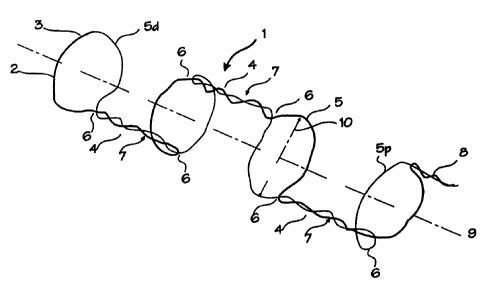
Figure 1 shows the stent in its expanded state. The stent
= 25 1 is comprised of a single wire 2 which is folded at a point
along the wire, such as mid-point 3 to form a length of double
wire comprising two wire segments. Several twisted sections 4
in the double wire are interspersed with several hoops 5 formed
by pulling the double wire apart into the hoop shape. The wire
5
CA 02289213 1999-11-03
WO 98/50102 PCT/US9S/08876
can also be made to form coils in place of the hoops. The
double wire is then bent at each junction 6 between the twisted
sections and the hoops to form an angle of about 90 between
each hoop and twisted section. The twisted sections create
alternately radially opposed struts, bridges or spines 7 between
successive hoops. The free ends 8 of the wire may be twisted
into a free spine as shown, or they may be joined together. The
hoops in this arrangement are aligned along a common axis 9
which defines the longitudinal axis of the stent, and they are
oriented approximately parallel to each other. The stent has an
unconstrained diameter defined by hoop diameter 10. The hoops
may be all the same overall diameter, or they may be of
different diameter, and it may be particularly useful to create
the hoops so that the size of the hoops increases from one end
of the stent to the other so as to better conform to the tapered
blood vessel. The struts may be all the same length or of
varying length. Although shown as being formed by intertwining
the tow wire segments, the struts maybe formed by welding the
segments to together, or by shaping the segments to run parallel
where the strength of the wire permits. Note that the stent may
be formed of two separate lengths of wire, but in this case a
free end strut at distal hoop 5d, or a joint provided elsewhere
along the stent, may be required.
Figure 2 shows the stent partially stretched out. In its
partially stretched condition, the hoops 5 have elongated into
reclining ellipses 11 oriented at an angle from the longitudinal
axis 9 of the stent 1. The angle is intermediate between the
longitudinal axis of the stent and the radius 12 of the stent.
Thus the overall diameter of the stent has been significantly
reduced by stretching along the longitudinal axis. The fact
that the struts are not radially aligned permits longitudinal
stretching or deformation of every hoop in the stent. As
illustrated, the each strut is radially opposed to the struts on
either side, meaning the each strut is on the opposite side of
the stent compared to the preceding or succeeding strut. When
the adjacent struts are 180 apart, maximum stretching of the
6
CA 02289213 1999-11-03
WO 98/50102 PCTIUS98/08876
hoops is achieved merely be pulling the ends of the stent.
Direct opposition, or opposition of exactly 1800, is not
requirJed to obtain the benefit of this construction, and it
suffices that the struts not be radially aligned.
Figure 3 shows the hoop stent elongated to its maximum
extent, in the condition in which it will be loaded onto the
delivery catheter. In its fully stretched condition, the hoops
5 have elongated into closed or nearly closed ellipses 13
oriented not at an angle from the longitudinal axis 9 of the
stent 1, but in line with the longitudinal axis. The angle is
close to the longitudinal axis of the stent and perpendicular to
the radius 12 of the stent. When stretched completely in the
longitudinal direction, the stent has an overall radial
thickness of only two wire thicknesses. This provides the
thinnest possible insertion diameter for the stent. The stent
is loaded in the distal end of a delivery catheter, and
delivered percutaneously to the deployment site. The stent may
be pushed out of the delivery catheter distal end, or it may be
held in place while the delivery catheter is withdrawn. Various
other deployment mechanisms may be used, such as the non-sliding
sheaths, zip cord sheaths and other embodiments. Where the
stent is made of a superelastic alloy (superelastic at body
temperature) it will revert to the open hoop configuration of
Figure 1 upon release from the catheter. Where the stent is
made of a shape memory alloy with a transition temperature
slightly above body temperature, reversion to the memorized
shape of Figure 1 will occur upon injection of warm fluid
through the catheter and onto the stent. The reversion will
occur between the austenite start temperature As and the
austenite finish temperature Af. Typically in this application,
the Af would be set to about 30 C plus or minus 5 C, so full
expansion occurs above the normal room temperature and below the
normal body temperature. If Af is a degree or so above body
temperature, hysteresis may be relied upon to ensure maintenance
of superelastic properties when the material is cooled to body
temperature.
7
CA 02289213 1999-11-03
WO 98/50102 PCT/US98/08876
The stent wires 2 may be made of a shape memory alloy such
as nitinol (or other shape memory material), pseudoelastic or
superelastic alloy such as nitinol (or other pseudoelastic or
superelastic material), spring metal such as stainless steel, or
other suitable materials. When made of shape memory nitinol or
superelastic nitinol, the stent may be trained to the shape
shown in Figure 1, and will revert to that shape either through
shape memory behavior at its chosen transition temperature, or
through superelastic behavior at body temperature. The
appropriate compositions and training regimens may be used to
obtain these characteristics. Spring materials such as
stainless steel may be used also, and fabricated so that the
shape of Figure 1 is the relaxed state of the material which is
regained elastically after stretching into the shape shown in
Figure 3. As with prior art stents, the stent may also be
deployed by inflating a balloon within the stent.
The configuration of the hoop stent shown in Figure 1 is
merely one of the many configurations that may be fabricated of
a single wire and deployed in very low profile configurations
equal to one or two wire thicknesses. Additional configurations
are shown in the remaining figures. These configurations are
designed to provide for partial occlusion or flow disruption of
aneurysms, which will promote thrombus formation and eventual
shrinkage of the aneurysm. While the support structure is
similar to the hoop stent discussed above, these stents use the
stent structure as a scaffolding mechanism for retaining flow
disrupting structures within or about the neck of the aneurysm.
A simple embodiment is shown in Figure 4. The flow
disrupting device 15 comprises a stent body which is formed of a
single wire 2, with the strut 7 extending between the distal and
proximal hoops. Rather than using overall straight
configuration for the strut, the strut is divided into two
smaller segments 17 and 18 which are disposed along the strut
line. These two struts support a flow disrupting structure 19
which may be integrally formed on the same wire, as shown. The
flow disrupting structure 19 in Figure 4 is a mere camel hump
8
CA 02289213 1999-11-03
WO 98/50102 PCTIUS98/08876
shape which protrudes axially outwardly from the imaginary
cylinder defined by the hoops 201 and 20r. The shape may be
any irregular shape which protrudes into the aneurysm sac and
avoids substantial protrusion into the lumen of the blood
vessel.
As the flow disrupting structure may not need to protrude
into the aneurysm sac, the flow disrupting device of Figure 5
may be used where protrusion into the aneurysm sac is
contraindicated. The flow disrupting device 21 comprises a
stent body 14 which is formed of a single wire 2, with the strut
7 extending between the distal and proximal hoops. As in Figure
4, the strut is divided into two smaller segments 22 and 23
which are disposed along the strut line. These two struts
support a flow disrupting structure which may be integrally
formed on the same wire, as shown. The flow disrupting segment
24 differs from that of Figure 4 in that the shape does not
protrude axially outwardly from the imaginary cylinder defined
by the hoops 201 and 20r. Instead, the flow disrupting segment
comprises a segment 24 of curves that conform to the imaginary
cylinder defined by the hoops 201 and 20r, and thus conform
generally to the curvature expected in the lumen of the blood
vessel in which the device is placed. When placed within a
blood vessel so that the flow disrupting segment bridges the
mouth of the aneurysm, the disruption of flow will result in
eventual thrombosis and diminishment of the aneurysm mass.
Another useful shape for the flow disrupting structure is
that of a coil, as illustrated in Figure 6. Coil tower 26
extends radially outwardly from the struts 27 and 28, and is
integrally formed from the single wire 29. The hoops 201 and
20r are formed in the manner as described in reference to Figure
1. The isometric view shown in Figure 7 provides another view
of the flow disrupting device, with each of the elements being
the same as in Figure 5. The coil tower 26 is sized and
dimensioned to fit within an aneurysm sac, while the hoops 201
and 20r are sized and dimensioned to expand into secure contact
with the surrounding blood vessel of normal or near normal
9
CA 02289213 1999-11-03
WO 98/50102 PCT/US98/08876
cy"lincirical cross section. The struts are of appropriate length
to extend from the aneurysm sac to the nearby lumen of normal or
near_normal cylindrical cross section. Figure 8 illustrates
another embodiment of the flow disrupting device in which the
flow disrupting structure comprises a flat coil 30 oriented so
that it does not protrude into the aneurysm space. The flat
coil may or may not conform to the cylindrical shape of the
ideal blood vessel wall.
A mandrel for forming the coiled flow disrupting structure
is illustrated in Figure 9. The mandrel is a tee shaped mandrel
with a post for forming the flow disrupting segment and a pair
of extensions for forming the struts and hoops. The length of
wire is folded over at point C, and the double wire is wrapped
around the mandrel tower 31 and held in recessed shaping groove
32. The pin 33 secures the wire to the tower. The doubled
wire is then separated so that one wire may be routed to the
right side mandrel 34 and wrapped around the mandrel in hoop
forming groove 35, while the second wire is routed to the left
side mandrel 36 and wrapped in hoop forming groove 37. Each
end of single wire may terminate near the endpoint 38 of the
hoop forming groove, after making substantially an entire wrap
around the mandrel, or each end may return to the center of the
device, near the tower for provide additional strength to the
struts. The mandrel may be made to disassemble at the junction
of the tee once the stent has been shape set on the mandrel.
Two types of aneurysms that permit different deployment
methods and different insertion configurations are the saccular
side wall aneurysm and the bifurcation aneurysm. In
intracranial clinical experience, the bifurcation aneurysm
appears predominant, and the basilar tip aneurysm is of great
clinical concern. In peripheral vessels, and in the carotid
arteries, saccular aneurysms are more common.
A first embodiment of insertion configurations applies to
bifurcation aneurysms (although we refer to the bifurcation
aneurysm in accordance with the common terminology in the art,
CA 02289213 1999-11-03
WO 98/50102 PCTIUS98/08876
it will be apparent that it may be used to treat any end-
approachable aneurysm). For insertion into the body, the device
may be stretched in the longitudinal direction to form a length.,:
of doubled wire, housed within an insertion catheter 39 as
illustrated in Figure 10. For treatment of bifurcation
aneurysms, the device is placed within the insertion catheter
with the wire bend 3 is disposed at the distal end of the
deployment configuration and the wire ends 8 at the proximal end
of the deployment configuration. The insertion catheter is
inserted into the body according to standard techniques to the
point where it is to be deployed. Figure 10 shows the distal
tip of the insertion catheter 39 approaching a basilar tip
aneurysm 40. The aneurysm occurs at the bifurcation of the
basilar artery 41 into the posterior cerebral arteries 421 and
42r. (This anatomical structure is located in the brain, and is
part of the Circle of Willis 43, which is shown for clarity.
The other arteries of the Circle of Willis include the posterior
communicating arteries 44, the anterior communicating arteries
45, the left and right middle cerebral arteries 46 and the left
and right anterior cerebral arteries 47. The internal carotid
arteries 48 also supply the Circle of Willis.) The wire bend 3
is placed at the distal end of the deployment configuration so
that this point of the device may be inserted into the aneurysm
space when the device is pushed or otherwise released from the
deployment catheter.
Upon release from the catheter, the distal end 49 of the
device will revert to its coiled shape. In Figure 11, the
device is partially deployed, and the coiled occluding structure
which is not restrained by the catheter has reverted to its coil
shape. The further release of the wire ends 8d and 8p will
allow the reversion of these portions of the wire to revert into
the hoops 201 and 20r and struts 27 and 28. Adjustments to the
position of the coil, the hoops or the struts can be made using
the insertion catheter to bump and push the parts into place.
= 35 The hoops should circumscribe the inner wall of the blood
vessel, as shown in Figure 12. After placement, the angle 50
11
CA 02289213 1999-11-03
WO 98/50102 PCTIUS98/08876
which appears between the struts and the coils is apparent.
This angle may be trained into the device before insertion by
annealing or training the wire to take on a shape which includes,
such an acute angle between the struts and the occluding
structure of the coil 26. On the other hand, the tension
resulting from the 90 angle shown in Figure 6, upon placement
in an environment requiring an installed angle of less that 90 ,
will result in tension which helps keep the occluding structure
26 firmly held within the aneurysm space.
For side wall aneurysms, or any aneurysm which can be
approached from the side, the occluding device may be inserted
fully stretched in a single wire configuration. As shown in
Figure 13, the occluding device 15 of Figure 4 is stretched into
a single long wire 2 with the free ends 8d and 8p of the wire at
the distal and proximal ends of the device, and the midpoint 3
in the longitudinal center of the device, housed within the
distal end of the insertion catheter 39. To deploy the device,
the insertion catheter 39 is inserted into the body and
navigated to the site of the aneurysm 51 within the blood vessel
52. As shown in Figure 13, the distal end of the device (which
corresponds to the distal free end 8d) is placed just distal to
the aneurysm 51. After the device is properly located, the
insertion catheter is removed (by pulling proximally, by peeling
or otherwise), the device will revert to its original shape.
The distal free end 8d will revert to its memorized hoop shape,
and the strut and occluding section will revert to the memorized
occluding shape, and finally the proximal free end 8p reverts to
its memorized hoop shape. Figure 14 shows the device partially
deployed, with the distal free end 8d reverted into distal hoop
5d and the wire near the midpoint reverted into the flow
disrupting structure 19. The proximal free end 8p is shown
still within the distal end of the insertion catheter, and will
revert to the shape of the proximal hoop 5p upon release.
Each of the occluding device embodiments and the stent
embodiments may be controlled with the various methods used for
stents and other devices made of nitinol. Nitinol is preferred
12
r--------
CA 02289213 1999-11-03
WO 98/50102 PCT/US98/08876
because its biocompatibility is well proven, and it is available
in numerous compositions with well-controlled transition
temperatures. (Other shape memory or pseudoelastic materials
may be used, and normally elastic stainless steel, Elgiloy, and
plastics may be used.) The nitinol used for the device may be
used in its shape memory formulation, with a transition
temperature just above body temperature, in which case the
device may be returned to its memorized shape upon the injection
of warm water (just above body temperature). Alternatively, the
nitinol used for the device may be used in its pseudoelastic
formulations, in which the nitinol is superelastic (also called
pseudoelastic) at body temperature, in which case the device
will automatically revert to its memorized shape when inside the
body. The superelastic device can be superelastically deformed
to fit within the insertion catheter so that it can be inserted
into the body, and it superelastically reverts to the memorized
shape when released from the catheter in the blood stream.
Common among the embodiments of the aneurysm occluding
devices is the desire that the occluding structure enhance
formation of thrombus within the aneurysm. To enhance this
function, the occluding structure may be coated with known
thrombogenic materials such as platinum. The hoops and struts
which remain outside the aneurysm sac and within the blood
stream must remain uncoated with such a thrombogenic coating,
and are preferably coated with an anti-thrombogenic coating such
as heparin or tin or many other such coatings. Thus the
occluding device will have segments of varying thrombo-active
coatings, depending on the desired characteristic of each
segment. The devices may also be coated with materials such as
tantalum, gold and platinum in order to enhance the visibility
of the devices under fluoroscopy. The device are clearly
visible under intravascular ultrasound which may be used to aid
in deployment and proper placement. While the devices will
provide for the primary treatment of aneurysms, they may also be
used in conjunction with embolic materials and GDC's in order to
13
CA 02289213 1999-11-03
WO 98/50102 PCT/US98/08876
hold these foreign materials within the aneurysm and prevent
their migration from the aneurysm into the blood stream.
-'~hus, while the preferred embodiments of the devices and
methods have been described in reference to the environment in
which they were developed, they are merely illustrative of the
principles of the inventions. Other embodiments and
configurations may be devised without departing from the spirit
of the inventions and the scope of the appended claims.
14