Note: Descriptions are shown in the official language in which they were submitted.
CA 02302883 2000-03-29
CONTROL METHOD FOR AN AUTOMATED
SURGICAL BIOPSY DEVICE
Field of the Invention
The present invention relates, in general, to a method of controlling the
excision of tissue in biopsy instruments and, more particularly, to a method
of
15 controlling the rotation and translation of a cutter in a biopsy
instrument.
Background of the Invention
The diagnosis and treatment of patients with cancerous tumors, pre-
20 malignant conditions, and other disorders has long been an area of intense
investigation. Non-invasive methods for examining tissue include palpation, X-
ray, magnetic resonance imaging (MRI), computed tomography (CT), and
ultrasound imaging. When a physician suspects that tissue may contain
cancerous
cells, a biopsy may be done using either an open procedure or a percutaneous
25 procedure. For an open procedure, a scalpel is used to create a large
incision in
the tissue to provide direct viewing and access to the tissue mass of
interest. The
entire mass (excisional biopsy) or a part of the mass (incisional biopsy) may
then
be removed. In most percutaneous biopsy procedures, a needle-like instrument
is
inserted through a very small incision to access the tissue mass of interest
and
30 obtain a tissue sample for later examination and analysis.
Aspiration and core sampling are two percutaneous methods for obtaining a
portion of tissue from within the body. In an aspiration procedure, tissue is
CA 02302883 2005-12-13
-2-
fragmented into pieces and drawn through a fine needle in a fluid medium. The
method is less intrusive than most other sampling techniques, however, it has
limited application since the structure of tissue excised by aspiration is
destroyed
leaving only individual cells for analysis (cytology) and not the tissue
structure for
analysis (pathology). In core biopsy, a core or fragment of tissue is obtained
in a
manner, which preserves both the cells and the structure for histological
examination. The type of biopsy used depends mainly on various factors, and no
single procedure is ideal for all cases. Core biopsy, however, is very useful
in a
number of conditions and is widely used by physicians.
Examples of core sampling biopsy instruments are described in U.S.
Patents 5,562,822 and 5,769,086 (both issued to Ritchart, et al), and in U.S.
Patent No. 6,007,497 issued December 28, 1999. Another
example of a core sampling biopsy instrument is the biopsy instrument now
marketed by Ethicon Endo-Surgery, Inc., Cincinnati, Ohio, under the trade name
MAMMOTOME. Each of these instruments is a type of image-guided,
percutaneous, coring, breast biopsy instrument, . which uses a vacuum for
retrieving tissue samples. A physician uses these instruments to capture
'"actively"
(using the vacuum) tissue prior to severing it from the body. In particular,
in
these biopsy instruments, tissue is drawn into a port at the distal end of a
piercing
element, hereinafter referred to as a piercer. A cutting element, hereinafter
referred to as a cutter, is rotated and advanced through a lumen of the
piercer past
the port. As the cutter advances through the port, it severs the tissue drawn
into
the port from the surrounding tissue. While the cutter is generally rotated
using
some type of motor, it may be advanced either manually or automatically. In
the
MAMMOTOME instrument, the surgeon manually moves the cutter back and
forth by lateral movement of a knob mounted on the outside of the instrument.
Once the cutter is in place, proximal to the tissue port, further lateral
movement of
the knob is prevented and the cutter is advanced through the tissue port to
sever
tissue by twisting the knob. This arrangement is advantageous because the
surgeon is able, through tactile and/or audible feedback, to determine.
whether the
cutter is effectively cutting tissue or if there is a problem, such as binding
or
CA 02302883 2000-03-29
-3-
stalling. The surgeon may then adjust the speed at which the cutter is moved
through the tissue, stop the cutter or back the cutter away from the tissue.
As described in U.S. Patents 5,562,822 and 5,769,086, the translation of
the cutter may be automated to facilitate the procedure. However, if the
procedure is automated as described in those references, the surgeon loses the
benefit of the tactile feedback, which results when the cutter is advanced
manually. It is generally desirable to ensure that the rotational speed of the
cutter
does not drop below a predetermined speed in order to sever cleanly the tissue
sample from surrounding tissue and to avoid damage to the tissue sample.
Automating translation of the cutter will, to some extent, eliminate the
tactile
feedback that the surgeon gets from moving the cutter manually. The
advantageous method of automatically measuring and controlling the rotation
and
translation of the cutter is not described for either of the devices in the
`822 or
`086 patents. This automatic control method could be used, for example, to
prevent the cutter from advancing when the opening is blocked by something
other
than tissue. Such an automatic control method could also be used to ensure
that
the cutter rotates at an optimal speed to ensure proper cutting of the tissue
and to
prevent the cutter rotational speed from dropping below a predetermined limit.
Another advantage of a core sampling biopsy device being used with an
automatic control method is that the operator would be able to perform the
surgical
procedure in less time than with a motorized device having no automatic
control
method. Since core sampling biopsy devices extract tissue samples from deep
within the body of the surgical patient, the penetrating element, or piercer,
for
accessing the tissue, is necessarily long. The driven element, or cutter, must
translate from the proximal end of the piercer to the distal end in order to
collect
the tissue sample. Then the cutter transports the tissue sample from the
distal end
of the piercer to the proximal end, which is outside the body of the -patient.
As
the cutter is actually cutting through tissue and collecting the tissue
sample, the
translational speed of the cutter should be maintained in an optimal range.
But for
all other portions of cutter translation, the translational speed of the
cutter may be
CA 02302883 2000-03-29
-4-
relatively high without detrimental effects. Thus the time required for
obtaining
each tissue sample might be reduced. Since many tissue samples may be
extracted
from the patient during a typical surgical procedure, the accumulated time
saved
could be significant, providing obvious benefit for both the surgeon and the
patient.
A core sampling, surgical instrument with a needle guiding stage of a
stereotactic mammography biopsy system is disclosed in U.S. Patent 5,830,219
by
Bird, et al. In this instrument, the driven element (hereinafter referred to
as a
cutter) translates to cut tissue captured in the distal end of the instrument.
This
instrument couples feedback from an optical encoder with a microprocessor to
calculate cutting resistance so that if the cutter encounters, for example, a
dense
tissue mass causing the cutter rotation to decrease, additional electrical
current is
automatically provided to the cutter motor to resume the desired cutter
rotational
speed. The encoder also provides information to the microprocessor to control
automatically the angular stroke of the cutter as it oscillates between the
clockwise
and counterclockwise directions. This closed loop cutter control method- is
described in `219 as being provided only for when the cutter is in the cutting
portion of its axial travel.
An alternate control method, however, to deal with an increase in cutting
resistance (due to encountering dense tissue or obstructions) is to slow
incrementally the translational speed of the cutter until a desired cutter
rotational
speed is resumed. If the cutter cannot penetrate the obstruction and the
desired
rotational speed cannot be resumed despite reducing translational speed, then
the
translation of the cutter could be completely halted and an error message, for
example, could be transmitted to the operator. This alternate control method
would have an advantage of preventing damage to the biopsy instrument because
the tissue sample is obtained less aggressively due to the slowed advance of
the
cutter through the tissue, or else the cutter translation is completely halted
if the
obstruction is impenetrable.
CA 02302883 2005-12-13
-5-
Another advantage for reducing the cutter translational speed rather than
increasing the cutter rotational speed in response to an increase in
rotational
resistance on the cutter is that smaller, less powerful, and less expensive
motors
could be used to drive the cutter. Both the translation motor for advancing
the
cutter and the rotation motor for rotating the cutter may be smaller because
the
overall rate at which work would be done on the tissue by the cutter could be
reduced. Using smaller, lighter weight motors would also facilitate their
incorporation into the handheld portion of the biopsy instrument. As described
in
pending Canadian Patent Application No. 2,287,087, the motors can be
remotely located in a separate control unit and operationally connected to the
handheld portion of the biopsy instrument by at least one rotatable shaft. In
such a
biopsy instrument, using small motors would be advantageous in allowing the
use
of small diameter, lightweight, rotatable shafts. In addition to the cost
savings
realized in the manufacture of the device, the biopsy instrument could be more
hand manipulatable by the operator during the surgical biopsy procedure.
It is also advantageous to use both types of responses to increased
rotational resistance on the cutter. That is, a single control method that
combines a
method for decreasing cutter translational speed and a method for increasing
cutter
rotational speed in response to decreasing cutter rotation may be used. For
example, if the cutter encounters an obstruction in tissue and the cutting
resistance
rises sharply, the electrical current to the cutter rotational motor may
automatically
be increased by a'predetermined amount. If the cutter rotational speed is
measured and compared to a desired, predetermined cutter rotation speecf, and
it is
determined that the cutter rotational speed is still not high enough, than the
cutter
translational speed may be decreased by a predetermined amount. These steps
could be repeated automatically until the tissue sample is obtained, or until
certain
operational thresholds (minimal translation speed, maximum current to rotation
motor, for example) are reached. By using such a combined method in response
to increasing rotational resistance on the cutter, the cutter rotational and
translational motors may be smaller than when using a method = to modify
cutter
rotation alone.
CA 02302883 2000-03-29
' -6-
When an operator uses a handheld instrument operationally connected to a
remotely located motor by a flexible, rotatable shaft, the operational
configuration
of the rotatable shaft may affect the efficiency of the mechanical energy
transmitted to the handpiece. For example, if it is necessary for the operator
to
~
hold the handheld instrument during the surgical procedure so that the
flexible,
rotatable phaft is sharply curved, the resistance to rotation of the rotatable
shaft is
higher than if the rotatable shaft was in a straight configuration. Also, when
the
operator manipulates the probe of the instrument to penetrate into the tissue
mass
of interest, a bending moment may be unavoidably applied to the piercer of the
probe, increasing the rotational resistance of the cutter which is constructed
coaxially in close alignment with the piercer. There also may be additional
mechanical losses, for example, due to wear or misalignment of power
transmission components. Therefore, it would be advantageous to be able to
measure the total, rotational resistance before the cutter encounters tissue,
so that
the cutter rotation may be increased to the desired, predetermined rotational
speed
for cutting tissue.
What is needed is a core sampling biopsy device having a control method
and apparatus that allows the cutter translational speed to be automatically
responsive to cutting resistance caused by obstructions or dense tissue
encountered
by the cutter. What is further needed is a core sampling biopsy device having
a
control method and apparatus that allows the cutter rotational speed to be
automatically responsive to total rotational resistance on the cutter before
and
during the cutting of tissue. What is further needed is a core sampling biopsy
device having a control method and apparatus that allows the cutter
translational
speed to be automatically responsive to cutter translational position so that
surgical
procedure time may be reduced.
Summary of the Invention
The present invention is directed to a method of automadcally controlling
the removal of at least one tissue sample from a surgical patient using a
biopsy
CA 02302883 2000-03-29
-7-
instrument. The biopsy instrument comprises an elongated piercer having a
piercer lumen extending therethrough. The piercer has a port for receiving and
transferring the tissue sample into the piercer lumen. The biopsy instrument
further comprises a cutter rotatably and translationally positionable relative
to the
piercer.
In one embodiment of the present invention, the method for using the
biopsy instrument comprises the steps of engaging tissue in the port;
translating the
cutter at a first, predetermined translational speed from a first position to
a second
position proximal to the port; measuring the translational speed of the
cutter;
translating the cutter at a second, predetermined translational speed from the
second position to a third position proximal to the port; and translating the
cutter
at a third, predetermined translational speed from the third position to a
fourth
position distal to the port.
In a further embodiment of the present invention, a method for using the
biopsy instrument comprises the steps of engaging tissue in the port;
translating the
cutter at a first, predetermined translational speed from a first position to
a second
position proximal to the port; measuring the translational speed of the
cutter;
translating the cutter at a second, predetermined translational speed from the
second position to a third position proximal to the port; translating the
cutter at a
third, predetermined translational speed from the third position to a fourth
position
distal to the port; rotating the cutter at a predetermined rotation speed;
measuring
the rotational speed of the cutter; and modifying the translational speed of
the
cutter when the rotational speed of the cutter varies from the predetermined
rotational speed by more than a first, predetermined differential rotational
speed.
In a yet further embodiment of the present invention, the method comprises
the steps of engaging tissue in the port; translating the cutter at a first,
predetermined translational speed from a first position to a second position
proximal to the port; measuring the translational speed of the cutter;
translating the
cutter at a second, predetermined translational speed from the second position
to a
CA 02302883 2007-09-27
8
third position proximal to the port; translating the cutter at a third,
predetermined
translational speed from the third position to fourth position distal to the
port; rotating the
cutter at a predetermined rotation speed; measuring the rotational speed of
the cutter; and
modifying the translational and rotational speeds of the cutter when the
rotational speed
of the cutter varies from the predetermined rotational speed by more than a
first,
predetermined differential rotational speed.
Another aspect of the present invention is a biopsy instrument for removing at
least one tissue sample from a surgical patient having an elongated piercer
having a
piercer lumen extending therethrough; a cutter translationally positionable
relative to the
piercer; and a port in the piercer for receiving and transferring the tissue
sample into the
piercer lumen. The instrument also has means for engaging tissue in the port
and a means
for measuring translational speed of the cutter. The instrument also has means
for
translating said cutter, the means for translating being adapted to translate
said cutter at a
first, predetermined translational speed from a first position to a second
position distal to
the first position and proximal to the port, the means for translating also
being adapted to
translate the cutter at a second, predetermined translational speed from the
second
position to a third position proximal to the port and distal to the second
position, the
means for translating also being adapted to translate the cutter at a third,
predetermined
translational speed from the third position to a fourth position distal to the
port.
Another aspect of the present invention is a biopsy instrument for removing at
least one tissue sample from a surgical patient having an elongated piercer
having a
piercer lumen extending therethrough. The instrument also has a cutter
rotatably and
translationally positionable relative to the piercer and a port in the piercer
for receiving
and transferring the tissue sample into the piercer lumen. There is also a
means for
engaging tissue in the port; means for measuring translation speed of the
cutter; a means
for translating the cutter at a first predetermined translational speed from a
proximal
position proximal to the port to a distal position distal to the port; means
for rotating the
cutter at a predetermined rotational speed; a means for measuring the
rotational speed of
CA 02302883 2007-09-27
8a
the cutter; and a means for modifying the translational speed of the cutter
when the
rotational speed of the cutter varies from the predetermined rotational speed
by more than
a predetermined differential rotational speed.
Another aspect of the present invention is a biopsy instrument for removing at
least one tissue sample from a surgical patient having an elongated piercer
having a
piercer lumen extending therethrough; a cutter rotatably and translationally
positionable
relative to the piercer; a port in the piercer for receiving and transferring
the tissue sample
into the piercer lumen; a means for engaging tissue in the port; a means for
measuring
translational speed of the cutter; and a means for translating the cutter, the
means for
translating being adapted to translate the cutter at a first, predetermined
translational
speed from a first position to a second position distal to the first position
and proximal to
the port, the means for translating also being adapted to translate the cutter
at a second,
predetermined translational speed from the second position to a third position
proximal to
the port and distal to the second position, and the means for translating also
being adapted
to translate the cutter at a third, predetermined translational speed from the
third position
to a fourth position distal to the port. The instrument also has a means for
rotating the
cutter at a predetermined rotational speed; a means for measuring rotational
speed of the
cutter; and a means for modifying the translational speed of the cutter when
the rotational
speed of the cutter varies from the predetermined rotational speed by more
than a
predetermined differential rotational speed.
Another aspect of the present invention is a use of the instrument described
above
for removing at least one tissue sample from the patient.
Brief Description of the Drawings
The novel features of the invention are set forth with particularity in the
appended
claims. The invention itself, however, both as to organization and methods of
operation,
together with further objects and advantages thereof, may best be understood
by
CA 02302883 2007-09-27
8b
reference to the following description, taken in conjunction with the
accompanying
drawings in which:
Figure 1 is an isometric view of the present invention, a biopsy instrument,
which
includes a handpiece for the collection of soft tissue;
Figure 2 is an isometric view of the handpiece showing a probe assembly prior
to
attachment to a holster;
Figure 3 is an exploded isometric view of the probe assembly illustrated in
Figure
2;
Figure 4 is an isometric view of the probe assembly of Figure 2 with the left
handle shell removed to reveal the internal components;
Figure 5 is an exploded isometric view of the holster illustrating a non-
encased
rotation sensor mounted on a screw drive shaft;
Figure 6A is a top view in section of the probe assembly and a distal portion
of
the holster, revealing a cutter in a first, fully retracted position;
CA 02302883 2000-03-29
-9-
Figure 6B is a top view in partial section of the distal end of the probe
assembly illustrating the cutter in the first, fully retracted position
wherein the port
on the distal end of the piercer is open;
Figure 7A is a top view in section of the probe assembly and a distal
portion of the holster, revealing the cutter in the third position wherein the
distal
end of the cutter is immediately proximal to the port;
Figure 7B is a top view in partial section of the distal end of the probe
assembly with the port on the distal end of the piercer open and the distal
end of
the cutter in the third position immediately proximal to the port;
Figure 8A is a top view in section of the probe assembly and a distal
portion of the holster illustrating the cutter in the fourth, fully deployed
position;
Figure 8B is a top view in partial section of the distal end of the probe
assembly illustrating the distal end of the cutter in the fourth position
distal to the
port at the distal end of the piercer;
Figure 9 is an isometric view of the probe assembly with the left handle
shell removed, showing the cutter in the first position, with a tissue sample
shown
deposited onto a tissue sampling surface;
Figure 10 is a partial top view of a further embodiment of the present
invention wherein a first and a second motor are contained within a handheld
holster rather than in a remotely located control unit as for the embodiment
of
Figure 5, and wherein the holster upper shell and the probe assembly upper
shell
have been removed to reveal the internal components;
Figure 11 is an isometric view of the holster and probe assembly lower
shells shown in Figure 10, wherein the holster lower shell includes a slot for
the
removable attachment to a latch on the probe assembly lower shell;
CA 02302883 2000-03-29
-10-
Figure 12 is a longitudinal section of the holster and probe assembly lower
shells of Figure 11, illustrating their removable attachment to each other;
Figure 13 is an exploded isometric view of a further elnbodiment of the
holster illustrated in Figure 5, wherein the further embodiment includes the
three
switches lieing mounted on a switch board electrically connected by a ribbon
cable
to the control cord (instead of the three switches being electrically
connected to the
control cord by discrete switch conductors as illustrated in Figure 5), and
wherein
the further embodiment includes an encased rotation sensor rather than the non-
encased rotation sensor of the embodiment illustrated in Figure 5;
Figure 14 is a schematic diagram of a control unit according to the present
invention;
Figure 15 is an enlarged diagram of an LCD display illustrated in Figure
14;
Figure 16A is the first of two portions of a divided schematic diagram of
the control unit components illustrated in Figure 14;
Figure 16B is the second of two portions of the divided schematic diagram
of the control unit components illustrated in Figure 14;
Figure 17A is a first portion of a flow chart pertaining to a first
embodiment and a second embodiment of a control method for the operation of
the
cutter, showing the control unit logic for when the cutter translates from the
first
position to the second position;
Figure 17B is a second portion of a flow chart pertaining to the first
embodiment and the second embodiment of the control method for the operation
of
the cutter, showing the control unit logic for when the cutter translates from
the
second position to the third position;
CA 02302883 2000-03-29
ll
Figure 17C is a third portion of a flow chart pertaining to the first
embodiment and the second embodiment of the control method for the operation
of
the cutter, showing additional control unit logic for when the cutter
translates from
the second position to the third position;
Fi4ure 17D is a fourth portion of a flow chart pertaining specifically to the
first control method embodiment for the operation of the cutter, showing the
control unit logic for when the cutter translates from the third position to
the
fourth position; and
Figure 17E is a fourth portion of a flow chart pertaining specifically to the
second control method embodiment for the operation of the cutter, showing the
control unit logic for when the cutter translates from the third position to
the
fourth position.
Detailed Description of the Invention
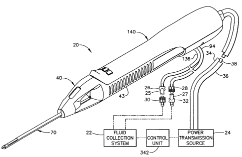
Figure 1 shows a core sampling biopsy instrument comprising a probe
assembly 40, a holster 140, a fluid collection system 22, a control unit 342,
and a
power transmission source 24. Probe assembly 40 is detachably connected to
holster 140. Together they constitute a lightweight, ergonomically shaped,
hand
manipulatable portion referred to as a handpiece 20. Probe assembly 40
includes a
piercer 70 extending distally from a hollow handle 43. Probe assembly 40 is
fluidly connected to fluid collection system 22 by a first vacuum tube 94 and
a
second vacuum tube 136. First and second vacuum tubes are detachably connected
to fluid collection system 22 by a first connector 27 and a second connector
25,
respectively. First connector 27 has a male portion 32 and a female portion 28
attached to first vacuum tube 94. Second connector 25 has a female portion 30
and a male portion 26 attached to second vacuum tube 136. Connector portions,
26, 28, 30, and 32 are attached in this manner to prevent the accidental
switching
of first and secorid tubes, 136 and 94, to fluid collection system 22. Holster
140
includes a first rotatable shaft 34, a second rotatable shaft 36, and a
control cord
CA 02302883 2000-03-29
= - !
12
38. First and second rotatable shafts, 34 and 36, are preferably flexible so
that the
operator may easily manipulate handpiece 20 with one hand. Control cord 38
operatively connects the handpiece 20 to power transmission source 24 and
control
unit 342.
Sirlce handpiece 20 is manipulated by the operator's hand rather than by an
electro-mechanical arm, the operator may steer the tip of handpiece 20 with
great
freedom towards the tissue mass of interest. The surgeon has tactile feedback
while doing so and can thus ascertain, to a significant degree, the density
and
hardness of the tissue being encountered. In addition, handpiece 20 may be
held
approximately parallel to the chest wall of the patient for obtaining tissue
portions
closer to the chest wall then may be obtained when using a instrument mounted
to
an electro-mechanical arm.
Those skilled in the art may appreciate that a mount or "nest" could be
provided to hold handpiece 20 securely to the movable arm of an X-ray
stereotactic table. This would provide the operator with the option to use
handpiece 20 to access the tissue mass within the surgical patient in much the
same
manner as was described earlier for using the MAMMOTOME instrument. This
versatility may be advantageous to the operator, for example, in a situation
where
the handheld imaging device was temporarily not available for use, and it
would
be necessary to use the X-ray stereotactic table.
Figure 2 shows holster 140 and probe assembly 40 separated. A pair of
tabs 144 project laterally from each side of a holster upper shell 142, and
insert
into right and left undercut ledges, 138 and 139 respectively, of hollow
handle 43
of probe assembly 40. A plurality of indentations 66 is provided on handle 43
to
improve the operator's grip on the instrument. A tube slot 162 in lower shell
156
of holster 140 provides clearance for first and second vacuum tubes, 94 and
136.
A cutter forward switch 146 for moving a cutter 96 (see Figure 3) in the
distal
direction, a cutter reverse switch 148 for moving cutter 96 in the proximal
direction, and a vacuum switch 150, are mounted in the distal portion of
holster
CA 02302883 2000-03-29
-13-
140 so that the operator can use handpiece 20 with a single hand. One-handed
operation allows the other hand to be free, for example, to hold an ultrasonic
imaging device. A ridge 152 on the distal end of holster 140 is provided to
assist
the operator in grasping handpiece 20 and in operating switches 146, 148, and
150.
Still in Figure 2, probe assembly 40 includes a window 58 so that a portion
of first vacuum tube 94 may be viewed. First and second vacuum tubes, 94 and
136, are made from a flexible, transparent or translucent material, such as
silicone
tubing. This enables visualization of the material flowing through the tubes,
94
and 136. By having window 58 in probe assembly 40, the operator can see the
flow in first vacuum tube 94 without needing to look away from the tissue into
which piercer 70 is inserted. A transverse opening 68 is provided in the
distal end
of hollow handle 43 which allows access from either side to a tissue sampling
surface 64. The tissue extracted from the surgical patient is retrieved by the
operator or by an assistant from tissue sampling surface 64.
Figure 3 is an exploded isometric view of probe assembly 40. Handle 43
is formed from a right handle shell 42 and a left handle shell 44; each
injection
molded from a rigid, biocompatible plastic such as polycarbonate. Upon final
assembly of probe assembly 40, left and right handle shells, 42 and 44, are
joined
together by ultrasonic welding along a joining edge 62, or joined by any of
several
other methods well known in the art. Probe assembly 40 comprises piercer 70
having an elongated, metallic piercer tube 74 and a piercer lumen 80. On the
side
of the distal end of piercer tube 74 is a port 78 for receiving the tissue to
be
extracted from the surgical patient. Joined alongside piercer tube 74 is an
elongated, tubular, metallic vacuum chamber tube 76 having a vacuum lumen 82.
Piercer lumen 80 is in fluid communication with vacuum lumen 82 via a
plurality
of vacuum holes 77 (see Figure 6B) located in the bottom of the "bowl" defined
by port 78. These vacuum holes 77 are small enough to remove the fluids but
not
large enough to allow excised tissue portions to be removed through first
vacuum
tube 94 (see Figure 2) which is fluidly connected to vacuum chamber 76. A
CA 02302883 2000-03-29
-14-
metallic, sharpened distal end 72 is attached to the distal end of piercer 70.
It is
designed to penetrate soft tissue such as the breast of a female surgical
patient. In
this embodiment, sharpened distal end 72 is a three-sided, pyramidal-shaped
point,
although the tip configuration may also have other shapes.
Still referring to Figure 3, the proximal end of piercer 70 is attached to a
union sleeve 90 having a longitudinal bore 84 through it, a widened center
portion
86, and a transverse opening 88 through widened center portion 86. Union
sleeve
90 is mounted between left and right handle shells, 44 and 42 respectively, on
a
pair of union sleeve ribs 50 (only the rib in the right handle shell is
visible)
projecting from each handle shell. An. elongated, metallic, tubular cutter 96
is
axially aligned within longitudinal bore 84 of union sleeve 90 and piercer
lumen
80 of piercer 70 so that cutter 96 may slide easily in both the distal and
proximal
directions. A pair of cutter guides 46 are integrally molded into each of
handle
halves, 42 and 44, to slidably retain cutter 96 in an co-axially aligned
position
with the proximal end of piercer tube 74. Cutter 96 has a cutter lumen 95
through
the entire length of cutter 96. The distal end of cutter 96 is sharpened to
form a
cutter blade 97 for cutting tissue held against cutter blade 97 as cutter 96
is
rotated. The proximal end of cutter 96 is attached to the inside of a cutter
gear
bore 102 of a cutter gear 98. Cutter gear 98 may be metallic or polymeric, and
has a plurality of cutter gear teeth 100, each tooth having a typical spur
gear tooth
configuration as is well known in the art.
Still in Figure 3, cutter gear 98 is driven by an elongated drive gear 104
having a plurality of drive gear teeth 106 designed to mesh with cutter gear
teeth
100. The function of drive gear 104 is to rotate cutter gear 98 and cutter 96
as
they translate in both longitudinal directions. Drive gear 104 is preferably
made
from a metal such as stainless steel. A distal drive axle 108 projects from
the
distal end of drive gear 104 and mounts into an axle support rib (not visible)
molded on the inside of left handle shell 44. A gear shaft 110 projects from
the
proximal end of drive gear 104 and is supported by a gear shaft support rib
(not
visible) also molded on the inside of left handle shell 44. A left cross pin
112 is
CA 02302883 2000-03-29
15-
attached to the proximal end of gear shaft 110 as a means for rotationally
engaging
drive gear 104.
Still referring to Figure 3, a carriage 124 is provided to hold cutter gear 98
and to carry cutter gear 98 as it is rotated in the distal and proximal
directions.
Carriage 1 124 is preferably molded from a rigid polymer and is cylindrically
shaped with a threaded bore 126 through it and with a carriage foot 130
extending
from its side. Foot 130 has a recess 128 formed into it for rotatably holding
cutter
gear 98 in the proper orientation for cutter gear teeth 100 to mesh properly
with
drive gear teeth 106. Carriage 124 is attached via threaded bore 126 to an
elongated screw 114, which is parallel to drive gear 104. Screw 114 has a'
plurality of conventional lead screw threads 116 and is preferably made from a
stainless steel. The rotation of screw 114 in one direction causes carriage
124 to
move distally, while the reverse rotation of screw 114 causes carriage 124 to
move
proximally. In turn cutter gear 98 moves distally and proximally according to
the
direction of the screw rotation, and cutter 96 is advanced or retracted. In
this
embodiment, screw 114 is shown with a right hand thread so that clockwise
rotation (looking from the proximal to distal direction) causes carriage 124
to
translate in the proximal direction. It is also possible to use a left-hand
thread for
screw 114 as long as provisions are made to do so in control unit 342. A
distal
screw axle 118 and a proximal screw shaft 120 project from the distal and
proximal ends, respectively, of screw 114. Distal screw axle mounts rotatably
in
a distal screw support 48 of right handle shell 42 while proximal screw shaft
120
mounts rotatably in a proximal screw support 54, also in right handle shell
42. A
right cross pin 122 is attached to the proximal end of screw shaft 120 as a
rotational engagement means.
At this point in the detailed description, it is important to point out that
during operation of the present invention, cutter 96 translates in either
direction
between a fully retracted position just proximal to tissue sampling surface 64
and a
fully deployed position just distal to port 78 (see Figure 4). There are key
intermediate positions along the length (about six inches for this particular
,-----""'-
CA 02302883 2000-03-29
16-
embodiment) of the cutter translation. When the distal end of cutter 96
reaches
each of these positions, important adjustments to either the cutter rotational
speed
(sometimes referred to simply as rotation speed) or the cutter translational
speed
(sometimes referred to simply as translation speed), or , both, are made
automatically. For the embodiment of the biopsy device described herein, there
are four ~ositions along the length of the cutter translation. At these
positions,
signals to control unit 342 are sent in order to make appropriate adjustments
to
cutter rotational speed and/or cutter translational speed. To facilitate
description
of the cutter positions, they are to be understood as actually the positions
of cutter
blade 97 on the distal end of cutter 96. These four cutter positions are the
following: a first position where cutter 96 is just proximal to the tissue
sampling
surface 64 (see Figure 6B); a second position where cutter 96 is just distal
to the
tissue sampling surface 64 (in Figure 6B, the cutter blade 97 would be located
to
the left of tissue sampling surface 64 instead of to the right); a third
position where
cutter 96 is just proximal to port 78 (see Figure 7B); and a fourth position
where
cutter 96. is just distal to port 78 (see Figure 8B). These four cutter
positions are
given by way of example although numerous other cutter positions may be used
in
the present invention for automatically signaling adjustments _to cutter
rotational
speed and/or translational speed. These four positions are sometimes referred
to
as a position one, a position two, a position three, and a position four. They
are
also referred to as a position 1, a position 2, a position 3, and a position
4.
It is possible to have more or less than the four cutter positions identified,
depending on what is programmed into control unit 342. For example, a fifth
position of the cutter 96 may be at a location about 2mm proximal to the port
78.
The rotation of the cutter 96 may then be accelerated to the appropriate speed
(1450 rpm, for example) slightly before the cutter 96 encounters tissue
prolapsed
into port 78. Likewise, a sixth position of the cutter 96 may be at a location
about
2mm distal to port 78 so that the cutter 96 is decelerated after it has
traversed the
entire length of the port 78.
CA 02302883 2000-03-29
-17-
Now referring again to Figure 3, the distal end of first vacuum tube 94 is
attached to a polymeric vacuum fitting 92 which inserts tightly into
transverse
opening 88 of the union sleeve 90. This allows the communication of fluids in
piercer lumen 80 to fluid collection system 22. First vacuum tube 94 is
contained
within the hollow handle 43 in an open space above screw 114 and drive gear
104,
and exits i the distal end of hollow handle 143 through an opening 57. Second
vacuum tube 136 is fluidly attached to the proximal end of an elongated,
metallic,
tubular tissue remover 132. Second vacuum tube 136 exits the hollow handle 43
alongside first vacuum tube 94 out the opening 57. A strainer 134 is attached
to
the distal end of tissue remover 132 to prevent the passage of fragmented
tissue
portions through it and into fluid collection system 22. Tissue remover 132
inserts
slidably into tubular cutter 96. During operation of the biopsy instrument,
tissue
remover 132 is always stationary and is mounted between a pair of proximal
supports 52 on the inside of the right and left handle shells, 42 and 44
respectively. When cutter 96 is fully retracted to the first position, the
distal end
of tissue remover 132 is approximately even with the distal end of cutter 96.
The
distal end of cutter 96 when at its first, fully retracted position, is
slightly distal to
a vertical wall 69 which is proximal and perpendicular to tissue sampling
surface
64.
In Figure 3, a right access hole 56 is shown in the proximal end of right
handle shell 43. Right access hole 56 provides access to the proximal end of
the
screw 114 for operational engagement to power transmission source 24.
Similarly, a left access hole (not shown) is provided in left handle shell 44
to
provide access to the proximal end of drive gear 104 for operational
engagement
with power transmission source 24.
Tissue remover 132 has two functions. First, it helps to evacuate fluids
contained in piercer lumen 80. This is accomplished by the attachment of
second
vacuum tube 136 to the proximal end of tissue remover 132. Since the distal
end
of tissue remover 132 is inserted into piercer lumen 80, piercer lumen 80 is
fluidly
connected to fluid collection system 22. Second, tissue remover 132 removes
CA 02302883 2000-03-29
-18-
tissue from cutter 96 as follows. When a tissue sample is taken, cutter 96
advances to the fourth position just distal to port 78, and a severed tissue
sample
200 (see Figure 9) is captured within cutter lumen 95 in the distal end of
cutter 96.
Then cutter 96 translates to the first position so that cutter blade, 97 is
just distal to
tissue sampling surface 64. At this position of cutter 96, the distal end of
tissue
remover 1132 (which is always stationary) is approximately even with the
distal end
of cutter 96. Therefore, any tissue portion of significant size contained
within
cutter lumen 95 is pushed out of cutter lumen 95 and onto tissue sampling
surface
64, as is shown in Figure 9. The operator or an assistant may then retrieve
tissue
sample 200.
Now turning to Figure 4, an isometric view of probe assembly 40 with left
handle shell 44 removed reveals the placement of the components described for
Figure 3. Part of first vacuum tube 94 has also been removed for clarity.
Carriage 124 is shown in the fully retracted position so that cutter 96 is
also at the
fully retracted or first position. Cutter blade 97 is slightly distal to
vertical wall
69 on handle 43. Foot 130 of carriage 124 is adapted to slide along a carriage
guide surface 60 on the inside bottom of hollow handle 43.
As shown in Figure 4, a cutter translational transmission 121 includes
carriage 124, screw 114, and screw shaft 120. A cutter rotational transmission
109 includes drive gear 104, cutter gear 98, and gear shaft 110.
Figure 5 is an exploded isometric view of holster 140. A holster upper
shell 142 and a holster lower shell 156 are each injection molded from a
rigid,
biocompatible plastic such as polycarbonate. Upon final assembly, the shells
are
joined together by screws (not shown) or other types of fasteners well known
in
the art, into a plurality of alignment holes 164. A gear drive shaft 180 and a
screw drive shaft 182 are contained within the proximal, enclosed portion of
holster 140. These shafts extend from a grommet 176 which has a groove 172 for
retainably mounting onto shell edge 170 of both holster upper and lower
shells,
142 and 156, respectively. Grommet 176 rotatably attaches first rotatable
shaft 34
CA 02302883 2000-03-29
-19-
to screw drive shaft 182 and second rotatable shaft 36 to gear drive shaft
180.
First rotatable shaft 34 rotatably inserts into a left bore 172 of grommet
176.
Second rotatable shaft 36 rotatably inserts into a right bore 178. Grommet 176
also provides a strain-relieved attachment of control cord 38 to }tolster 140.
Sthl referring to Figure 5, gear drive shaft 180 is supported rotatably upon
a pair of gear drive mounts 160 formed into a first wall 166 and a second wall
168
of the inside of holster shells, 142 and 156. Screw drive shaft 182 is
likewise
supported rotatably on screw drive mounts 158. A left coupler 184 is attached
to
the distal end of drive gear shaft 180 and has a left coupler mouth 192 for
rotational engagement with left cross pin 112 attached to gear shaft 110. When
probe assembly 40 shown in Figure 4 is attached to holster 140, gear shaft 110
becomes rotatably engaged to gear drive shaft 180. This may be seen more
clearly
in Figure 6A. Similarly, screw drive shaft 182 has a right coupler 186 with a
mouth 194, which rotatably engages with cross pin 122 of screw shaft 120. Each
of the left and right couplers, 184 and 186, have a coupler flange, 188 and
190,
which rotatably insert into thrust slots 159 formed into the corresponding
portions
of drive mounts 158 and 160. Coupler flanges, 188 and 190, bear the
translational
loading of drive shafts, 180 and 182.
Still referring to Figure 5, holster 140 further includes an non-encased,
rotation sensor 198 for providing an electronic signal to control unit 342 to
be
described later. A suitable example of an non-encased rotation sensor 198 is
an
optical encoder, Part Number HEDR-81002P, available from the Hewlett-Packard
Corporation. In this first embodiment, non-encased rotation sensor 198 is
mounted within the inside of holster upper shell 142 and in a position
directly
above screw drive shaft 182. A fluted wheel 199 is attached to screw drive
shaft
182 and extends in front of a light emitting diode contained within non-
encased
rotation sensor 198. As fluted wheel 192 rotates, the interrupted light beams
are
electronically detected and transmitted back to control unit 342 to provide
information about the rotational speed of screw drive shaft 182. By counting
the
number of screw rotations from the beginning of operation, the instantaneous
axial
CA 02302883 2000-03-29
-20-
translation position and speed in either direction of the cutter 96 may be
calculated
by control unit 342. Non-encased rotation sensor leads 196 pass through
grommet
176 and are part of the bundle of conductors within control cord 38.
1
Holster 140 shown in Figure 5 has forward, reverse, and vacuum switches,
146, 148,1 and 150 respectively, mounted on the inside of holster upper shell
142.
Switches 146, 148, and 150 are electronically connected to a plurality of
conductors 193 contained in control cord 38. Vacuum switch 150 operates fluid
communication with fluid collection system 22 and also sets control unit 342
to
respond to various commands as described later. Reverse switch 148 operates
the
movement of cutter 96 in the proximal direction and sets control unit 342 to -
respond to various commands. Forward switch 150 operates the movement of
cutter 96 in the distal direction and sets control unit 342 to respond to
various
commands. The physical locations of switches, 146, 148, and 150 on handpiece
20 are not restricted to the locations depicted in Figure 2. Other embodiments
of
handpiece 20 of the present invention may incorporate certain ergonomic or
other
considerations, and switches 146, 148, and 150 may be located elsewhere. In
addition, switches 146, 148, and 150 may be of varying shapes and colors, or
have varying surface treatments, so as to distinguish from one another, and to
assist the operator in differentiating each one from the others either by
tactile or
visual identification.
As already described, Figures 6A through 8A depict three of the four
positions of the cutter 96 during the operation of the present invention as
embodied
in the prior Figures 1-5. The three positions are most easily distinguished by
observing the relative positions of the carriage 124 (which moves together
with
cutter 96) and cutter blade 97 on the distal end of cutter 96.
In Figures 6A and 6B, cutter 96 is at the first position. Carriage 124
begins its translation on the proximal ends of drive gear 104 and screw 114.
Cutter blade 97 is shown to be immediately proximal to tissue sampling surface
CA 02302883 2000-03-29
-21-
64. In the first position, tissue sample 200 may be retrieved from tissue-
sampling
surface 64 (see Figure 9).
In Figures 7A and 7B, cutter 96 is at the third position, Carriage 124 is
shown to have translated to the intermediate position that is a short distance
from
the distal lends of screw 114 and drive gear 104. Cutter blade 97 is shown by
hidden lines to be located just proximal to port 78. Vacuum holes 77 are open
to
port 78 so that soft tissue adjacent to port 78 can be pulled into port 78
when first
vacuum tube 94 is fluidly connected to the vacuum of fluid collection system
22.
Figures 8A and 8B show cutter 96 at the fourth position. Carriage 124 is
located near the distal ends of screw 114 and drive gear 104. Cutter blade 97
is
shown now (by hidden lines) to be distal to port 78 and to be covering vacuum
holes 77. The tissue pulled into port 78 will have been severed by the
rotating,
advancing cutter blade 97 and stored inside cutter lumen 95 of the distal end
of
cutter 96.. When cutter 96 retracts back to the first position as shown in
Figures
6A and 6B, tissue sample 200 may be retrieved as shown in Figure 9.
Figure 10 shows a further embodiment of the present invention, including
an integrally motorized holster 221. The main difference from the embodiment
of
holster 140 shown in Figure 5 is that integrally motorized holster 221
contains a
first brushless, electric motor 234 and a second, brushless electric motor
236. A
suitable example for first and second brushless, electric motors, 234 and 236,
is
Part Number B0508-050, available from Harowe Servo Controllers, Incorporated.
In the embodiment of Figure 10, rotatable shafts 34 and 36 have been
eliminated
so that only a control/electrical power cord 232 is required to electrically
connect
integrally motorized holster 221 to power transmission source 24 and control
unit
342 (see Figure 1). A holster lower shell 222 has a first wall 242 and a
second
wall 244, which are spaced apart and adapted to support the pair of brushless,
electric motors, 234 and 236, in a side-by-side arrangement. The use of
brushless, electric motors, 234 and 236, eliminates the need for a separate
rotation
sensor to be mounted in the drive train of one or both of a screw 206 and a
drive
CA 02302883 2000-03-29
-22-
gear 204 as was described for holster 140 shown in Figure 5. As for holster
140 of
Figure 5, when a probe assembly 202 is attached to integrally motorized
holster
221, a right coupler 238 rotationally engages a right cross pin 214 of a screw
shaft
210. A left coupler 240 rotationally engages a left cross pin 216 of a gear
shaft
212. An attachment slot 233 in the holster shell 222 retains a grommet 230
having
a grommet groove 231. Fastener holes 228 are provided to fasten holster lower
shell 222 to a holster upper shell (not shown) using screws or other types of
fasteners well known in the art.
Another difference of integrally motorized holster 221 shown in Figure 10
from holster 140 shown in Figure 5 is that probe assembly 202 comprises a
lower
shell 208 and an upper shell (not shown). Hollow handle 43 of holster 140
shown
in Figure 5, however, is divided vertically into left and right shells, 44 and
42
respectively. This arrangement facilitates the mounting of brushless motors,
234
and 236, and additional features described next.
Figure 11 shows an isometric view of probe lower shell 208 and holster
lower shell 222 of integrally motorized holster 221 illustrated in Figure 10.
The
view in Figure 11 is upside-down with respect to the view in Figure 10 in
order to
show a probe latch 220 molded into probe lower shell 208. Probe latch 220 is a
cantilever beam and can be deflected downwards by a force applied to a latch
ramp surface 223. Probe latch 220 further comprises a latch projection 219 for
insertion into a holster slot 224 as probe assembly 202 is inserted into
integrally
motorized holster 221. Ramp surface 220 is deflected downwards by interaction
with an inside surface 225 of holster shell 222 and retainably snaps into a
slot key
226 when probe assembly 202 is fully inserted into integrally motorized
holster
221. By engaging probe latch 220 in this way, the left and right couplers, 240
and
238, rotationally engage to drive shaft 212 and gear shaft 210, respectively,
as
shown in Figure 10. To remove probe assembly 202 from integrally motorized
holster 221, the operator presses on projection 219 while pulling them apart.
Figure 12 shows a longitudinal section through the center axis of probe lower
shell
CA 02302883 2000-03-29
-23-
208 and holster lower shell 222 of Figure 11 for when they are fully attached
together.
Figure 13 is an exploded isometric view of a further embodiment of the
present invention that includes a switchboard 274 integrally mounted inside of
a
switch board-modified holster 251. Switch board-modified holster 251 may be
used with probe assembly 40 shown in Figures 1-4. A first rotatable shaft 264
and
a second rotatable shaft 266 are each attached by a grommet 262 to a drive
shaft
258 and a screw shaft 260, respectively. Rotatable shafts, 264 and 266, are
preferably flexible too, in order for switch board-modified holster 251,
together
with probe assembly 40 (see Figure 2), to be easily manipulatable with one
hand.
An encased rotation sensor 268 is shown mounted on a screw shaft 260. A
suitable example for encased rotation sensor 268 is a miniature optical
encoder,
which is commercially available as Model Number SEH17 from CUI Stack,
Incorporated. It is electrically connected to a switchboard 274 which mounts
to
the inside of the holster upper shell 252. Switchboard 274 also has a ribbon
cable
270 containing a plurality of conductors for conveying electronic information
to
and from control unit 342. Switch board 274 has mounted on its distal end,
three
switches, 276, 278, and 280, for operation of the present invention in the
same
manner as described for holster 140 of Figure 5: a vacuum switch 280 for
fluidic
connection to the vacuum of fluid collection system 22; a forward switch 276
for
the forward movement of cutter 96; and a reverse switch 278 for the reverse
movement of cutter 96. Switches 276, 278 and 280 project through three switch
openings 254 of holster upper shell 252. A holster lower shell 256 attaches to
upper shell 252 as in the other embodiments to enclose the components of the
proximal portion of holster 251. It is well known in the art that controls for
a
surgical instrument such as described in the embodiments herein may be
incorporated into a foot operable mechanism in order to free the hands of the
operator.
CA 02302883 2000-03-29
-24-
Figure 14 is a schematic diagram which illustrates the interconnection of
the electro-mechanical components of the biopsy device to control unit 342.
Figure 14 illustrates the biopsy device illustrated in Figure 1 and comprises
control
unit 342, fluid collection system 22, power transmission source 24, and
handpiece
20 (see Figure 1). A more detailed schematic diagram illustrating the elements
of
control unit 342 is shown in Figures 16A and 16B and will be described later.
All
of the components of Figure 14 may be packaged into a portable, wheeled unit,
and moved from room to room such as in a physician's office. Handpiece 20 (see
Figure 1), as described earlier, may be mounted to a stereotactic table
already in
the room, or handheld and used in combination with a handheld imaging device
such as a handheld ultrasonic imager. Each time the biopsy device is used for
a
new patient, a new sterile probe assembly 40 may be used in handpiece 20.
In particular, Figure 14 illustrates the interconnection of switchboard
modified holster 251 with control unit 342, and the connection of power
transmission source 24 to control unit 342. In the embodiment of the invention
illustrated in Figure 14, power transmission source 24 comprises a rotation
motor
338 and a translation motor 340. Rotation motor 338 and translation motor 340
transmit rotational power to switchboard-modified holster 251 via first and
second
rotatable shafts, 264 and 266, respectively. An example of a motor which is
suitable for either rotation motor 338 or translation motor 340 is available
from
Micro Motors Electronics, Incorporated, as DC Micro Motors Series 3863, with
integral, miniature optical encoder, Part Number SHE 17.
By having encased rotation sensor 268, as shown in Figure 14, mounted in
switchboard modified holster 251, it is possible for control unit 342 to
calculate
the amount of twisting along the length of first rotatable shaft 266 by
comparing
the output of the encoder of rotation motor 338 to the output of encased
rotation
sensor 268. Since the number of revolutions of rotatable shaft 266 is used to
determine where cutter 96 is located axially, this twisting could cause
significant
error, especially if rotatable shaft 266 is very long. This error could
result, for
example, in cutter 96 not stopping immediately when translation motor 340 is
CA 02302883 2000-03-29
-25-
turned off, because first rotatable shaft 266 is continuing to "unwind". As a
result,
control unit 342 uses the signals from the rotation sensor of translation
motor 340
and rotation sensor 268 to calculate accurately the axial position of cutter
96.
Second rotatable shaft 264 runs parallel to first rotatable shaft 266 between
control un4t 342 and holster 251. The mechanical efficiency of either shaft in
transmitting rotation from the respective motor to holster 251 varies to some
degree with the orientation of the rotatable shaft. If for example, it is
necessary
during the surgical procedure for the operator to drape first and second
rotatable
shafts, 266 and 264, so that they are bent significantly, then there will be
more
frictional energy losses than if the shafts were straight. In one embodiment
of the
present invention, if the initial current supplied to rotation motor 338 is
not
sufficient to attain a predetermined cutter rotational speed, the current to
rotation
motor 338 increases until a desired rotational speed is reached. The rotation
sensor integrated into rotation motor 338 provides feedback signals to control
unit
342, so that the compensating current can be supplied to rotation motor 338.
Once the desired rotational speed is reached, the current to rotation motor
338 is
"locked" until the cutter 96 reaches position four at the end of its
translation. This
electrical compensation occurs for each time cutter 96 translates between the
second and third positions, before cutter 96 begins to cut tissue. This allows
for
variations in the way rotatable shafts, 264 and 266, are oriented for each
time the
operator positions the biopsy instrument for collecting a tissue sample.
Referring now to fluid collection system 22 shown in Figure 14, fluid
collection system 22 comprises a first valve 314, a second pinch valve 316, a
fluid
collection canister 318, a regulator valve 322, a pressure sensor 328, and a
vacuum pump 330. These components are interconnected to each other, control
unit 342, and probe assembly 40 (Figure 1) as follows. First vacuum tube 94
comes from probe assembly 40 (Figure 1), and.is attached to a first vacuum Y-
connector 302 which is fluidly connected to a first upper line 306 and a first
lower
line 308. The two lines, 306 and 308, pass through first pinch valve 314. An
example of a suitable, commercially available, three-way pinch valve for this
CA 02302883 2000-03-29
-26-
application is Model Number 373 12-7 15, available from Angar Scientific
Company, Incorporated. Pinch valve 314 closes either the upper line 306 or the
lower line 308, but never both lines simultaneously. Lower line 308 provides a
vent to atmospheric pressure. Upper line 306 attaches to fluid collection
canister
318. Similarly, second vacuum line 136 from probe assembly 40 attaches to a
second Y-Connector 304 which is fluidly connected to a second upper line 310
and
a second lower line 312. The first and second vacuum Y-connectors, 302 and
304, may be molded from a rigid polymer such as polycarbonate. Second upper
line 310 passes through a second pinch valve 316, which is identical to the
first,
and to the canister 318. Second lower line 312 passes through second pinch
valve
316 and vents to the atmosphere. Again, only one or the other of the two
lines,
310 and 312, may be pinched closed at any time.
Still referring to fluid collection system 22 of Figure 14, a main vacuum
line 320 attaches the canister 318 to electrically powered vacuum pump 330. An
example of a suitable vacuum pump for this application is available as WOB-L
PISTON Series 2639 from Thomas Compressors and Vacuum Pumps,
Incorporated. Main vacuum line 320 passes through regulator valve 322 to
adjust
electronically the vacuum pressure supplied to canister 318. An example of a
commercially available regulator valve for this application is model number
VSONC6S11VHQ8 from Parker Hannifin Corporation, Pneutronics Division.
Pressure sensor 328 is fluidly attached to main vacuum line 320 at a sensor
connection 324. The signal from pressure sensor 328 is sent to an A/D
converter
396 of control unit 342. An example of a commercially available, compensated
pressure sensor for this application is model number SDX15 from SenSym,
Incorporated.
In Figure 14 control unit 342 is shown to include the elements inside the
drawn box, a liquid crystal display (LCD) 334, and a touchscreen 336. Figures
16A and 16B together form a detailed schematic of the elements of control unit
342. Figures 14, 16A, and 16B may be referred to concurrently for the
description of the elements of control unit 342. At the heart of control unit
342 is
CA 02302883 2000-03-29
-27-
a microprocessor 408. An example of a suitable microprocessor 408 is 40 MHz,
32-bit microprocessor, available from Motorola, Incorporated as Part Number
XCF5206EFT40. Microprocessor 408 is designed to perform logic operations that
may be translated into simple electromechanical actions. LCD 334 prompts and
informs the operator during the operation of the biopsy device. An suitable
example or LCD 334 is 640 x 480 color TFT-LCD display available from Sharp
Electronics Corporation as part number LQ64D343. A resistive touch screen 336
covers LCD 334 for the user interface. An example of a suitable touch screen
336
is available from Dynapro Thin Film Products, Incorporated as Part Number
95638. LCD 334 is electronically connected to a touch screen controller 402 in
control unit 342.
Interfacing with microprocessor 408 is an oscillator 540, an EPROM 542,
and a voltage supervisor 541. Oscillator 540 is available, for example, as
Part
Number ASV-40.000000-PCSA (40 megahertz) from Abracon Corporation. A
suitable example for EPROM 542 is Part Number AT27BV4096-15JC available
from Atmel Corporation. A suitable example for voltage supervisor 541 (for a
2.93-volt supply) is available as Part Number TLC773ID from Texas Instruments,
Incorporated.
Touch screen controller 402 allows control unit 342 to respond to the
user's touch by interpreting touch inputs. Other more conventional devices,
such
as mechanical switches, may be used instead of touch screen controller 402 for
controlling control unit 342. Touch screen controller 402, however, is easy to
keep clean and is intuitive for the operator to use. Touch screen controller
402
comprises a microcontroller 511, an A-D converter 512, a multiplexer-
demultiplexer 513, and an EEPROM 514. A suitable example for microcontroller
511 is 8-bit micro-controller Part Number 95705 from Microchip Technology,
Incorporated. A suitable example for A-D converter 512 is 10-bit serial A-D
converter Part Number TLV1543CDW from Texas Instruments, Incorporated. A
suitable example for multiplexer-demultiplexer 513 is dual 4-to-1 line analog
multiplexer-demultiplexer Part Number MC74HC4052D from Motorola,
CA 02302883 2000-03-29
-28-
Incorporated. A suitable example for EEPROM 514 is 1K-bit serial EEPROM
Part Number 93AA46SN from Microchip Technology, Incorporated.
A LCD controller 404 is provided to interface between microprocessor
408 and LCD 334. LCD controller 404 reduces the burden of microprocessor 408
by efficiently controlling display parameters such as color, shading, screen
update
rates, and it typically accesses the memory chips of microprocessor 408
directly.
LCD controller 404 comprises a 25-megahertz oscillator 539 that is available,
for
example, as part number ASV-25.000000-PCSA from Abracon Corporation.
LCD controller 404 also comprises an LCD/CRT controller 508 that is available,
for example, as part number SED1354FOA from Seiko Epson Corporation, and a
1-meg x 16-bit, 60 nanosecond, EDO DRAM 507 that is available, for example,
as part number MT4LC1M16E5TG-6 from Micron Technology, Incorporated.
LCD controller 404 further comprises a pair of 16-bit drivers, 509 and 510, of
the
non-inverting, buffer-line type, that are available, for example, as part
number
74ACTQ16244SSCX from National Semiconductor Corporation.
A miniature annunciator 332 is provided with control unit 342 in order to
provide the operator with audible feedback "beeps" upon each activation of an
icon control on the LCD 334. An example of a suitable annunciator for this
application is model number EAS-45P104S from Matshusita Electric Corporation
of America (Panasonic Division). Annunciator 332 interfaces with
microprocessor
408 by an oscillator 400 which converts the digital input signal from
microprocessor 408 to an analog, periodic output signal, thus controlling the
audio
frequency of the connector 332. The volume of the sound coming from
annunciator 332 is controllable, as will be described later. Referring to
Figure
16B, oscillator 400 comprises a 62dB audio attenuator 517 that is available,
for
example, as Part Number LM1971M from National Semiconductor Corporation.
Oscillator 400 further comprises an operational amplifier 516 that may be
identical, for example, to operational amplifier 530 already described.
Oscillator
515 further comprises a power audio amplifier 515 that is available, for
example,
as part number LM486M from National Semiconductor Corporation.
CA 02302883 2000-03-29
-29-
Still referring to control unit 342 shown in Figures 14, 16A and 16B, a
first motor controller and driver 390 interfaces with translation motor 340
and
with microprocessor 408. Translation motor 340 is operationally connected to
second rotatable shaft 266. Controller and driver 390 converts digital input
signals
from microprocessor 408 into analog motor input signals for controlling motor
rotational ldirection and speed. Closed loop digital speed control of
translation
motor 340 is also achieved within controller and driver 390 using feedback
signals
from encased rotation sensor 268 in holster 251 and rotation sensor integrated
within translation motor 340. First motor controller and driver 390 comprises
a
first H-bridge motor driver 552 and a first motor controller 523. A suitable
example of a first H-bridge motor driver is available as Part Number LMD18200T
from National Semiconductor Corporation. A suitable example of a motor
controller is available as Part Number LM629M-8 from National Semiconductor
Corporation.
Still referring to Figures 14, 16A, and 16B, rotation motor 338 drives
first rotatable shaft 264. Rotation motor 338 interfaces with microprocessor
408
through second controller and driver 406 which comprises a second H-bridge
motor driver 551 and a second motor controller 522. Second H-bridge motor
driver 551 may be identical to first H-bridge motor driver 552, already
described.
Second motor controller 522 may be identical to first motor controller 523,
already described. Microprocessor 408 via second controller and driver 406
continually calculates and updates the rotational positions of cutter 96, as
well as
the rotational speed and acceleration, using feedback signals from the
rotation
sensor integrated within rotation motor 338.
Still referring to control unit 342 shown in Figures 14, 16A, and 16B, a
serial controller 380 is electronically connected to switchboard 274 by ribbon
cable
270 and control cord 265. Ribbon cable 270 is contained within holster 251.
Control cord 265 runs along, and may be attached to, first rotatable shaft 264
and
second rotatable shaft 266. Serial controller 380 coordinates information
exchange
across the serial communication link between switchboard 274 and
microprocessor
CA 02302883 2000-03-29
-30-
408. An optional card reader 382 may be provided in control unit 342 for
reading
data from memory card in order to facilitate future software upgrades and
servicing. A serial port 384 is provided for the bi-directional data exchange
in a
serial transmission mode, again to facilitate future software upgrades and
servicing. Serial controller 380 includes a quad differential line receiver
524 that
is available, for example, as Part Number DS90C032TM from National
Semiconductor Corporation. Serial controller 380 further includes an ESD
(electrostatic discharge) over-voltage protection array 525 that is available,
for
example, as Part Number SP723AB from Harris Semiconductor Products.
A first PWM (pulse width modulation) driver 386 interfaces first pinch
valve 314 with microprocessor 408. First PWM driver 386 converts a digital
input signal from microprocessor 408 to an analog output signal having a wave
of
fixed frequency and amplitude, but varying duty cycle. To drive the solenoid
in
pinch valve 314, PWIv1 driver 386 is used when the duty cycle is high to
initially
move the solenoid. Once pinch valve 314 is actuated, the duty cycle is reduced
to
a level, which maintains valve position, thus minimizing power requirements. A
second PWM driver 388 similarly interfaces a second pinch valve 316 with
microprocessor 408. A suitable example for both first PWM driver 386 and
second PWM driver 388 is FET (60 volt, 3.5 amp, 0.10 ohm, N-channel dual)
Part Number NDS9945 available from Fairchild Semiconductor Corporation.
Referring to Figure 16B, a first EPLD (Erasable Programmable Logic
Device) 521 interfaces with LCD controller 404, PWM driver 388, PWM driver
386, an FET 554, oscillator 400, a first 8 MHz. oscillator 538, serial
controller
380, and microprocessor 408 (via the path represented by the encircled "A"). A
suitable example for first EPLD 521 is available as Part Number
EPM7256ATC144-7 from Altera Corporation. FET 554 may be identical, for
example, to FET 556 of second PWM driver 388. First oscillator 538 is
available, for example, as Part Number ASL-8.000000-PCSA from Abracon
Corporation.
CA 02302883 2000-03-29
-31-
A second EPLD 520 interfaces microprocessor 408 with serial port 384,
first controller and driver 390, second controller and driver 406, touch
screen
controller 402, RAM 392, flash memory 398, and oscillator 540. EPLD 520 is
capable of operating at 166.7 megahertz and is available, for example, as Part
Number EPM7256ATC144-7 from Altera Corporation.
A third PWM driver 394 interfaces with regulator valve 322 and A/D
converter 396. PWM driver 394 comprises a voltage reference device 526
comprising a first operational amplifier and a voltage reference. PWM driver
394
further comprises a second operational amplifier 527, a third operational
amplifier
528, a fourth operational amplifier 529, a fifth operational amplifier 530, a
sixth
operational amplifier 531, and a seventh operational amplifier 532. The
operational amplifier in voltage reference device 526, and operational
amplifiers
527, 528, 529, 530, 531, and 532 are more descriptively referred to as "Quad
Rail-to-Rail Operational Amplifiers". A suitable example for each is available
as
Part Number LMC64841M from the National Semiconductor Corporation. PWM
driver 394 further comprises a first FET (Field Effect Transistor) 553. A
suitable
example of FET 553 is available as Part Number NDS9945 (60 volt, 3.5 amp,
0.10 ohm, N-channel dual) from Fairchild Semiconductor Corporation.
A RAM (Random Access Memory) memory device 392 is provided with
microprocessor 408, and inherently loses stored data when power is removed. A
flash memory device 398, on the other hand, is provided with microprocessor
408
to store data even without power, but it has slower access time than RAM
memory
device 392. RAM memory device 392 comprises four EDO DRAM devices, 547,
548, 549, and 550. These devices may be identical and a suitable example of
each
is available as Part Number MT4LC1M16E5TG-6 from Micron Technology,
Incorporated. Flash memory device 398 comprises four RAM devices which may
be identical and a suitable example of each is available as Part Number
AM29LV800BT-70REC from Advanced Micro Devices, Incorporated.
CA 02302883 2000-03-29
-32-
Serial port 384 comprises a dual, universal, asynchronous
receiver/transmitter 533 available, for example, as part number ST16C2552C744
from Exar Corporation. Serial port 384 further comprises a first driver-
receiver
534 and a second driver-receiver 535, each more descriptively called a
"TIA/EIA-
232, 3x5 driver-receiver" and available, for example, as Part Number
DS14C33~MSA from National Semiconductor Corporation. Serial port 384
further includes a first transient suppressor 536 and a second transient
suppressor
537, each a bi-directional, 24 volt, 300 watt unit available, for example, as
Part
Number SMDA24C-8 from General Semiconductor, Incorporated.
Location for an optional card reader 382 interfacing with microprocessor
408 is also shown in Figure 16A. Card reader 382 may be used in future
embodiments of the biopsy device to program control unit 342 with alternate
values, for example, of the desired translation and rotation speeds of the
cutter 96.
An A/D converter 396 converts voltage signals from pressure sensor 328
into digital signals which are transmitted to microprocessor 408, and used by
microprocessor 408 to maintain a desired vacuum pressure in fluid collection
system 22. A suitable example of A/D converter 396 is ADC-DAC, 8-bit, 12C
bus interface available as Part Number PCF8591AT from Philips Electronics N.V.
The biopsy device is provided with a conventional, 48-volt DC power
supply used in combination with standard DC-to-DC converters and electrical
voltage regulators in order to supply reduced voltages to the components of
control
unit 342.
The microprocessor 408 may be used to monitor the output value of second
controller and driver 406 PID filter such that if the output of it exceeds a
predefined maximum value, the translational speed of cutter 96 is reduced a
set
amount by sending an updated speed command to first controller and driver 390.
This closed-loop system insures that the desired cutter rotational speed is
maintained by decreasing the translational speed of cutter 96. This automatic
CA 02302883 2000-03-29
-33-
adjustment to cutter translational speed occurs when cutter rotational
resistance
becomes abnormally high. Cutter rotational resistance is the combination of
cutting
resistance (when the cutter 96 encounters obstructions, very dense tissue, or
calcified lesions, for example) and mechanical resistance (when the operator,
for
example, manipulates piercer 70 into tissue with enough force to place a
significan~ bending moment on piercer 70 so that cutter 96 binds inside
piercer
lumen 80.) Rather than attempting to maintain cutter translational speed by
ramping up cutter rotational speed, the cutter translational speed is
decreased in
order to reduce the cutter rotational resistance. In one embodiment of the
present
invention, this is accomplished in the following manner. While in the sampling
mode and with cutter 96 advancing toward the third position (proximal to port
78),
when cutter 96 reaches a predetermined translational position, microprocessor
408
sends a signal to second controller and driver 406 to initiate cutter
rotation. The
rotational speed of cutter 96 follows a predefined speed profile which insures
that
the cutter rotational speed is at a predetermined Q (also referred to as
predetermined rotational speed) revolutions per minute (rpm) when cutter 96
reaches the third position. When cutter 96 reaches the third position,
microprocessor 408 sends a signal to first controller and driver 390 to
advance
cutter 96 at a predetermined translation speed T (also referred to as a third,
predetermined translation speed) inches per second (in/sec). Cutter 96 then
progresses through port 78 at predetermined translation speed T in/sec while
rotating at velocity Q rpm. While advancing through port 78, cutter 96
rotational
speed is monitored by second controller and driver 406, using signals from the
rotation sensor integrated within rotation motor 338. If the rotational speed
is
greater than Q rpm, electrical current to translation motor 340 is increased.
If the
cutter rotational speed is less than Q rpm, electrical current to translation
motor
340 is decreased.
If it is desired to control the speed of either translation motor 340 or
rotation motor 338 in response to increased cutter rotation resistance, such
as in a
further embodiment of the present invention, one way to do so is to generate
an
error signal based on the difference between the desired speed (translation or
CA 02302883 2000-03-29
-34-
rotation, depending on which motor is controlled) and the actual speed. The
error
signal is then input into a proportional, integral, and derivative (PID)
digital filter,
which is part of the respective controller and driver, either first controller
and
driver 390, or second controller and driver 406. The sum of these three terms
is
used to generate the pulse width modulation (PWM) signal. First and second
controller and driver, 390 and 406, each generate the error signal and the PWM
signal. A PWM signal is input to first controller and driver 390 to generate
an
analog output signal to drive translation motor 340. Similarly, a PWM signal
is
input to the second controller and driver 406 to generate an analog output
signal to
drive rotation motor 338.
Next is described the operator interface for the biopsy device according to
the present invention. Figure 15 is an enlarged view of LCD 334 having a
display
area 344, and touch screen 336, both of which are part of control unit 342 of
Figure 14. In one embodiment of the present invention, twelve separate
operating
modes are available to a user. A control switch for each operating mode is
displayed graphically on LCD 334 in the form of icons, 346, 348, 350, 352,
354,
356, 358, 360, 362, 364, 366, and 368. The user may initiate a particular
operation by pressing touch screen 336 in the region of the appropriate icon
at the
appropriate time during the surgical procedure to electronically control the
operation of the biopsy device.
Each mode of operation is utilized for a particular portion of the general
biopsy procedure. The "Prime" mode of operation is selected when the operator
is preparing the instrument for use. When an operator activates the "Prime"
mode
of operation by, for example, touching the LCD 334 in the region of icon 346,
display area 344 indicates the status as being "Prime Mode". Cutter 96 then
translates to the third position just proximal to port 78. Once cutter 96 is
in the
third position, display area 344 instructs the operator to apply saline to
port 78 and
to depress vacuum switch 150 as needed to draw saline into piercer 70 and
through
probe assembly 40. The operator may observe the flow of saline through window
CA 02302883 2000-03-29
-35-
58. Finally, first pinch valve 314 and second pinch valve 316 are each set to
respond to vacuum switch 150.
The "Insert" mode of operation is next selected when -the operator is
preparing to insert the piercer into tissue. When an operator activates the
"Insert"
mode of bperation by, for example, touching LCD 334 in the region of Icon 348,
display area 344 indicates the status as being "Insert Mode". Cutter 96 then
translates to the fourth position, just distal to port 78. Once cutter 96
translates to
the fourth position, the display indicates that the instrument is ready to
insert.
The "Verify" mode of operation is selected when the operator wants to
verify whether or not cutter 96 is at the fourth position. When an operator
activates the "Verify" mode of operation by, for example, touching LCD 334 in
the region of Icon 350, display area 344 indicates the status as being "Verify
Mode". If cutter 96 is not at the fourth position, translation motor 340 is
set to
respond to forward switch 146 on handpiece 20. Then display area 344 instructs
the operator to close port 78 by pressing forward switch 146 on handpiece 20.
When the operator presses forward switch 146, cutter 96 translates to the
fourth
position. Translation motor 340 is then set to respond to reverse switch 148
on
handpiece 20. If cutter 96 is already at the fourth position when the "Verify"
mode is selected, then second motor 340 is set to respond to reverse switch
148.
Then display area 344 instructs the operator to open port 78 by pressing
reverse
switch 148 on handpiece 20. When the operator presses reverse switch 148,
cutter
96 translates to the third position just proximal to port 78. Then translation
motor
340 is set to respond to forward switch 146.
The "Sample" mode of operation is selected when the operator desires to
extract a portion of tissue from the surgical patient. When the operator
activates
the "Sample" mode of operation by, for example, touching LCD 334 in the region
of icon 352, display area 344 indicates the status as being ""Sample Mode".
Cutter 96 then translates to the third position, which is just proximal to
port 78.
Then translation motor 340 is set to respond to forward switch 146. Once
cutter
CA 02302883 2005-12-13
-36-
96 is in the third position, display area 344 instructs the operator to take a
tissue
sample by pressing forward switch 146 on handpiece 20. When forward switch
146 is pressed, first pinch valve 314 and second pinch valve 316 are opened,
and
rotation motor 338 is activated to rotate cutter 96 at the appropriate speed.
Then
cutter 96 translates to the fourth position, severing the tissue portion
prolapsed into
port 78 as' cutter 96 moves distally. During translation to the fourth
position, the
operator may abort the sampling stroke, prior to cutter 96 reaching the
midpoint of
port 78, by pressing any one of the forward, reverse, or vacuum switches, 146,
147, or 150 respectively, on handpiece 20. At this point cutter 96 translation
is
halted and display area 344 instructs the operator to either continue sampling
by
pressing forward switch 146 or reverse switch 148 to return to the first,
fully
retracted position. Once cutter 96 reaches the fourth position, rotation motor
338
is deactivated and cutter 96 stops rotating. Then first pinch valve 314 is
activated
to close first upper line 306. Next display area 344 instructs an operator to
retrieve a tissue sample by pressing reverse switch 148 on handpiece 20.
Translation motor 340 is set to respond to reverse button 148 on handpiece 20.
When the operator presses reverse switch 148, cutter 96 translates to the
first,
fully retracted position, just distal to sampling surface 64. Then second
pinch
valve 316 is activated to close the vacuum for tissue remover 132. A "smart-
vacuum" is also activated and a plurality of vacuum pulses (0.5 seconds on and
0.5 seconds off) are supplied to second vacuum tube 136. A detailed
description
of the "smart vacuum" is provided in U.S. Patent No. 6,017,316 issued January
25, 2000.
Display area 344 then instructs the operator to remove the tissue sample 200.
If
there was no sample extracted, that is, the severed tissue sample remained at
the
distal end of piercer 70 and was not deposited onto tissue sample surface 64,
the
operator is instructed to select "Dry Tap". The operator is also instructed to
select
"Remove Air/Blood" if required to remove excessive fluids in the patient and
in
probe assembly 40. The operator is finally instructed to press forward switch
146
on handpiece 20 to extract the next sample. Next, translation motor 340 is set
to
respond to forward switch 146 on handpiece 20. When forward switch 146 is
pressed by the operator, the "smart-vacuum" is stopped and first and second
pinch
CA 02302883 2005-12-13
37
valves, 314 and 316, are activated to open. Rotation motor 338 is activated to
rotate cutter 96, which then translates to the fourth, fully distal position.
Then
cutter 96 rotation is stopped and first pinch valve 314 is closed to stop the
vacuum
to vacuum pressure chamber tube 76 supplied by first vacuum tube 94.
The "Mark" mode of operation is selected when the operator desires to
implant a metallic marker within the surgical patient at the location from
which the
tissue sample 200 was extracted. When the operator activates the "Mark" mode
of
operation by, for example, touching LCD 334 in the region of icon 354, display
area 344 indicates the status as being "Marker Mode" and also prompts the
operator to select "Dry Tap" if required. Then the operator is instructed to
press
vacuum switch 150 on handpiece 20 to activate the "Mark" mode. A marking
instrument which may be used in combination with the present invention for
marking tissue is commercially available under the trade name MICROMARK
from Ethicon Endo-Surgery, Inc., Cincinnati, Ohio. A complete description of
the MICROMARK applier and clip, and the method of its use, is included in U.S.
Patent No. 5,941,890 issued August 24, 1999 and U.S. Patent No. 6,261,302
issued July 17,
2001.
When the operator presses vacuum switch 150, cutter 96 translates to
the first position just proximal to tissue sampling area 64. Display area 344
then
instructs the operator to insert the MICROMARK instrument, to press vacuum
switch 150 on handpiece 20 when ready to deploy, and to deploy the marker.
Then
when vacuum switch 150 is pressed, first pinch valve 314 is activated to the
open
position for five seconds to supply vacuum to the port 78 through vacuum
chamber
76. Next display area 344 instructs the operator to reposition the MICROMARK
instrument if marker deployment was not complete, to press vacuum switch 150
on the handpiece 20 when ready to deploy the marker, to deploy the marker, and
if the marker deployment is complete, to remove the MICROMARK instrument.
The "Remove" mode of operation is selected when the operator is ready to
remove piercer 70 from within the tissue of the surgical patient. When the
operator activates the "Remove" mode of operation by, for example, touching
CA 02302883 2000-03-29
38
LCD 334 in the region of icon 356, display area 344 indicates the status as
being
"Remove Mode". Cutter 96 translates to the fourth, fully distal position and
closes port 78. Display area 344 instructs the operator that the instrument is
ready
to remove.
Th6 "Remove Air/Blood" mode of operation is selected when the operator
desires to remove any fluids present near the distal end of piercer 78 and
within
probe assembly 40. When the operator activates the "Remove Air/Blood" mode
of operation by, for example, pressing LCD 334 in the region of icon 360,
Display area 344 indicates the status as being "Remove Air/Blood Mode". Cutter
96 then translates to the third position just proximal to the port 78. First
pinch
valve 314 and second pinch valve 316 are each set to respond to vacuum switch
150 on handpiece 20. Display area 344 then instructs the operator to remove
the
air/blood by pressing vacuum switch 150 on handpiece 20. When vacuum switch
150 is pressed, first pinch valve 314 and second pinch valve 316 are activated
to
open for five seconds. When they are closed, cutter 96 then translates to the
first,
fully retracted position just proximal to tissue sampling surface 64. Then the
"Remove Air/Blood" mode is automatically exited and the previous mode selected
is automatically reset.
The "Dry Tap" mode of operation is selected for when the operator had
attempted to extract a tissue portion from the surgical patient using the
"Sample"
mode of operation, but a tissue sample 200 was not deposited onto tissue
sample
surface 64. This may occur when the tissue sample 200 is properly severed from
the surgical patient, but remained in the distal end of piercer 78. When the
operator activates the "Dry Tap" mode of operation by, for example, touching
LCD 334 in the region of icon 358, display area 344 indicates the status as
being
"Dry Tap Mode". Cutter 96 then translates to the third position just proximal
to
port 78. Then second pinch valve 316 is activated to open for 0.5 seconds and
to
close for 0.5 seconds three times in order to pulse the vacuum supplied to
tissue
remover 132 through second vacuum tube 136. Cutter 96 then translates to the
first, fully retracted position just distal to tissue sampling surface 64. The
"Dry
CA 02302883 2000-03-29
-39-
Tap" mode of operation is then exited and the previously selected mode of
operation is automatically selected.
The "Flush" mode of operation is selected when the operator desires to
clear any obstructions (tissue fragments, etc.) on the distal end of tissue
remover
132 to eriable the passage of fluids through it. When an operator activates
the
"Flush" mode of operation by, for example, touching LCD 334 in the region of
icon 362, Display area 344 indicates the status as being "Flush Mode". Cutter
96
then translates to the first, fully retracted position, thus exposing the
distal end of
tissue remover 132. Then control unit 342 is set to respond to vacuum switch
150, which when pressed by the operator, causes the "Flush" mode of operation
to
be exited and the previously selected mode of operation to be automatically
reset.
Before pressing vacuum switch 150, however, the operator may temporarily
disconnect second connector 304, inject fluid such as saline into second
vacuum
tube 136 using a syringe, and reconnect second connector 304.
The "Inject" mode of operation is selected when the operator desires to
inject a fluid, such as a local anesthetic, into the tissue surrounding the
distal end
of piercer 78. When the operator activates the "Inject" mode of operation by,
for
example, touching LCD 334 in the region of icon 364, Display area 344
indicates
the status as being "Inject Mode". Cutter 96 then translates to the second
position
just proximal to port 78. Then control unit 342 is set to respond to vacuum
switch
150 on the handpiece 20. Next LCD display instructs the operator to inject the
fluid into second vacuum tube 136, and to press vacuum switch 150 again once
the
injection is complete. When the operator has completed the injection into
second
vacuum tube 136, reconnected it to fluid collection system 22, and pressed
vacuum switch 150, cutter 96 translates to the first, fully retracted
position. At
that point, the "Inject" mode of operation is exited, and the previously
selected
mode of operation is automatically reset.
The "Manual" mode of operation is selected when the operator desires to
perform cutter positioning and/or vacuum functions manually. When the operator
CA 02302883 2000-03-29
-40-
activates the "Manual" mode of operation by, for example; touching LCD 334 in
the region of icon 366, Display area 344 indicates the status as being "Manual
Mode." In this mode, translation motor 340 is set to respond to forward switch
146 and reverse switch 148 for the duration of switch depression. The operator
at
any point between the first, fully retracted position, and the fourth, fully
distal
position may halt translation of cutter 96. Additionally, first pinch valve
314 and
second pinch valve 316 are each set to respond to vacuum switch 150.
An alternative way of selecting the operating mode is available to the
operator. By a rapid double clicking of vacuum switch 150 on handpiece 20, the
unit is placed in a "Scroll" mode of operation. Display area 344 instructs
the=
operator to press forward switch 146 or reverse switch 148 to move to the
desired
operational mode. Upon reaching the selected mode, it is actuated by pressing
vacuum switch 150. This way of selecting the operating mode is especially
useful
to an operator who does not have an assistant (with clean hands) to use touch
screen 336 while the operator is manipulating the imaging device and handpiece
20.
Each time one of the available operating modes is selected, Display area
344 provides written and graphic information to prompt the user as to the
correct
usage of the instrument and the next operational steps. A mode indicator
display
370 includes a representation of the probe assembly 40 showing the
instantaneous
position of cutter 96, referred to as a cutter position indicator 373. Mode
indicator display 370 also shows activation of a front vacuum indicator 372
(corresponding with first vacuum tube 94), and activation of a rear vacuum
indicator 371 (corresponding with the second vacuum tube 136).
Figures 17A, 17B, 17C, and 17D illustrate a flow diagram of a first
control method embodiment according to the present invention, wherein the
cutter
96 has four distinct positions. Figures 17A, 17B, 17C, and 17E illustrate a
flow
diagram of a second control method embodiment according to the present
invention, wherein the cutter 96 also has four distinct positions. The steps
of the
CA 02302883 2000-03-29
-41-
control method are represented in the flow chart. Even though each box may
represent more than one step, or may only be part of a step, each box is
referred
to simply as a step. Progression of the steps occurs generally in the
direction of
the arrows connecting the boxes. The first and second control method
embodiments may be used with any of the biopsy instrument embodiments shown
in Figures 5, 10, and 13. In the following description of the first and second
control method embodiments, however, the biopsy instrument embodiment shown
in Figure 13, also shown in Figure 14, will be referred to.
The steps for the first and second control method embodiments are identical
for Figures 17A, 17B, and 17C. Please see Figure 16 for references to elements
of
control unit 342. Referring first to Figure 17A, step 410 represents the
beginning
of the control method. When either the "manual" or "sample" modes of operation
are activated, the serial communication loop is completed through
microprocessor
408 and EPLD 520 to receive the switch interface data for the initiation of
controller.523 to enable H-bridge motor driver 552 to send the appropriate
current
to translation motor 340. Cutter 96 begins to move distally from the first
position,
but does not rotate. At step 412, cutter 96 continues to translate from
position one
to position two. At step 414, a signal is read from the encoder of translation
motor 340 and compared with a first, predetermined translation speed R
programmed in controller 523. The value of R may vary for different
embodiments, but a preferred value for R is approximately in the range of 0.67
inches/second. A difference between R and the actual translation speed of the
cutter 96 is calculated and in step 416, the difference is compared to a
programmed value called RD (also referred to as a first predetermined
differential
translation speed), which represents an allowable differential for the value
R. A
preferred value for RD is approximately in the range of 0.02 inches/second. If
the
calculated difference between R and the actual speed is greater than RD, then
current to translation motor 340 is stopped (step 418) and the error is
reported on
the display area 344. If the translation speed difference is less than RD,
then the
current to translation motor 340 is adjusted to reduce the translation speed
difference, as represented by step 420. The cutter 96 continuously adjusts
END-563
CA 02302883 2000-03-29
-42-
translation speed in this way until cutter 96 reaches position two, as shown
in step
422. The number of translation motor 340 rotations are counted by rotation
sensor
268, so that the number of screw rotations of screw 114 (see Figure 9) can be
determined, thus providing the position of carriage 124 (see Figure 9), and
finally
the position of cutter 96. Correction for twisting of rotatable shaft 266 is
calculated' at this point of the control method by using the signals from both
rotation sensor 268 and the encoder integral to translation motor 340.
The control method next progresses to step 424 of Figure 17B for
continuing the translation of the cutter 96 from position two to position
three.
Next in step 426 if cutter 96 has reached a predetermined intermediate
position
between two and three, then the control method progresses to step 428. The
location of the predetermined intermediate position is based on the actual
translation speed and predetermined cutter rotation speed Q. This is to allow
a
sufficient amount of time for cutter 96 to accelerate from zero to the
predetermined rotation speed Q before cutter 96 has reached position 3, where
cutting of tissue begins. The actual translation speed is compared to a
second,
predetermined translation speed S, which is programmed into the controller
523.
The difference is calculated in step 428 and compared in step 430 to a value
SD,
the 'allowable differential, (also referred to as a second, differential
translation
speed) which is programmed in controller 523. A preferred value for SD is
approximately in the range of 0.02 inches/second. Value S may vary for
different
embodiments, but a preferred value of predetermined translation speed S is
approximately in the range of 1.45 inches/second. The second predetermined
translation speed S is much higher than first predetermined translation speed
R
because the distance traversed by the cutter 96 is much greater between
positions 2
and 3 than between positions 1 and 2. Reducing the time to operate the device
during the sample mode is important in reducing the overall duration of the
surgical procedure because several samples may be taken from the tissue of the
patient. In addition, between positions two and three, sharp distal end 72 (of
cutter 96) is protected by piercer 70. Sharp distal end 72, however, is
exposed
between positions one and two, making it advantageous to move slower between
CA 02302883 2000-03-29
-43-
positions one and two. In step 430, if the calculated difference between S and
actual speed is greater than SD, than controller 523 signals H-bridge motor
driver
552 to stop the current to translation motor 340 in step 432, thus stopping
the
advancement of cutter 96. If the difference between S and the actual speed is
not
greater than SD, then the current is adjusted to reduce the speed difference,
as
indicated in step 434. The control method next progresses to step 424 to
continue
the translation of cutter 96 from position two to three. For step 434, if
cutter 96
has not reached position three, the adjustments to translation motor speed
continue
as already described. If cutter 96 has reached position three, then the
control
method progresses to step 436 of Figure 17C.
Now referring to Figure 17C, cutter 96 is still translating from position two
to position three. At step 436 microprocessor 408 and EPLD 520 initiate second
controller 522 to enable second H-bridge driver 551 to supply the appropriate
current to rotation motor 338 to rotate at a predetermined rotation speed of
Q.
The value of Q may vary for different embodiments, but a preferred value for Q
is
approximately 1350 revolutions per minute (rpm). It has been found through
experimentation that at this speed, the tissue is normally cut cleanly and
without
tearing, resulting in a good core tissue sample. In step 438 the difference
between
predetermined rotation speed Q and the actual rotation speed is calculated in
controller 522 using the signal from the integral encoder of rotation motor
338.
Then in step 440 the current to rotation motor 338 is adjusted to reduce the
rotation speed difference. As before, the actual translation speed is compared
to
predetermined translation speed S, which is programmed into controller 523.
The
difference is calculated in step 444 and compared in step 446 to a value SD,
the
allowable differential translation speed, also referred to as a second,
predetermined
differential translation speed, which is programmed in controller 523. In step
452,
if the calculated difference between S and actual speed is greater than SD,
than
controller 523 signals H-bridge motor driver 552 to stop the current to
translation
motor 340 and rotation motor 338, thus stopping the advancement and rotation
of
cutter 96. If the difference between S and the actual speed is not greater
than SD,
then the current to translation motor 340 is adjusted to reduce the speed
difference,
CA 02302883 2000-03-29
-44-
as indicated in step 448. In step 450, if cutter 96 has not reached position
three,
then control goes back to step 438. If cutter 96 has reached position three,
then
the control method proceeds to step 454, where first controller 523 initiates
first
H-bridge driver 552 to modify the current to translation motor 340 to change
the
cutter translation speed to a new commanded translation speed. A preferred
initial
value for the commanded translation speed is approximately in the range of
0.28
inches/second. It was found through experimentation that this value for
initial
commanded translation speed provides a clean core tissue sample. The initial
commanded translation speed is slower than the speeds during the other
segments
of the cutter 96 journey because a slower speed is desired for severing tissue
between positions 3 and 4 along the length of the port 78. The subsequently
commanded translation speeds may be less due to increased cutter rotational
resistance, as is described next. After reaching step 454, the control method
may
continue either as an embodiment identified as "First Control Method"
(encircled
letter "C") or as a further embodiment identified as "Second Control Method"
(encircled letter "D"). The control method of the present invention is not
limited
to these two embodiments; they ai-e provided as examples of the method
according
to the present invention.
Now referring to Figure 17D which describes the last portion of the first
control method embodiment, in step 456 cutter 96 continues to translate
distally
from position three towards position four. In step 458, the actual rotation
speed of
cutter 96 is compared to a lowest allowable rotation speed QL (also referred
to as
a predetermined minimal rotation speed). A preferred value for QL is about
1200
rpm, although this value may vary. The integral encoder of rotation motor 338
sends a signal to be compared with the programmed value for QL in second
controller 522. If the actual rotation speed is less than QL, then both
rotation
motor 338 and translation motor 340 are stopped (step 460) and the error is
reported on display area 344. If rotation speed is greater than QL, than the
control method proceeds to step 461 to calculate W, where W equals
predetermined rotation speed minus actual rotation speed. In step 462, if the
predetermined rotation speed minus the actual rotation speed is less than QD,
a
CA 02302883 2000-03-29
-45-
predetermined differential rotation speed, then the control method proceeds to
step
470. A preferred value for QD is approximately in the range of 200 rpm. If W
is
not less than QD, the commanded translational speed is decreased by a
predefined
amount as specified in step 464. An approximate value for the predefined
amount
is in the range of .06 in/sec. Then the control method progresses to step 466.
If
commanded translation speed is less than a predetermined minimal translation
speed, TL, then the control method proceeds to step 468. An approximate value
for TL is in the range of .06 in/sec. If not, then the control method proceeds
to
step 470 where, as in previous steps, the commanded translation speed is
compared to the actual translation speed as measured by the integral encoder
on
translation motor 340. If the calculated difference in step 472 is greater
than an
allowable TD (also referred to as a third, predetermined differential
translation
speed) in step 450, then current to translation motor 340 and rotation motor
338 is
stopped and the error is reported in step 468. If the difference is not
greater than
TD, then the current to translation motor 340 is adjusted in step 474 to
reduce the
speed difference in the same manner as before. A preferred value of TD is
approximately in the range of 0.01 inches/second although this value may vary
in
other embodiments. In step 476, if cutter 96 has reached position four (the
most
distal position of the cutter) then rotation and translation motors, 338 and
340
respectively, are stopped (step 468), and cutter 96 stops immediately,
regardless of
the translational position. If cutter 96 has not reached position four, then
the
control method goes back to step 456 and the adjustments to translation and
rotation speeds continue as before.
Now referring to Figure 17E which illustrates the last portion of the second
control method embodiment, cutter 96 translates from position three to four.
In
step 456, cutter 96 continues to translate from position three to four. In
step 458,
actual rotation speed is compared to QL. The difference between the
predetermined rotation speed Q and the actual rotation speed is calculated in
controller 522 using the signal from the integral encoder of rotation motor
338.
If the actual rotation speed is less than QL which is programmed into
controller
522, then current to rotation motor 338 and translation motor 340 is stopped.
CA 02302883 2000-03-29
-46-
Otherwise, in step 459 the difference is calculated between Q and the actual
rotation speed. In step 463, current is adjusted to rotation motor 338 to
reduce the
difference between Q and the actual rotation speed. Then in step 465, if the
current to rotation motor 338 is greater than a value X, the control method
proceeds to step 464 where the commanded translation speed is decreased by a
predefined amount. The value for X depends on the specifications of the
particular motor used. For the example provided in the description of Figure
14, a
preferred value of X is approximately in the range of 3.5 amps. Then the
control
method continues through the same steps as described for the first control
method
embodiment of Figure 17D. If in step 465 the current to rotation motor 338 is
not
greater than X, then the second control method embodiment proceeds to step 470
and continues through the same steps as described for the first control method
of
Figure 17D.
When the operator activates cutter reverse switch 148 (see Figure 2), cutter
96 translates proximally back to position one at a fourth predetermined
translation
speed. A value of a fourth predetermined translational speed is approximately
in
the range of 1.45 inches/second in this embodiment, although this speed may
vary
for other embodiments. The severed tissue portion is deposited on the tissue
sampling surface 64 and may be retrieved by the operator as described earlier.
The above control method is repeated for each time a tissue sample is
extracted
using the "manual" or "sample" modes.
While preferred embodiments of the present invention have been shown
and described herein, it will be obvious to those skilled in the art that such
embodiments are provided by way of example only. Numerous variations,
changes, and substitutions will now occur to those skilled in the art without
departing from the spirit and scope of the appended claims.