Note: Descriptions are shown in the official language in which they were submitted.
CA 02306561 2000-04-06
WO 99/16366 PCT/US98/20622
1
BUBBLE DETECTION
The United States Government has rights in this invention pursuant
to Contract No. W-7405-ENG-48 between the United States Department of Energy
and the University of California for the operation of Lawrence Livermore
National
5 Laboratory.
The present invention relates to the use of lasers to produce acoustic
signals in liquid media, and more specifically; it relates to systems for
diagnosing
10 the presence of a gas bubble in liquid media.
In U.S. Patent No. 4,986,659, titled "Method For Measuring The
Size And Velocity Of Spherical Particles Using The Phase And Intensity Of
Scattered Light," an improved apparatus and method for determining the change
in
1 S the effective cross-section of a sample volume defined by two crossed
laser beams
is disclosed. A laser generation means is provided for generating a pair of
coherent
laser beams and means are provided to change the separation, intersection
angle,
and focused diameter of the beams. These beams are directed along an axis, and
are
caused to cross the axis at a given angle to define an interference pattern
20 constituting a sample volume. A collection apparatus for sensing the
scattering of
light caused by particles, droplets, bubbles, or the like within the sample
volume is
provided. In the presently preferred embodiment, the collection apparatus is
disposed at preferred off axis angles including off axis backscatter with the
angle
predetermined, and the angle defined by the direction of beam propagation. The
25 collected scattered light is directed onto photo-detectors which are
coupled to a
signal phase determining means, for measuring the relative phase between the
CA 02306561 2000-04-06
WO 99/16366 PCT/US98/206Z2
2
signals produced by each photo-detector and a signal amplitude determining
means
to measure the relative amplitude of the signals produced as the particle,
drop,
bubble, or the like passes through the sample volume. Sizing means are coupled
to
/the signal phase and amplitude determination means for determining the size
of the
5 particle, drop, bubble, or the like from phase and amplitude changes in the
received
signals. Methods and apparatus are disclosed for determining the change in the
effective cross-section of the sample volume due to size variations of
particles
passing through the interference pattern. The velocity of the particle drop,
bubble,
yr the like is determined using well known laser Doppler anemometry
techniques.
10 U.S. Patent No. 5,263,361, titled "Apparatus For Leak Testing A
Fluid Containing Chamber Utilizing A Laser Beam" is directed to a method and
apparatus for leak testing a fluid containing chamber wherein the chamber is
pressurized with a gas and is submerged in a liquid. The bubbles of gas rising
finm
the submerged chamber are directed past a plurality of a predetennined
locations
15 that are each in optical communication with a photoelectric detector. The
signals
fiom the detectors are counted and when the number of bubbles exceeds a
predetermined number, a signal is activated indicating a leaking container. By
grouping a number of adjacent photoelectric detectors into a predetermined
set, the
apparatus can discriminate between random bubbles rising from the chamber
20 surface as it is submerged and a number of bubbles all originating from a
given
location indicating a leak. The photoelectric detectors may be positioned in
the
liquid adjacent the predetermined locations or positioned out of the liquid
and
coupled to the predetermined locations by fiber optic cables. Alternatively, a
laser
beam can be directed across the predetermined location and received by a
detector
25 on the opposite side of the laser source. When a bubble interrupts the
laser beam,
a signal is generated.
U.S. Patent No. 4,662,749, titled "Fiber Optic Probe And System For
Particle Size And Velocity Measurement" discloses a system for the
simultaneous
measurement of the size and velocities of bubbles or drops in a multiphase
process
30 environment wherein light passing through a Ronchi grating is projected
onto a
measurement volume within the multiphase process stream by a coherent fiber
optic
CA 02306561 2000-04-06
WO 99/16366 PCT/US98/20622
3
bundle and a gradient index imaging lens. Drops or bubbles passing through the
measurement volume reflect or refi~act light which is sensed by velocity and
size
sensor fiber optic bundles disposed opposite the imaging lens and the sensed
signal
is coupled to signal processing means which convert the light signal to
electrical
5 signals. The appropriate size velocity measurements are made using one or
more
of the visibility techniques, phase lag techniques or transit time techniques.
U.S. Patent No. 5,473,136, titled "Method And Apparatus For The
Machining Of Material By Means Of A Laser" discloses a method for the
machining of material using a laser with detection of the material to be
machined,
10 where laser light is directed at the material via a laser optical system
and the light
re-emitted by the material is guided to a first detector arrangement which
measures
the intensity of the light and behind which there is connected an evaluation
circuit
for controlling the laser power or energy. The energy fed to the material via
the
laser optical system is measured, and the detector arrangement supplies to the
15 evaluation circuit a display signal which indicates the beginning of the
dielectric
breakdown. The evaluation circuit reduces the power of the laser and/or
interrupts
the laser pulse if no display signal has as yet occurred at a predetermined
time at
which a predetermined energy was fed to the material.
20 It is an object of the present invention to provide an optical based
method of detecting the presence of a vapor bubble.
It is another object of the invention to pmduce a signal which
indicates the presence of a vapor bubble.
Still another object of the invention is to provide a feedback system
25 for control of laser pulses used for bubble formation.
A light source such as a laser is coupled into an optical fiber and
transmitted to the desired origin of bubble formation. The light reflected
back into
the distal fiber tip is monitored as it returns and is emitted out of the
proximal end
of the fiber. As a bubble forms at the distal end, the amount of reflected
light
30 increases as the index of refraction mismatch increases (ng~~ - n~ > ng,,~ -
n,;q,~~.
CA 02306561 2000-04-06
WO 99/16366 PCT/US98/20622
4
This signal can yield information about the bubble and irradiated material
such as
time of bubble formation and collapse, size of bubble, absorption
characteristics of
the material, and mechanical characteristics of the material. This data can be
used
in a feedback control system for optimizing irradiation conditions. The
invention
5 may be used in a variety of applications including remote detection of
cavitation or
vaporization of target material as a result of laser irradiation. It may be
used in
hospitals in conjunction with laser based methods of stroke treatment and can
be
used for remote bubble detection in a variety of experiments where bubbles are
formed, particularly at the end of an optical fiber.
10
Figure 1 shows an embodiment of the present invention.
Figure 2A shows the typical output from the detector of Figure 1
during bubble formation and collapse.
Figures 2B-E show the bubble growth and collapse at times of 5 ws,
15 55 ~s, 85 ps and 100 ~s respectively.
Figure 3 shows a flowchart of the logic control elements of an
embodiment of the feedback system of the invention.
Figure 4 shows data on bubble lifetime.
Figure 5 shows data on bubble diameter versus energy/pulse.
20 Figure 6A shows a sketch of an application the present invention in
an optical fiber-based opto-acoustic thrombolysis catheter.
Figure 6B depicts the ultrasonic dissolution of a blockage using an
adjunct fluid.
Figures 7A-C depict the thermo-elastic operation as a method of
25 bubble formation.
Figures 8A-C depict the superheated vapor expansion mode as a
method of bubble formation.
CA 02306561 2000-04-06
WO 99/16366 PCT/US98/20622
5
Although this invention may be used for a variety of bubble
detection applications, it is discussed in light of medical applications,
where a
bubble is formed at a remote location within the body. Many bubble detection
S methods exist but are impractical for this application. Optical methods have
been
used to detect bubbles, often collecting light from the side opposite to the
emission
signal. In the present invention, light may be delivered and collected from
the same
optical fiber, eliminating the need to cross an occlusion and allowing for
remote and
minimally invasive access. Further, the same optical fiber used for delivering
10 therapeutic radiation can be used for the bubble detection mechanism.
This invention incorporates a beam from a light source, such as a
HeNe laser beam or diode laser beam, that is coupled into an optical fiber via
a lens
and directed to the site of bubble formation. This diagnostic beam can use the
same
optical fiber used by a second laser beam for bubble generation. Some of the
15 diagnostic light emerging from the distal end of the fiber will be coupled
back into
the fiber through reflection and scattering. The light reflected directly back
into the
fiber is dependent on the change in refi~active index between the fiber, nl,
and the
material at the distal end, n2, where the fi~action of reflected light R=~ (n2-
nl)/(n2+nl)~2. In addition, some light is scattered back into the fiber,
depending on
20 the optical properties, (scattering coefficient, absorption coefficient,
and
anisotropy), of the material at the distal end. This reflected and scattered
feedback
light is measured at the proximal end of the same fiber, allowing remote
access to
the treated area. As a bubble develops, the intensity of the feedback light
changes.
The DC level of the measured signal depends on the material at the output of
the
25 fiber. The AC component of the signal con~esponds to the bubble dynamics.
Time
of growth and collapse, and the size of the generated bubble or bubbles, can
be
determined. Because the feedback signal is dependent on the material's optical
properties, feedback signals at multiple wavelengths can be used as a method
for
identifying different types of tissuc. This information can be incorporated
into a
30 feedback system, as discussed above, that controls and adjusts the
irradiation
parameters of the treatment laser.
CA 02306561 2000-04-06
WO 99/16366 PCT/US98/20622
6
An embodiment of the present invention is shown in Figure 1. A
laser system provides a laser beam 10 for bubble generation. This beam is
reflected
from a dichroic mirror or beamsplitter 12, passes through beamsplitter 14 (or
a
mirror 14 with a hole), and is focused by lens 16 into the proximal end of
fiber optic
5 18. The distal end of this fiber is positioned for the delivery of laser
light into a
medium, such as near a thrombus within the vasculature. A second laser system
provides a Iaser beam 20 for bubble detection. Laser beam 20 passes through
beamsplitters 12 and 14 and is focused by lens 16 into fiber optic 18. As
laser beam
10 forms bubbles in the liquid medium, laser beam 20 is variably reflected
(Fresnel
10 reflection) by the fiber-bubble interface at the distal end of fiber optic
18. This
reflected light propagates back toward the proximal end of fiber optic 18, to
exit and
be collected by lens 16. A portion of this collected beam is reflected by
beamsplitter 14, and is passed through poiarizer 28, focused by lens 22, and
passed
through filter 24 onto grating 26. Other surfaces within this system also
generate
15 back reflected light, e.g., the dominant cause of unwanted back reflected
light is the
focusing lens 16 and the proximal surface of fiber optic 18. A properly
oriented
linear polarizes 28 rejects the linearly polarized reflected laser Iight from
these
surfaces while h~ansmitting the randomly polarized light emerging from the
optical
fiber 18. A component of laser beam 10 also propagates back toward
beamsplitter
20 14, to be focused by lens 22 and passed through polarizes 28. Filter 24
eliminates
a portion of this bubble generating light. Grating 26 spatially separates the
two
wavelengths produced one each by Iaser beam 10 and laser beam 20. Detector 30
is generally positioned to receive light only from laser beam 20.
Figure 2A shows the signal emitted by the detector 30. This signal
25 is delivered to the logic control electronics 50 which provide the feedback
information, as shown in Figure 3. The magnitude and temporal history of the
light
arriving at the detector, and thus the detector output signal, yield important
information about the status of bubble formation and material properties at
the distal
tip of the fiber. A typical output from the detector during bubble formation
and
30 collapse is shown in Figure 2A. Once a bubble forms at the fiber tip, the
detector
signal increases in magnitude as more light is reflected back into the fiber.
Figure
CA 02306561 2000-04-06
WO 99/16366 PCT/US98/20622
7
2B shows the formation of a bubble 40 at the tip of fiber 18 at 5 ps. A simple
determination of bubble or no bubble can be made by comparing the signal
before
the therapeutic laser is fired and shortly thereafter, for example 10 ps. A
trigger
signal 52, as shown in Figure 3, to the therapeutic laser 54 (and its
associated
S power supply 56) can also be delivered to the logic control electronics 50
of the
feedback device. This signal can trigger the feedback system to obtain a
sample 58
from the detector 30 output immediately before the laser 54 is pulsed. A
second
sample 62 is taken at a predetermined delay 60 and compared to the first, as
shown
in block 64. If a preset threshold is surpassed, as shown at block 66, one can
10 assume a bubble was formed. If no bubble was formed, either there was
insufficient
laser energy supplied or insufficient absorption due to a diluted sample or
improper
placement of the fiber tip, for example. This information could be used to
temporarily turn ofl'the laser, as shown at block 68, preventing useless
delivery of
energy. As shown at block 70, the laser trigger and laser gate signal from the
laser
15 computer control 72 and the logic control signal must be present before the
bubble
generating laser 54 can fire.
One can see from the sample trace (Fig. 2A) that the lifetime of the
bubble can be determined from the duration of the increased detector signal.
The
detector signal can be sampled at multiple times to determine when the signal
20 returns to baseline. Figures 2C-E show the bubble 40 growth and collapse at
times
of 55 ps, 85 ps and 100 ps respectively. Alternatively a timing circuit can be
triggered upon surpassing a positive edge threshold and terminated upon a
negative
edge. This will yield data on the lifetime of a bubble which directly
correlates to
maximum bubble diameter (Fig. 4).
25 Referring to Figure 5, the bubble size is a function of the energy
density (laser energy, spot size, and penetration depth) and the material
properties.
As the intensity of the reflected light depends on the index of refraction
difference
between the fiber and the surrounding media, tissue discrimination may be
achieved
by analyzing the detector signal. Biological tissues have indices of
refraction that
30 vary between approximately 1.33 to 1.5. Depending on the choice of optical
fiber
material (n=1.4-1.5), the percentage of reflected light due solely to Fresnel
CA 02306561 2000-04-06
WO 99/16366 PCT/US98/20622
8
reflection at the fiber tip can be made to vary between 0 and 0.3%. By
monitoring
the detector signal, having a prior calibration curve (for index of
refi~action), and
prior knowledge of potential tissues encountered, a user can distinguish which
material is immediately proximal to the fiber tip. The use of additional
probing
5 wavelengths would make tissue discrimination easier as different wavelengths
can
have dramatically different optical properties (index of refraction,
absorption,
scattering, anisotropy) in tissues. The returned and detected signals from two
or
more probing wavelengths can be ratioed to give an indication of material
type. For
example, to discriminate whether a probe is immersed in blood or proximal to
an
10 artery wall, a wavelength strongly absorbed by blood (blue wavelength) and
a
wavelength poorly absorbed by both (red) may be used. When the fiber is
immersed in blood, the ratio of the red light to the strongly absorbed and
less
scattering blue light should be greater than when the fiber is abutting the
vessel. In
this manner, intelligent choices for laser wavelengths can be made with
respect to
15 ~ the likely target tissues and calibration curves could be generated. A
'smart' laser
system could be provided these data to determine which tissue is being
irradiated
and alter the irradiation parameters (wavelength, pulse duration,
energy/pulse,
power, etc.) to achieve a desired effect or prevent undesirable consequences.
A
laser could be tuned to match the strongest absorption of the target material
or could
20 be disabled when an inappropriate target is present. A computer could be
used to
interpret this data and control the laser or these tasks could be performed by
timing,
level detection, and logic circuits.
In one embodiment of the invention, a feedback system may be
included in a laser-based method of disrupting thrombus as a treatment for
stroke.
25 The treatment laser may consist of a pulsed laser. As minimal thermal
energy could
initiate complications and further damage, irradiation should be limited to
the extent
possible. If the treatment laser is not producing the desired effect it should
be
prevented from continued operation. The present feedback system, incorporating
a continuous-wave low-power laser, monitors the status at the distal end of
the fiber
30 optic delivery system. If no significant change in detector signal is
observed
immediately prior to, and several microseconds after, the treatment pulse,
then the
CA 02306561 2000-04-06
WO 99/16366 PCT/US98/20622
9
feedback system blocks delivery of the treatment laser. After a duration,
another
treatment pulse is given and the bubble monitor probes for positive indication
of a
bubble. When a bubble is detected, it is assumed the laser is interacting
properly
with the target media and the treatment is allowed to continue. In this
manner,
5 wasteful and potentially damaging deposition of heat is prevented.
Applications envisioned for this invention include any method or
procedure where the detection of vapor or cavitation bubbles is desirable.
Applications may include bubble diagnostic and/or feedback mechanism during:
~ Laser-based treatment (e.g. Optical Acoustic Thrombolysis) of
10 vascular occlusions that lead to ischemic stroke. This technology can lyse
thrombus
and lead to reperfusion of the affected cerebral tissue.
~ Laser-based treatment (e.g. Optical Acoustic Thmmbolysis) of
cerebral vasospasm. This technology can relax vaso-constriction leading to
restoration of normal perfusion and therefore prevent further transient
ischemic
15 attacks or other abnormal perfusion situations.
~ Laser-based treatment (e.g. Optical Acoustic Thrombolysis) of
cardiovascular occlusions. This technology can lyse thrombus or remove
atherosclerotic plaque finm arteries.
~ Laser-based treatment (e.g. Optical Acoustic Thrombolysis) of
20 stenoses of the carotid arteries.
~ General restoration of patency in any of the body's luminal
passageways wherein access can be facilitated via percutaneous insertion of
optical
fibers and subsequent vaporization driven ablation.
~ Any vaporization or cavitation based procedure using lasers or other
25 means of generating vapor bubbles.
An embodiment of the invention incorporates a catheter containing
an optical fiber. The optical fiber is coupled at the proximal end to a high
repetition
rate laser system which inj~ts pulses of light along the beampath of laser
beam 110
as described in Figure 1. The light emerging from the fiber at the distal end
is
30 absorbed by the fluid surrounding the catheter. This fluid may be blood, a
biological saline solution containing all absorbing dye, a thrombolytic
CA 02306561 2000-04-06
WO 99/16366 PCT/US98/20622
10
pharmaceutical or thrombus itself. The optical fiber fimctions as a means of
energy
transmission such that the optical energy produced by the laser is delivered
to the
end of the fiber. The high repetition rate laser light emerging finm the
distal end
of the fiber optic has a pulse frequency within the range of 10 Hz to 100 kHz,
a
5 wavelength within the range of 200 nm to 5000 nm and an energy density
within
the range of 0.01 J/cm2 to 4 J/cm2, or up to SO J/cm2, if dictated by a small
optical
fiber diameter. The energy applied is maintained below 5 milli-Joules, and
preferably less than one milli-Joule. In one embodiment, the pulse frequency
is
within the range of 5 kHz to 25 kHz. Alternately, a lower end of the pulse
10 fi~equency range may be 100 Hz, with an upper end of the range being 100
kHz.
Lysis of thrombus, atherosclerotic plaque or any other occluding
material in the tubular tissue is facilitated by an ultrasonic radiation field
created in
the fluids near the occlusion. As an adjunct treatment, a working channel
which
surrounds or runs parallel to the optical fiber may be used to dispense small
1 S quantities of thrombolytic drugs to facilitate fiuther lysis of any
significantly sized
debris (>S~m dia. particles) left over fibm the acoustic thrombolysis process.
The
conversion of optical to acoustic energy may proceed through several
mechanisms
that may be thermoelastic, thermodynamic or a combination of these. Figure 6A
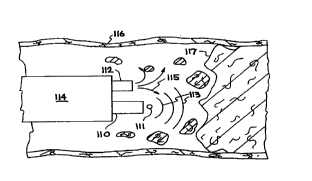
shows an optical fiber 110 with a parallel working channel 112, where both the
fiber
20 110 and the working channel 112 are both located within a catheter 114
which has
been inserted into a blood vessel 116. The distal end of fiber 110 is placed
near
thrombus 118 and/or stenotic plaque 120 within blood vessel 116. In Figure 6B,
fiber 110 delivers laser light to produce a collapsing cavitation bubble 111
and the
resulting expanding acoustic wave 113. A parallel working channel 112 in
catheter
25 114 delivers an adjunct fluid 115 to aid in the removal of occlusion 117
from inside
blood vessel 116.
As depicted in Figures 7A-C, in the thermoelastic mode, through
fiber optic 121, each laser pulse 122 delivers a controlled level of energy in
the fluid
124 which creates a large thermoelastic stress in a small volume of the fluid.
The
30 expanding direction of this stress is indicated by arrows 125 in figure 7A.
The
volume of fluid 124 which is heated by the laser pulse 122 is determined by
the
CA 02306561 2000-04-06
WO 99/16366 PCT/US98120622
11
absorption depth of the laser light in the fluid 124, and must be controlled
to
produce a desired size. For example, an appropriate size may be the fiber
diameter,
or a distance comparable to some fi~action of the vessel containing the
occlusion.
This can be adjusted by controlling the laser wavelength or the composition of
the
5 fluid such that most of the laser energy is deposited in a fluid depth of
the desired
size. The laser pulse duration is ideally short enough to deposit all of the
laser
energy into the absorbing fluid in a time scale shorter than the acoustic
transit time
across the smallest dimension of absorbing region. This is an isochoric
(constant
volume) heating process. For an absorption volume of approximately 100 pm in
10 diameter the acoustic transit time is approximately 70 ns, so the
deposition time
must be significantly less than this, e.g., around 10 ns.
The absorbing fluid responds thermoelastically to the deposition of
energy such that a region of high pressure is created in the fluid in the
heated
volume. The boundary of the high pressure zone decays into a pattern of
acoustic
1 S waves: a compression wave propagates away from the energy deposition
region
(diverging wave front) and a rarefaction wave propagates towards the center of
the
energy deposition region (converging wave front). When the rarefaction wave
converges on the center of the initial deposition region, it creates a region
126 of
tensile stress that promotes the formation of a cloud of cavitation bubbles
which
20 coalesce to form a larger bubble 130. Eventually, the cavitation bubble
collapses
(132), resulting in an expanding acoustic wave 133. Collapse and subsequent
rebound of the cavitation bubble will generate acoustic impulses in the
surrounding
fluid, which will carry off a portion of the energy of the cavity. The
collapse and
rebound processes take place on a time scale governed principally by the fluid
25 density and the maximum size of the initial cavity. The first collapse and
rebound
will be followed by subsequent collapse and rebound events of diminishing
intensity until the energy of the cavity is dissipated in the fluid.
Subsequent laser
pulses are delivered to repeat or continue this cycle and generate an
ultrasonic
radiation field at a frequency or frequencies determined by the laser pulse
30 frequency.
CA 02306561 2000-04-06
WO 99/16366 PCT/US98/20622
12
To summarize, a device operating through the first mode produces
an ultrasonic radiation field in the fluid by: (i) depositing laser energy in
a volume
of fluid comparable to the fiber dimension in a time scale of duration less
than the
acoustic transit time across this dimension (as controlled by choice of laser
5 wavelength and absorbing fluid as the case may be); (ii) controlling the
laser energy
such that the maximum size of the cavitation bubble is approximately the same
as
the fiber diameter; and (iii) pulsing the laser at a repetition rate such that
multiple
cycles of this process generate an acoustic radiation field in the surrounding
fluid;
resonant operation may be achieved by synchronizing the laser pulse repetition
rate
10 with the cavity lifetime. Typical operation ieads to a fluid-based
transducer that
cycles at 1-100 kHz with a reciprocating displacement of 100-200 pro (for
typical
optical fiber dimensions). This displacement is very similar to that found in
mechanically-activated ultrasound angioplasty devices.
In the superheated vapor expansion mode, as shown in Figures 8A-C,
15 in fiber optic 141, each laser pulse 140 delivers a controlled level of
energy in the
fluid within an absorption depth which is very small compared to the
characteristic
size of the vessel containing the catheter, or even small compared to the
fiber
diameter. The absorption depth may also be small compared to the distance that
a
sound wave travels in the duration of the laser pulse. The laser energy
deposits a
20 sufficient level of energy to heat most of the fluid within the absorption
depth well
above the vaporization temperature of the fluid at the ambient pressure: In
the
process of depositing the laser energy, a thermoelastically-generated acoustic
wave
is launched in the fluid, which propagates out from the heated region. On time
scales longer than 1 ps, the superheated fluid 142 undergoes vaporization,
which
25 creates a bubble of vapor. As the fluid vaporizes, its volume 144 increases
by a
large factor.
The laser pulse duration need not be restricted to times as short as
in the thermoelastic mode since the bubble expansion is nearly an isobaric
process;
however, the laser pulse duration should be shorter than the bubble expansion
time,
30 and it should be much shorter than a typical thermal relaxation time for
the
superheated region. (According to the Rayleigh bubble collapse theory the
bubble
CA 02306561 2000-04-06
WO 99/16366 PCT/US98/20622
13
lifetime in water is approximately 25 ps for a 50 ~m diameter bubble; thermal
relaxation occurs on a few hundred microsecond time scale, so the laser pulse
should be several microseconds or less in duration). The vapor bubble expands
up
to a maximum radius which depends on the vapor pressure initially created in
the
5 fluid and the fluid properties. At the maximum bubble radius, the vapor
pressure
in the expanded bubble has dropped to well below the ambient pressure and the
bubble 146 undergoes collapse, resulting in an expanding acoustic wave 148.
Rebound and subsequent collapse events may take place following the first
collapse.
The bubble expansion and collapse couples acoustic energy into the fluid.
10 Subsequent laser pulses are delivered to repeat or continue this cycle and
generate
an ultrasonic radiation field at a frequency or frequencies determined by the
laser
pulse frequency. Similar to the first mode, a resonant operation may be
achieved
by matching the laser pulse period to the lifetime of the vapor bubble.
To summarize, a device operating through the second mode produces
15 an ultrasonic radiation field in the fluid by: (i) depositing laser energy
in a small
volume of fluid (as controlled by choice of laser wavelength and absorbing
fluid as
the case may be); (ii) controlling the laser energy such that the maximum size
of the
vapor bubble is such that the bubble does not damage the surrounding tissues;
and
(iii) pulsing the laser energy at a repetition rate such that multiple cycles
of the
20 bubble generation and collapse process generates an acoustic radiation
field in the
surrounding fluid. Unlike the first mode, the delivery time is not a
significant issue,
so longer pulse duration lasers (up to several les) may be useful.
For either mode of operation the laser wavelength, laser pulse
duration and laser absorption depth must be precisely controlled such that an
25 adequate acoustic response is obtained with a minimum of laser pulse
energy. For
the first mode this entails matching the absorption volume to a characteristic
dimension of the system such as the fiber diameter or some fraction of the
vessel
diameter, and using a short laser pulse (less than 20 ns). For the second mode
this
entails depositing the laser energy in a very small absorption depth to
achieve a
30 sufficient level of superheat in a small fluid mass such as can be
accommodated by
CA 02306561 2000-04-06
WO 99/16366 PCT/US98/20622
14
a small energy budget and without creating a vapor bubble so large as to be
damaging to the surrounding tissues.
These opto-acoustic modes of coupling laser energy into acoustic
excitations in tissues include a number of features. Low to moderate laser
pulse
5 energy combined with high repetition rate avoids excessive tissue heating or
intense
shock generation. Localized absorption of the laser energy occurs. Laser
energy
may interact thermoelastically or thermodynamically with the ambient fluids.
An
acoustic radiation field is generated by repeated expansion and collapse of a
bubble
at the tip of the fiber. Resonant operation may be achieved by matching the
laser
10 pulse period to the lifetime of the generated bubble. Soft fibrous
.occlusions
(thrombus) may be disrupted by generating the bubbles directly within the
thrombus.
Control and/or manipulation of the spatial and temporal distribution
of energy deposited in the fluid at the fiber tip, as shown in Figure 1 and
Figure 3,
15 can be used to modify the near field acoustic radiation pattern, for
example, to
concentrate acoustic energy on an object in proximity to the fiber, or to
distribute
the acoustic radiation more uniformly. Techniques based on this strategy will
be
most successfi~I for a special case of thermoelastic response (first mode)
where the
laser pulse duration is short and the fluid absorption is also relatively
strong, such
20 that the laser energy is deposited in a thin layer adjacent to the surface
of the fiber
tip. For example, by forming a concave surface on the fiber tip, the optical
energy
is deposited in the fluid in a similar shaped distribution. Acoustic waves
emitted
from this concave distribution will tend to focus to a point at a distance R
from the
fiber tip, where R is the radius of curvature of the concave surface. A planar
fiber
25 tip will generate an initially planar acoustic wavefront in proximity to
the fiber tip.
A convex fiber tip will produce a diverging spherical wavefront which will
disperse
the acoustic energy over a larger solid angle. Another means of modifying the
near
field radiation pattern may be to use a fiber bundle through which the laser
energy
is delivered, and control the temporal distribution of deposited laser energy.
The
30 laser energy may be arranged to arnve at individual fiber strands in the
catheter tip
at different times, which, in combination with the different spatial positions
of these
CA 02306561 2000-04-06
WO 99/16366 PCT/US98/20622
15
individual strands, can be adjusted to control the directionality and shape of
the
acoustic radiation pattern, similar to phased-array techniques used in radar.
Commercial fibers are usually jacketed to protect them from the
environment. "Bare" or unjacketed fibers are available. It is helpful to use
coatings
5 on fibers to make them slide more easily through catheters. A variable
diameter
optical fiber allows for greater physical strength at the proximal end and
greater
access at the distal end. This can be accomplished through modifying existing
fibers (stripping the protective sheath from around the core) or by making
custom
fibers. Custom fabrication can be accomplished by varying the extrusion or
draw
10 rate for the fiber. Glass or plastic composition can be changed as a
function of
drawing the fiber so that greater control of the fiber from a distal end is
achieved
without sacrificing optical quality. One particular instance of this is to
treat the tip
so that it is "soft," so the end will not jam in the catheter sheath. Also,
shape
memory in the tip allows steering of the fiber when it protrudes from the
distal end
15 of the catheter sheath.
The pulsed laser energy source used by this invention can be based
on a gaseous, liquid or solid state medium. Rare earth-doped solid state
lasers, ruby
lasers, alexandrite lasers, Nd:YAG lasers and Ho:YLF lasers are all examples
of
lasers that can be operated in a pulsed mode at high repetition rate and used
in the
20 present invention. Any of these solid state lasers may incorporate non-
linear
frequency-doubling or frequency-tripling crystals to produce harmonics of the
fimdamental lasing wavelength. A solid state laser producing a coherent beam
of
ultraviolet radiation may be employed directly with the invention or used in
conjunction with a dye laser to produce an output beam which is tunable over a
25 wide portion of the ultraviolet and visible spectrum. Tunability over a
wide
sp~trum provides a broad range of flexibility for matching the laser
wavelength to
the absorption characteristics of the fluids located at the distal end of the
catheter.
The output beam is coupled by an optical fiber to the surgical site through,
for
example, a percutaneous catheter. In operation, a pulsed beam of light drives
the
30 ultrasonic excitation which removes and/or emulsifies thrombus or
atherosclerotic
CA 02306561 2000-04-06
WO 99/16366 PCT/US98/20622
16
plaque with less damage to the underlying tissue and less chance of
perforating the
blood vessel wall than prior art devices.
Various other pulsed lasers can be substituted for the disclosed laser
sources. Similarly, various dye materials and configurations can be used in
the dye
laser. Configurations other than a free-flowing dye , such as dye-impregnated
plastic films or cuvette-encased dyes, can be substituted in the dye Laser.
The dye
laser can also store a plurality of different dyes and substitute one for
another
automatically in response to user-initiated control signals or conditions
encountered
during use (e.g. when switching from a blood-filled field to a saline field or
in
10 response to calcific deposits). Suitable dyes for use in the dye laser
components of
the invention include, for example, P-terphenyl (peak wavelength 339); BiBuQ
(peak wavelength: 385); DPS (peak wavelength: 405); and Coumarin 2 (peak
wavelength: 448).
In yet another embodiment the pulsed light source may be an optical
15 parametric oscillator (OPO) pumped by a frequency-doubled or fi~equency-
tripled
solid-state laser. OPO systems allow for a wide range of wavelength tenability
in
a compact system comprised entirely of solid state optical elements. The laser
wavelength in OPO systems may also be varied automatically in response to user-
initiated control signals or conditions encountered during use.
20 Catheters, useful in practicing the present invention, can take various
forms. For example, one embodiment can consist of a catheter having an outer
diameter of 3.5 millimeters or less, preferably 2.5 millimeters or less.
Disposed
within the catheter is the optical fiber which can be a 400 micron diameter or
smaller silica (fused quartz) fiber such as the model SG 800 fiber
manufactured by
25 Spectran, Inc. of Sturbridge, Mass. The catheter may be mufti-lumen to
provide
flushing and suction ports. In one embodiment the catheter tip can be
constructed
of radio-opaque and heat resistant material. The radio-opaque tip can be used
to
locate the catheter under fluoroscopy.
Changes and modifications in the specifically described
30 embodiments can be carried out without departing from the scope of the
invention,
which is intended to be limited by the scope of the appended claims.